Review
. 2020;16(3):212-220.
doi: 10.2174/1573403X15666190819144336.

Exercise Training as a Mediator for Enhancing Coronary Collateral Circulation: A Review of the Evidence

Affiliations
Review

Exercise Training as a Mediator for Enhancing Coronary Collateral Circulation: A Review of the Evidence

Thomas Nickolay et al. Curr Cardiol Rev. 2020.

Abstract

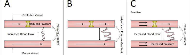
Coronary collateral vessels supply blood to areas of myocardium at risk after arterial occlusion. Flow through these channels is driven by a pressure gradient between the donor and the occluded artery. Concomitant with increased collateral flow is an increase in shear force, a potent stimulus for collateral development (arteriogenesis). Arteriogenesis is self-limiting, often ceasing prematurely when the pressure gradient is reduced by the expanding lumen of the collateral vessel. After the collateral has reached its self-limited maximal conductance, the only way to drive further increases is to re-establish the pressure gradient. During exercise, the myocardial oxygen demand is increased, subsequently increasing coronary flow. Therefore, exercise may represent a means of driving augmented arteriogenesis in patients with stable coronary artery disease. Studies investigating the ability of exercise to drive collateral development in humans are inconsistent. However, these inconsistencies may be due to the heterogeneity of assessment methods used to quantify change. This article summarises current evidence pertaining to the role of exercise in the development of coronary collaterals, highlighting areas of future research.

Keywords: Chronic total occlusion; angiogenesis; arteriogenesis; artery; collateral; coronary; shear force.


Figures

Fig. (1)
Fig. (1)
Angiography images of a patient with a Chronic Total Occlusion (CTO) of the Left Anterior Descending artery (LAD). (A): contrast injected into the left coronary artery demonstrates severe stenosis in the proximal part of the LAD, contrast flow stops abruptly at the point of complete occlusion in the mid-LAD. (B): contrast injected into the right coronary demonstrating retrograde filling of the LAD via multiple collateral channels, both septal (i) as well as epicardial (ii, iii) channels can be identified. (C): contrast injected into the left coronary artery after successful revascularisation of the LAD. Two stents have been implanted (one in the proximal LAD and a second at the site of the occlusion). (A higher resolution / colour version of this figure is available in the electronic copy of the article).
Fig. (2)
Fig. (2)
Pressure gradient of a developing collateral. Early after the development of arterial stenosis, low pressure distal to the stenosis draws flow through the collateral and increases shear stress on the endothelium (A). Collateral development reaches a plateau; collateral diameter increases, reducing collateral shear stress (B). An increase in donor artery pressure during exercise training reintroduces a pressure gradient across the collateral; fluid shear stress again increases across the collateral endothelium (C). (A higher resolution / colour version of this figure is available in the electronic copy of the article).

References

    1. Zimarino M., D’Andreamatteo M., Waksman R., Epstein S.E., De Caterina R. The dynamics of the coronary collateral circulation. Nat. Rev. Cardiol. 2014;11(4):191–197. doi: 10.1038/nrcardio.2013.207. - DOI - PubMed
    1. Werner G.S., Surber R., Ferrari M., Fritzenwanger M., Figulla H.R. The functional reserve of collaterals supplying long-term chronic total coronary occlusions in patients without prior myocardial infarction. Eur. Heart J. 2006;27(20):2406–2412. doi: 10.1093/eurheartj/ehl270. - DOI - PubMed
    1. Cui K., Lyu S., Song X., et al. Effect of coronary collaterals on prognosis in patients undergoing primary percutaneous coronary intervention for acute st-segment elevation myocardial infarction: A Meta-analysis. Angiology. 2018;69(9):803–811. doi: 10.1177/0003319718768399. - DOI - PubMed
    1. Baroldi G., Mantero O., Scomazzoni G. The collaterals of the coronary arteries in normal and pathologic hearts. Circ. Res. 1956;4(2):223–229. doi: 10.1161/01.RES.4.2.223. - DOI - PubMed
    1. Fulton W.F.M. The time factor in the enlargement of anastomoses in coronary artery disease. Scott. Med. J. 1964;9(1):18–23. doi: 10.1177/003693306400900104. - DOI - PubMed

MeSH terms