

Effect of Recombinant Activated Coagulation Factor VII on Hemorrhage Expansion Among Patients With Spot Sign-Positive Acute Intracerebral Hemorrhage: The SPOTLIGHT and STOP-IT Randomized Clinical Trials
- PMID: 31424491
- PMCID: PMC6704754
- DOI: 10.1001/jamaneurol.2019.2636
Effect of Recombinant Activated Coagulation Factor VII on Hemorrhage Expansion Among Patients With Spot Sign-Positive Acute Intracerebral Hemorrhage: The SPOTLIGHT and STOP-IT Randomized Clinical Trials
Abstract
Importance: Intracerebral hemorrhage (ICH) is a devastating stroke type that lacks effective treatments. An imaging biomarker of ICH expansion-the computed tomography (CT) angiography spot sign-may identify a subgroup that could benefit from hemostatic therapy.
Objective: To investigate whether recombinant activated coagulation factor VII (rFVIIa) reduces hemorrhage expansion among patients with spot sign-positive ICH.
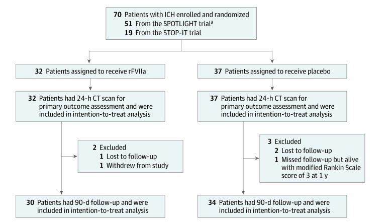
Design, setting, and participants: In parallel investigator-initiated, multicenter, double-blind, placebo-controlled randomized clinical trials in Canada ("Spot Sign" Selection of Intracerebral Hemorrhage to Guide Hemostatic Therapy [SPOTLIGHT]) and the United States (The Spot Sign for Predicting and Treating ICH Growth Study [STOP-IT]) with harmonized protocols and a preplanned individual patient-level pooled analysis, patients presenting to the emergency department with an acute primary spontaneous ICH and a spot sign on CT angiography were recruited. Data were collected from November 2010 to May 2016. Data were analyzed from November 2016 to May 2017.
Interventions: Eligible patients were randomly assigned 80 μg/kg of intravenous rFVIIa or placebo as soon as possible within 6.5 hours of stroke onset.
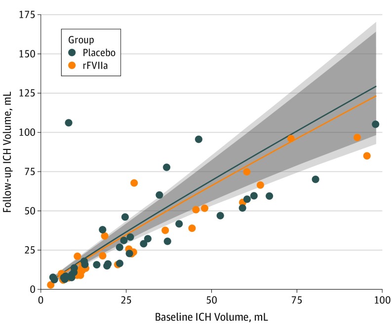
Main outcomes and measures: Head CT at 24 hours assessed parenchymal ICH volume expansion from baseline (primary outcome) and total (ie, parenchymal plus intraventricular) hemorrhage volume expansion (secondary outcome). The pooled analysis compared hemorrhage expansion between groups by analyzing 24-hour volumes in a linear regression model adjusted for baseline volumes, time from stroke onset to treatment, and trial.
Results: Of the 69 included patients, 35 (51%) were male, and the median (interquartile range [IQR]) age was 70 (59-80) years. Baseline median (IQR) ICH volumes were 16.3 (9.6-39.2) mL in the rFVIIa group and 20.4 (8.6-32.6) mL in the placebo group. Median (IQR) time from CT to treatment was 71 (57-96) minutes, and the median (IQR) time from stroke onset to treatment was 178 (138-197) minutes. The median (IQR) increase in ICH volume from baseline to 24 hours was small in both the rFVIIa group (2.5 [0-10.2] mL) and placebo group (2.6 [0-6.6] mL). After adjustment, there was no difference between groups on measures of ICH or total hemorrhage expansion. At 90 days, 9 of 30 patients in the rFVIIa group and 13 of 34 in the placebo group had died or were severely disabled (P = .60).
Conclusions and relevance: Among patients with spot sign-positive ICH treated a median of about 3 hours from stroke onset, rFVIIa did not significantly improve radiographic or clinical outcomes.
Trial registration: ClinicalTrials.gov identifier: NCT01359202 and NCT00810888.
Conflict of interest statement
Figures

Comment in
-
Is Hyperselection of Patients the Right Strategy?JAMA Neurol. 2019 Dec 1;76(12):1426-1427. doi: 10.1001/jamaneurol.2019.0213. JAMA Neurol. 2019. PMID: 31424478 No abstract available.
References
-
- Davis SM, Broderick J, Hennerici M, et al. ; Recombinant Activated Factor VII Intracerebral Hemorrhage Trial Investigators . Hematoma growth is a determinant of mortality and poor outcome after intracerebral hemorrhage. Neurology. 2006;66(8):1175-1181. doi: 10.1212/01.wnl.0000208408.98482.99 - DOI - PubMed
Publication types
MeSH terms
Substances
Associated data
Grants and funding
LinkOut - more resources
Full Text Sources
Other Literature Sources
Medical
