Case Reports
. 2019 Dec:132:104577.
doi: 10.1016/j.nbd.2019.104577. Epub 2019 Aug 16.

Factors in the disease severity of ATP1A3 mutations: Impairment, misfolding, and allele competition

Affiliations
Case Reports

Factors in the disease severity of ATP1A3 mutations: Impairment, misfolding, and allele competition

Elena Arystarkhova et al. Neurobiol Dis. 2019 Dec.

Abstract

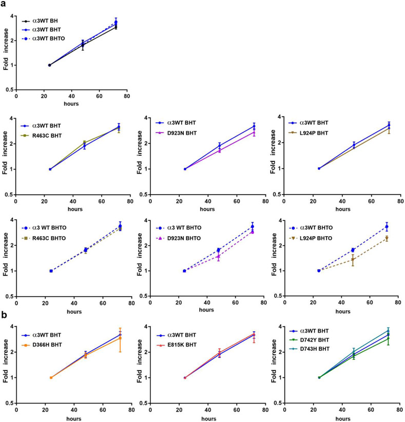
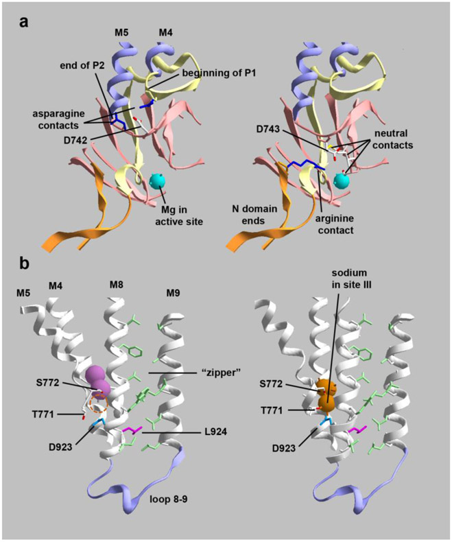
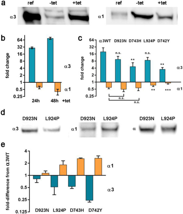
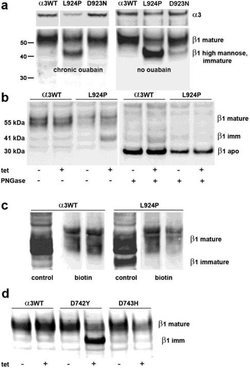
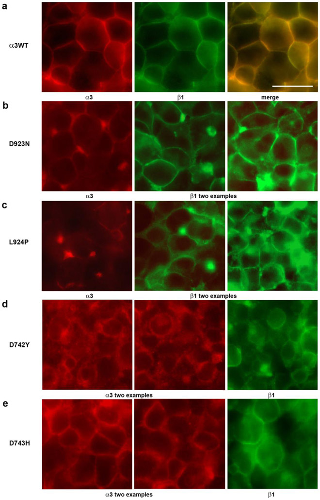
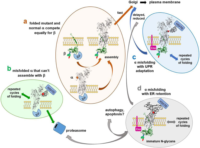
Dominant mutations of ATP1A3, a neuronal Na,K-ATPase α subunit isoform, cause neurological disorders with an exceptionally wide range of severity. Several new mutations and their phenotypes are reported here (p.Asp366His, p.Asp742Tyr, p.Asp743His, p.Leu924Pro, and a VUS, p.Arg463Cys). Mutations associated with mild or severe phenotypes [rapid-onset dystonia-parkinsonism (RDP), alternating hemiplegia of childhood (AHC), or early infantile epileptic encephalopathy (EIEE)] were expressed in HEK-293 cells. Paradoxically, the severity of human symptoms did not correlate with whether there was enough residual activity to support cell survival. We hypothesized that distinct cellular consequences may result not only from pump inactivation but also from protein misfolding. Biosynthesis was investigated in four tetracycline-inducible isogenic cell lines representing different human phenotypes. Two cell biological complications were found. First, there was impaired trafficking of αβ complex to Golgi apparatus and plasma membrane, as well as changes in cell morphology, for two mutations that produced microcephaly or regions of brain atrophy in patients. Second, there was competition between exogenous mutant ATP1A3 (α3) and endogenous ATP1A1 (α1) so that their sum was constant. This predicts that in patients, the ratio of normal to mutant ATP1A3 proteins will vary when misfolding occurs. At the two extremes, the results suggest that a heterozygous mutation that only impairs Na,K-ATPase activity will produce relatively mild disease, while one that activates the unfolded protein response could produce severe disease and may result in death of neurons independently of ion pump inactivation.

Keywords: Ataxia; Cytopathology; Dystonia; Epilepsy; Mutation validation; Neurodegeneration; Phenotype-genotype relationship.


Conflict of interest statement

Declaration of interest

E.A., T.L., F.M., P.F., C.S., J.F.C., S.D., L.J.O., and K.J.S. declare no competing interests. I.U.H. has performed research for Allergan, Boston Scientific, Great Lakes Neurotechnology, and Pfizer. His conflicts of interest are managed by Wake Forest School of Medicine. R.S-P. has support from the NIH (U01-NS094148), the Bigglesworth Family Foundation, and consults for Denali. S.B. has support from the Michael J. Fox Foundation and consults for Denali. A.B. performs research funded by NINDS, Revance, and Ipsen, and consults for Revance and Ipsen. Her conflicts of interest are managed by Wake Forest School of Medicine.

Figures

Figure 1.
Figure 1.. Growth rates of isogenic cell lines expressing wild type or mutated ATP1A3.
In all cases, cells had been maintained in the indicated mixture of drugs. BH: blasticidin and hygromycin, antibiotics used to ensure the stability of the constructs. In this condition only endogenous ATP1A1 is expressed. BHT: BH with tetracycline to induce the exogenous ATP1A3 construct. BHTO: BHT with 3 μM ouabain to inhibit the activity of endogenous ATP1A1. Cells were trypsinized and replated at time zero. (a) These four cell lines all had enough ATP1A3 activity to support life in the survival assay and were grown with and without ouabain. R463C cells had a growth rate similar to WT, but D923N and L924P cells both consistently exhibited lags in growth after plating. (b) These four cell lines died when treated with ouabain, and so they were grown without it so that endogenous α1 was active during growth. [color optional if redone with distinctive lines]
Figure 2.
Figure 2.. Cutaway images of crystal structures showing the positions and interactions of mutated residues.
(a) D742 and D743 are adjacent aspartates at a junction between protein domains. They are on an unstructured link between the second part of the P domain (P2) and transmembrane span M5. (left) D742 (colored in CPK) has polar interactions with two asparagines (dark blue), one at the beginning of P1 and one at the end of P2. Substitution with a bulky tyrosine (D742Y) may impede the folding of the two parts of the P domain. (right) D743 (also colored in CPK) points down toward the active site where Mg+2 is positioned. D743 interacts with 3 local residues in P2 and with an arginine (dark blue) located at the beginning of the N domain; D743 and R586 do not appear to form a strong salt bridge. Substitution with hydrophilic histidine may not affect protein folding but may impact the active site. (b) D923N (blue) and L924P (magenta) are adjacent residues that also point in different directions. (left) K+-bound conformation (2 pink spheres); (right) Na+-bound conformation (3 orange spheres). D923 on M8 is close to T771 and S772 on M5, residues that bind the third sodium ion. The empty sodium binding site is marked with a dashed circle in the K+ conformation. T771 and S772 produce AHC when mutated, while D923N produces RDP or AHC. D923N is well-known to have residual activity and to reduce affinity for sodium (Einholm et al., 2010; Holm et al., 2016). L924 (magenta) has a distinct structural role. While most transmembrane spans in polytopic membrane proteins intersect each other at angles, M8 and M9 are very parallel. They are held together by a zipper-like structure of opposing residues (light green). L924 (magenta) interacts with two opposing leucines in the zipper near its base where the M8-M9 hairpin establishes the position of the 8-9 loop, an important part of the structure. Substitution of L924 with a proline will disrupt the helix at that position, so it is likely to cause misfolding and may displace both D923 and the 8-9 loop. The structures are PDB codes 2ZXE (Shinoda et al., 2009) and 3WGU (Kanai et al., 2013). [color essential]
Figure 3.
Figure 3.. Competition between exogenous and endogenous Na,K-ATPase α subunits in isogenic cell lines.
(a-c) Acute addition of tetracycline produced changes in expression of α3 and α1 as estimated by western blot quantification. (a) A representative blot where ref is a reference sample of mouse brain microsomes, and −tet and +tet are lysates of α3WT-expressing cells. (b) Quantification of replicates for the response of α3WT over 24 and 48 h, expressed as fold-change. The 30- and 65- fold increase of α3 is relative to a low level of leakiness of the promoter or the endogenous ATP1A3 gene, and α1 in turn dropped by ~30 and 60%. (c) 24 h induction experiments where α3WT, D923N, D743H, L924P, and D742Y were compared (averaged from 3-5 experiments). Significance by two-tailed t-test of <0.01, **; of <0.001, ***. Error bars show standard deviations. The reduction of α1 was not significantly different for WT, D923N, and D743H. (d, e) Chronic addition of tet produced reduced α3 and increased α1 at steady state, i.e. after >5 passages in tet-containing medium. (d) A representative blot showing that the level of total α subunit was equal in D923N cells and L924P cells, as detected by a pan -specific antibody 9A7, while the two cell lines differed in the levels of α3 and α1. (e) The steady state results for the four isogenic cell lines were normalized to the levels of α3 and α1 in α3WT (= 1.0). Shown are averages of 3-5 biological replicates in chronic tetracycline. [Color optional]
Figure 4.
Figure 4.. Retention of β in endoplasmic reticulum.
(a) Cells were grown continuously in tet with or without continuous ouabain to inhibit α1. Blots were cut, and blot pieces were stained separately for α3 and β1. The expression of α3 was reduced for L924P relative to α3WT and D923N, as also seen in Fig. 2. The majority of β subunit migrated at 55 kDa in the α3WT and D923N cells, while more than half migrated at ~40 kDa in L924P, the position of β with high-mannose N-glycans in the ER. (b) On the left, L924P cells did not accumulate immature β until tet induction of mutant α3. On the right, removal of all N-glycans with PNGase F reduced both the mature and immature N-glycan forms of β to the mobility of the intact apoprotein, 30 kDa. (c) When cells were biotinylated with an impermeable reagent, only the mature form of β was recovered with streptavidin beads, not the immature N-glycan form, consistent with its residence in the ER. (d) D742Y but not D743H accumulated immature β when α3 was induced with tet. [gray scale]
Figure 5.
Figure 5.. Cytopathology in Flp-In cells expressing ATP1A3 mutations.
(a) α3WT cells, like the parent cell line, packed tightly when confluent and adopted a polygonal appearance. Both α3 and β1 stain was mainly at the surface. (b and e) Both D923N cells and D743H cells were often almost as healthy as α3WT, but also showed some blebs and mislocalization. (c) In L924P cells, stain for both subunits was more irregular, with blebs and occasional cytoplasmic localization. (d) In D742Y cells there was deterioration of the packing of cells accompanied by intracellular localization of stain and blebbing. The length bar is 20 μm. [Color essential]
Figure 6.
Figure 6.. Alternative fates of ATP1A3 mutations.
There are four ways that Na,K-ATPase mutations can navigate the biosynthetic pathway: (a) unimpaired folding and biosynthesis; (b) irremediable misfolding followed by proteasomal degradation (ERAD); (c) successful chaperone-assisted folding, which delays biosynthesis; and (d) failed chaperone-assisted folding. The chaperones illustrated (BiP/GRP-78, calnexin, and HSP70) are just representative of different chaperone classes. The factor that makes the consequences of Na,K-ATPase mutations difficult to predict is the competition that must occur between mutant and normal ATP1A3 alleles for β subunit. In (a), well-folded mutant and normal α subunits should compete equally well for β and achieve a 50:50 ratio in patient neurons. In (b), misfolded α3 incapable of binding to β will be degraded by ERAD. In this case only WT α3 subunits will succeed in reaching the plasma membrane, leading to a mild phenotype. Mutant α3 degradation may also increase the pool of β subunit available to nascent chains from the normal allele. In (c), association of mutant α3 with β will lead to delayed folding (Tokhtaeva et al., 2010) and potentially to a less-than-50:50 ratio of mutant to normal α3 at the membrane. This could lessen the effect of gain-of-toxic-function mutations. In (d), prolonged retention of β subunit in the ER could effectively sequester β subunit and lead to further reduction of Na,K-ATPase expression, protein aggregation, and the triggering of autophagy and apoptosis. Any given mutation could lie between these four extremes. Superimposed on the outcomes for (a, c, and d) will be any functional effects of mutation on the activity or kinetics of the folded and trafficked protein. [Color essential]

References

    1. Anderson TR, Huguenard JR, Prince DA, 2010. Differential effects of Na+-K+ ATPase blockade on cortical layer V neurons. Journal of Physiology 588, 4401–4414. - PMC - PubMed
    1. Anselm IA, Sweadner KJ, Gollamudi S, Ozelius LJ, Darras BT, 2009. Rapid-onset dystonia-parkinsonism in a child with a novel ATP1A3 gene mutation. Neurology 73, 400–401. - PMC - PubMed
    1. Ashmore LJ, Hrizo SL, Paul SM, Van Voorhies WA, Beitel GJ, Palladino MJ, 2009. Novel mutations affecting the Na, K ATPase alpha model complex neurological diseases and implicate the sodium pump in increased longevity. Hum.Genet 126, 431–447. - PMC - PubMed
    1. Azarias G, Kruusmagi M, Connor S, Akkuratov EE, Liu X-L, Lyons D et al., 2013. A specific and essential role for Na,K-ATPase α3 in neurons co-expressing α1 and α3. Journal of Biological Chemistry 288, 2734–2743. - PMC - PubMed
    1. Benarroch EE, 2011. Na+,K+-ATPase: Functions in the nervous system and involvement in neurologic disease. Neurology 76, 287–293. - PubMed

Publication types

MeSH terms

Substances

Supplementary concepts