

Functions and the Emerging Role of the Foetal Liver into Regenerative Medicine
- PMID: 31426422
- PMCID: PMC6721721
- DOI: 10.3390/cells8080914
Functions and the Emerging Role of the Foetal Liver into Regenerative Medicine
Abstract
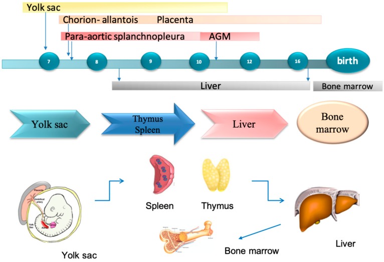
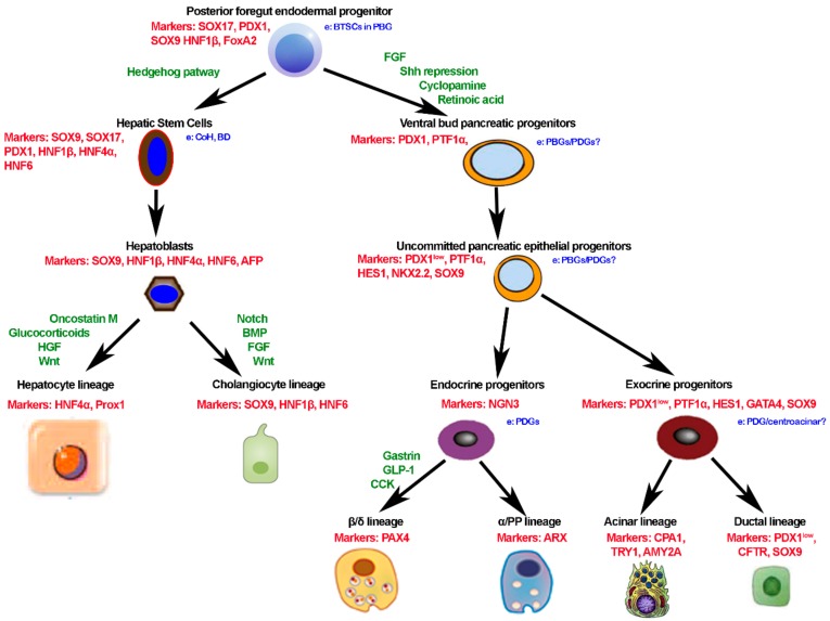
During foetal life, the liver plays the important roles of connection and transient hematopoietic function. Foetal liver cells develop in an environment called a hematopoietic stem cell niche composed of several cell types, where stem cells can proliferate and give rise to mature blood cells. Embryologically, at about the third week of gestation, the liver appears, and it grows rapidly from the fifth to 10th week under WNT/β-Catenin signaling pathway stimulation, which induces hepatic progenitor cells proliferation and differentiation into hepatocytes. Development of new strategies and identification of new cell sources should represent the main aim in liver regenerative medicine and cell therapy. Cells isolated from organs with endodermal origin, like the liver, bile ducts, and pancreas, could be preferable cell sources. Furthermore, stem cells isolated from these organs could be more susceptible to differentiate into mature liver cells after transplantation with respect to stem cells isolated from organs or tissues with a different embryological origin. The foetal liver possesses unique features given the co-existence of cells having endodermal and mesenchymal origin, and it could be highly available source candidate for regenerative medicine in both the liver and pancreas. Taking into account these advantages, the foetal liver can be the highest potential and available cell source for cell therapy regarding liver diseases and diabetes.
Keywords: foetal liver; foetal liver embryogenesis; foetal organoids; foetal stem cells; regenerative medicine.
Conflict of interest statement
The authors declare no conflict of interest.
Figures
References
-
- Lautt W.W. Morgan & Claypool Life Sciences; San Rafael, CA, USA: 2009. Hepatic Circulation: Physiology and Pathophysiology. - PubMed
-
- Wladimiroff J.W., vd Wijngaard J.A., Degani S., Noordam M.J., van Eyck J., Tonge H.M. Cerebral and umbilical arterial blood flow velocity waveforms in normal and growth-retarded pregnancies. Obstet Gynecol. 1987;69:705–709. - PubMed
Publication types
MeSH terms
LinkOut - more resources
Full Text Sources
Other Literature Sources
Medical
