Review
. 2019 Aug 18;12(16):2629.
doi: 10.3390/ma12162629.

Biomedical Application of Electroactive Polymers in Electrochemical Sensors: A Review

Affiliations
Review

Biomedical Application of Electroactive Polymers in Electrochemical Sensors: A Review

Damilola Runsewe et al. Materials (Basel). .

Abstract

Conducting polymers are of interest due to their unique behavior on exposure to electric fields, which has led to their use in flexible electronics, sensors, and biomaterials. The unique electroactive properties of conducting polymers allow them to be used to prepare biosensors that enable real time, point of care (POC) testing. Potential advantages of these devices include their low cost and low detection limit, ultimately resulting in increased access to treatment. This article presents a review of the characteristics of conducting polymer-based biosensors and the recent advances in their application in the recognition of disease biomarkers.

Keywords: biosensor; conducting polymer; electroactive polymer.


Conflict of interest statement

The authors declare no conflict of interest.

Figures

Figure 1
Figure 1
The structure of polyacetylene, showing the backbone containing conjugated double bonds.
Figure 2
Figure 2
Chemical structures of some common conducting polymers: (a) polyacetylene, (b) polyaniline (PANI), (c) polypyrrole (PPy), (d) polythiophene (PT), (e) poly(3,4-ethylenedioxythiophene) (PEDOT), and (f) poly(phenylene vinylene) (PPV).
Figure 3
Figure 3
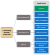
Conducting polymer properties with corresponding applications; the focus of this review is CP-based sensors, highlighted in green.
Figure 4
Figure 4
Removal of an electron from neutral PEDOT (top) results in formation of the PEDOT polaron (middle). Removal of a second electron results in the formation of the PEDOT bipolaron (bottom). These processes are reversible.
Figure 5
Figure 5
Electrochemical sensing setup.
Figure 6
Figure 6
Time dependence of the potential change for a CP/POM electrode in the presence of 0.1 mM solutions of different antioxidants (a–c) [35]. Reprinted from J. Electroanal. Chem., Vol. 828, Y. Tanaka, T. Hasegawa, T. Shimamura, H. Ukeda, and T. Ueda, “Potentiometric evaluation of antioxidant capacity using polyoxometalate-immobilized electrodes,” pp. 102–107, Copyright 2018, with permission from Elsevier.
Figure 7
Figure 7
(a) A carbazole-based CP electrode’s potentiometric response to Hg2+ ions is easily differentiated from its response to other metal ions. (b) The same CP can also be used as a colorimetric sensor for Hg2+ [36]. Reprinted from The Analyst, Vol. 142, R. Ayranci, D. O. Demirkol, S. Timur, and M. Ak, Rhodamine-based conjugated polymers: Potentiometric, colorimetric and voltammetric sensing of mercury ions in aqueous medium,” pp. 3407–3415, Copyright 2017, with permission from the Royal Society of Chemistry.
Figure 8
Figure 8
(a) Optimal electrode configuration, with PEDOT deposited on a glassy carbon electrode, coated with CNTs in chitosan, which are then coated with superoxide dismutase (SOD). (b) Example of determination of relative antioxidant capacity of wine; increasing amounts of KO2 are added to the SOD/CNT/PEDOT/GCE sensor in the absence (circles) and presence (triangles) of red wine (8 µL added to 2 mL solution of 0.1 M sodium phosphate buffer containing 0.05 M NaCl (pH 7.0) at an applied potential of −0.3 V vs. Ag/AgCl [43]. Reprinted from Sensors and Actuators B: Chemical, Vol. 236, R M. Braik, M. M. Barsan, C. Dridi, M. Ben Ali, and C. M. A. Brett, “Highly sensitive amperometric enzyme biosensor for detection of superoxide based on conducting polymer/CNT modified electrodes and superoxide dismutase,” pp. 574–582, Copyright 2016, with permission from Elsevier.
Figure 9
Figure 9
(a) Biosensing electrode with immobilized urease. (b) Interferents glucose (Glu), cholesterol (Cho), ascorbic acid (AA), uric acid (UA), and lactic acid (LA) have minimal amperometric response relative to urea [44]. Reprinted from Enzyme and Microbial Technology, Vol. 102, M. Dervisevic, E. Dervisevic, M. Senel, E. Cevik, H. B. Yildiz, and P. Camurlu, “Construction of ferrocene modified conducting polymer based amperometric urea biosensor,” pp. 53–59, Copyright 2017, with permission from Elsevier.
Figure 10
Figure 10
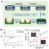
(a) Schematic illustration of sensor design. PANI is electrochemically deposited across the 20–60 nm gap between gold electrodes. Then, GOx is immobilized on the PANI surface. (b) Scanning electron micrograph of the sensor. (c) Drain current changes in response to successive 1 µL additions of 40 mM glucose. Eg = 35 mV vs. saturated calomel electrode (SCE) in 20 µL McIlvaine buffer, 0.5 M Na2SO4 at pH 5. (d) Corresponding calibration plot of drain current change vs. glucose concentration [50]. Reprinted with permission from E. S. Forzani, H. Zhang, L. A. Nagahara, I. Amlani, R. Tsui, and N. Tao, “A conducting polymer nanojunction sensor for glucose detection,” Nano Letters Vol. 4, pp. 1785–1788, Copyright 2004, American Chemical Society.
Figure 11
Figure 11
(a) Incorporation of some species, such as avidin, enables detection of other species, such as biotin, due to changes in conductivity/resistance. (b) Scanning electron micrograph of a 200-nm wide avidin-containing PPy nanowire. (c) While a PPy nanowire that does not contain avidin (A) shows no resistance change when biotin is added in 10 mM NaCl, avidin-containing PPy nanowires (B) and (C) exhibit a resistance change when biotin is added [52]. Reprinted with permission from K. Ramanathan, M. A. Bangar, M. Yun, W. Chen, N. V. Myung, and A. Mulchandani, “Bioaffinity sensing using biologically functionalized conducting-polymer nanowire,” Journal of the American Chemical Society, Vol. 127, no. 2, pp. 496–497, Copyright 2005, American Chemical Society.
Figure 12
Figure 12
(a) Linear sweep voltammograms for various concentrations of ACV in the range of (bottom to top) 0.03–10.00 μM at 100 mV s−1 in pH 7.0 phosphate buffer solution after 160 s accumulation time, and (b) the corresponding linear calibration curve of peak current vs. ACV concentration [58]. Reprinted from Materials Science and Engineering C, Vol. 53, S. Shahrokhian, M. Azimzadeh, and M. K. Amini, “Modification of glassy carbon electrode with a bilayer of multiwalled carbon nanotube/tiron-doped polypyrrole: Application to sensitive voltammetric determination of acyclovir,” pp. 134–141, Copyright 2015, with permission from Elsevier.
Figure 13
Figure 13
(a) A comparison of square wave voltammograms of 50 µM each of dopamine (DA) and serotonin (5-HT) on bare SPC (curve a), graphene-coated SPC (curve b), and CP/graphene-coated SPC (curve c) in phosphate buffer at pH 7.2 at a scan rate of 100 mV s−1 using an Ag/AgCl reference electrode. (b) Square wave voltammetry in pH 7.2 phosphate buffer with 5-HT concentration held constant at 50 µM while DA concentration was increased from 15 to 80 µM. (c) Square wave voltammetry in a pH 7.2 phosphate buffer with the DA concentration held constant at 20 µM while the 5-HT concentration was increased from 5 to 90 µM [60]. Reprinted from Sensors and Actuators B: Chemical, Vol 239, M. Raj, P. Gupta, R. N. Goyal, and Y. Shim, “Graphene/conducting polymer nano-composite loaded screen printed carbon sensor for simultaneous determination of dopamine and 5-hydroxytryptamine,” pp. 993–1002, Copyright 2017, with permission from Elsevier.
Figure 14
Figure 14
(a) SPP-PEDOT:PSS containing immobilized GOx is a selective amperometric sensor for glucose. Response to exposure to other sugars can be seen in the inset. (b) Image showing SPP-PEDOT:PSS micropatterned electrodes on the flexible fibroin substrate [70]. Reprinted from Biosensors and Bioelectronics, Vol. 81, R. K. Pal, A. A. Farghaly, C. Wang, M. M. Collinson, S. C. Kundu, and V. K. Yadavalli, “Conducting polymer-silk biocomposites for flexible and biodegradable electrochemical sensors,” pp. 294–302, Copyright 2016, with permission from Elsevier.
Figure 15
Figure 15
(a) The P3-TMA IL-1β sensor could be regenerated with some loss in activity for up to five cycles. (b) When stored dry at 4 °C, the sensor retained over 80% of its activity after 5 weeks [78]. Reprinted from Sensors and Actuators B: Chemistry, Vol. 270, E. B. Aydın, M. Aydın, and M. K. Sezgintürk, “Highly sensitive electrochemical immunosensor based on polythiophene polymer with densely populated carboxyl groups as immobilization matrix for detection of interleukin 1β in human serum and saliva,” pp. 18–27, Copyright 2018, with permission from Elsevier.
Figure 16
Figure 16
(a) A PPy/polythionine hydrogel was prepared containing gold nanoparticles and GOx as the doping agent. (b) Square wave voltammograms (SWV) showing the sensor response to NSE at concentrations from 0 ng mL−1 to 100 ng mL−1 in a 0.1M pH 6.5 phosphate buffer solution with 5 mM glucose. (c) Calibration plot with error bars, showing SWV peak current vs. logarithmic values of NSE concentrations [79]. Reprinted from Sensors and Actuators B: Chemistry, Vol. 254, H. Wang and Z. Ma, “A cascade reaction signal-amplified amperometric immunosensor platform for ultrasensitive detection of tumour marker,” pp. 642–647, Copyright 2018, with permission from Elsevier.
Figure 17
Figure 17
(a) Preparation of a microporous PANI:PSS/anti-AFP immunosensor. (b) Chronocoulometry of the reduction of 0.2 mM K3Fe(CN)6 with 0.1 M KCl revealed that the macroporous PANI:PSS (porous PANI/GCE c) electrode exhibited enhanced electroactivity relative to the thin film sensor (planar PANI/GCE b) or a bare glassy carbon electrode (GCE a). Inset shows the relationship between Q and t½, which can be used to calculate effective electrode surface area. (c) Calibration curves for the AFP detection for porous Ab/PANI/GCE a and planar Ab/PANI/GCE b reveal the enhanced sensitivity of the porous immunosensor for AFP. Insets show scanning electron micrographs of the two electrode materials [82]. Reprinted from Sensors and Actuators B: Chemistry, Vol. 255, S. Liu, Y. Ma, M. Cui, and X. Luo, “Enhanced electrochemical biosensing of alpha-fetoprotein based on three-dimensional macroporous conducting polymer polyaniline,” pp. 2568–2574, Copyright 2018, with permission from Elsevier.
Figure 18
Figure 18
Basic design of a DNA sensor based on a conducting polymer [86]. Reprinted from Biomaterials, Vol. 30, H. Peng, L. Zhang, C. Soeller, and J. Travas-Sejdic, “Conducting polymers for electrochemical DNA sensing,” pp. 2132–2148, Copyright 2009, with permission from Elsevier.
Figure 19
Figure 19
(a) DNA immobilization onto azido-PEDOT-modified gold electrodes via click chemistry. (b) Amperometric sensor response to increasing concentrations of target DNA, and (c) the corresponding calibration curve [92]. Reprinted with permission from Biosensors and Bioelectronics, “Label-free electrochemical DNA sensor using “click”-functionalized PEDOT electrodes,” T. Galán, B. Prieto-Simón, M. Alvira, R. Eritja, G. Götz, P. Bäuerle, and J. Samitier, Vol. 74, pp. 751–756, Copyright 2015, with permission from Elsevier.
Figure 20
Figure 20
Systematic evolution of ligands using exponential enrichments (SELEX) isolation strategy, applied to RNA aptamer preparation [102]. Reprinted from Analyst, Vol. 141, C. Jin, L. Qiu, J. Li, T. Fu, X. Zhang, and W. Tan, “Cancer biomarker discovery using DNA aptamers,” pp. 461–466, Copyright 2016, with permission from the Royal Society of Chemistry.
Figure 21
Figure 21
Thrombin sensors are fabricated as shown: (a) The interdigitated microelectrodes of the FET gate are treated with aminosilane. (b) Immobilization of the CPPy-NTs onto the substrate via amide linkages induced with the condensing agent 4-(4,6-dimethoxy-1,3,5-triazin-2-yl)-4-methylmorpholinium chloride (DMT-MM). (c) Thrombin aptamers are bonded to the CPPy-NTs via amide linkages. (d) SEM image of CPPy-NTs deposited onto the interdigitated microelectrode substrate. (e) Sensing ability of the CPPy-NT FET sensors. Real-time responses of CPPy nanotube FET sensors measured at a source-drain voltage of −15 mV (CPNT-1, circles) and −10 mV (CPNT-2, diamonds): current changes are apparent upon consecutive additions of a 90 nM target (thrombin, T) and nontarget (BSA, B) protein solutions added at times indicated by the arrows. No change in current occurred when the aptamer-free control CPPy-NT FET was used (squares) [104]. Reprinted from ChemBioChem, Vol. 9, H. Yoon, J. Kim, N. Lee, B. Kim, and J. Jang, “A Novel Sensor Platform Based on Aptamer-Conjugated Polypyrrole Nanotubes for Label-Free Electrochemical Protein Detection,” pp. 2634–641, Copyright 2008 Wiley-VCH Verlag GmbH & Co. KGaA, Weinheim.
Figure 22
Figure 22
(a) Differential pulse voltammetry (DPV) response of the aptasensor to a series of ATP solutions with concentrations from 10−13 to 10−6 M at a working voltage of 0.18 V vs. SCE. The current response decreases as ATP concentration increases due decreased ionic content at the electrode surface. (b) The corresponding response plot and (inset) calibration plot for the aptasensor (R2 = 0.998) [109]. Reprinted with permission from Springer Microchimica Acta, “A glassy carbon electrode modified with graphene oxide, poly(3,4-ethylenedioxythiophene), an antifouling peptide and an aptamer for ultrasensitive detection of adenosine triphosphate,” Z. Li, J. Yin, C. Gao, L. Sheng, and A. Meng, Vol. 186, Copyright 2019.
Figure 23
Figure 23
(a) Fabrication of a FET sensor. (b) Normalized current change response to varying concentrations of three forms of PDGF or (c) equal concentrations of PDGF-BB or a series of interfering agents, demonstrating high sensitivity and selectivity for PDGF-BB [112]. Reprinted with permission from Springer Journal of Materials Chemistry B, “Multidimensional hybrid conductive nanoplate- based aptasensor for platelet-derived growth factor detection,” J. S. Lee, W. Kim, S. Cho, J. Jun, K. H. Cho and J. Jang, Vol. 4, pp. 4447–4454, Copyright 2016.
Figure 24
Figure 24
(a) Design of siallyllactose-based PEDOT sensor for detection of H1N1 virus. (b) Potential changes as a function of time upon the sequential addition of H1N1 virus solutions of varying concentrations to PEDOT-only (control) or siallyllactose-granfted PEDOT. HAU—hemagglutinin units. (c) Potential changes as a function of H1N1 virus concentration. * p < 0.05 [119]. Reprinted with permission from ACS Applied Materials and Interfaces, “Specific Recognition of Human Influenza Virus with PEDOT Bearing Sialic Acid-Terminated Trisaccharides,” W. Hai, T. Goda, H. Takeuchi, S. Yamaoka, Y. Horiguchi, A. Matsumoto and Y. Miyahara, Vol. 9, pp. 14162–14170, Copyright 2017 with permission from the American Chemical Society.
Figure 25
Figure 25
(a) Preparation of conductive MIP-based biosensor for the detection of cTnT via the electropolymerization of pyrrole and carboxylated pyrrole in the presence of cTnT, followed by cTnT removal using oxalic acid. (b) Response of an MIP sensor (black dots, I) versus a non-imprinted sensor (prepared in the absence of cTnT), demonstrating enhanced sensitivity for MIP systems [120]. Reprinted with permission from Biosensors and Bioelectronics, “An ultrasensitive human cardiac troponin T graphene screen-printed electrode based on electropolymerized-molecularly imprinted conducting polymer,” B. V. M. Silva, B. A. G. Rodríguez, G. F. Sales, M. D. P. T. Sotomayor, and R. F. Dutra, Vol. 77, pp. 978–985, Copyright 2016 with permission from Elsevier.

References

    1. Huang W.S., Humphrey B.D., MacDiarmid A.G. Polyaniline, a Novel Conducting Polymer. Morphology and Chemistry of Its Oxidation and Reduction in Aqueous Electrolytes. J. Chem. Soc. Faraday Trans. 1 Phys. Chem. Condens. Phases. 1986;82:2385–2400. doi: 10.1039/f19868202385. - DOI
    1. Nambiar S., Yeow J.T.W. Conductive Polymer-Based Sensors for Biomedical Applications. Biosens. Bioelectron. 2011;26:1825–1832. doi: 10.1016/j.bios.2010.09.046. - DOI - PubMed
    1. Rivers T.J., Hudson T.W., Schmidt C.E. Synthesis of a Novel, Biodegradable Electrically Conducting Polymer for Biomedical Applications. Adv. Funct. Mater. 2002;12:33–37. doi: 10.1002/1616-3028(20020101)12:1<33::AID-ADFM33>3.0.CO;2-E. - DOI
    1. Abidian M.R., Kim D.H., Martin D.C. Conducting-Polymer Nanotubes for Controlled Drug Release. Adv. Mater. 2006;18:405–409. doi: 10.1002/adma.200501726. - DOI - PMC - PubMed
    1. Collier J.H., Camp J.P., Hudson T.W., Schmidt C.E. Synthesis and Characterization of Polypyrrole-Hyaluronic Acid Composite Biomaterials for Tissue Engineering Applications. J. Biomed. Mater. Res. 2000;50:574–584. doi: 10.1002/(SICI)1097-4636(20000615)50:4<574::AID-JBM13>3.0.CO;2-I. - DOI - PubMed

LinkOut - more resources