. 2019 Oct;18(10):2058-2077.
doi: 10.1074/mcp.RA119.001716. Epub 2019 Aug 19.

Vascular Endothelial Receptor Tyrosine Phosphatase: Identification of Novel Substrates Related to Junctions and a Ternary Complex with EPHB4 and TIE2

Affiliations

Vascular Endothelial Receptor Tyrosine Phosphatase: Identification of Novel Substrates Related to Junctions and a Ternary Complex with EPHB4 and TIE2

Hannes C A Drexler et al. Mol Cell Proteomics. 2019 Oct.

Abstract

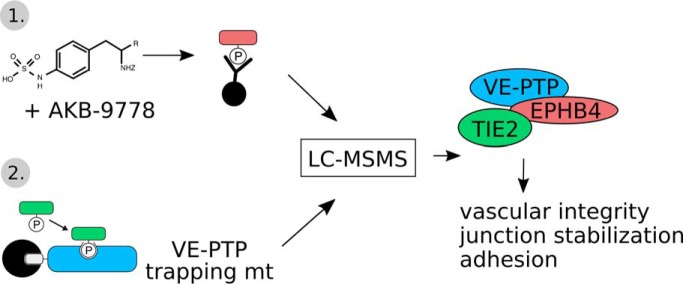
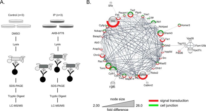
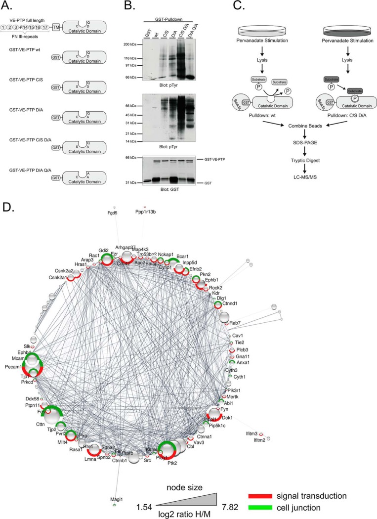
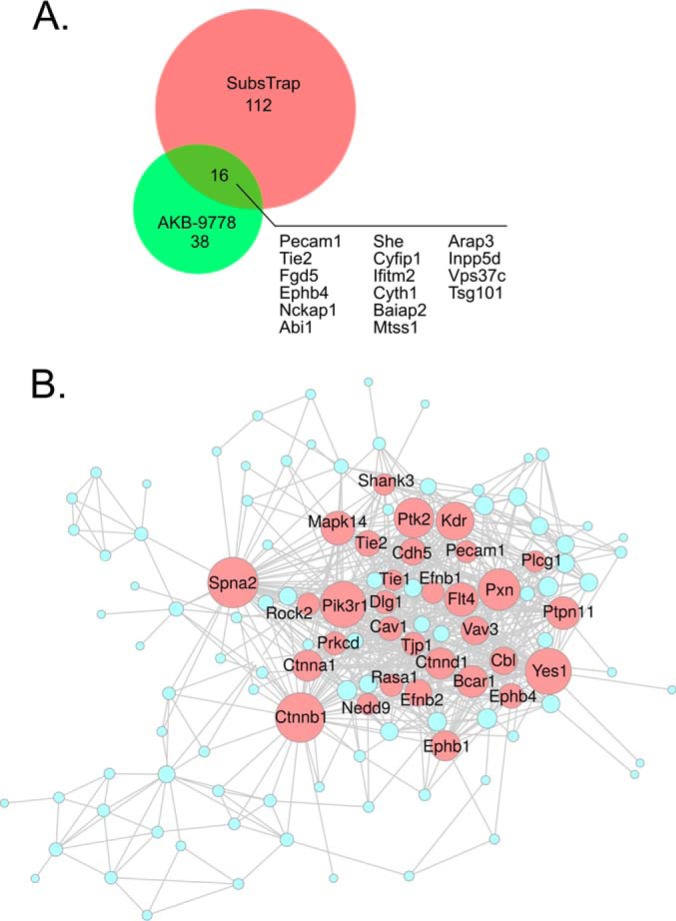
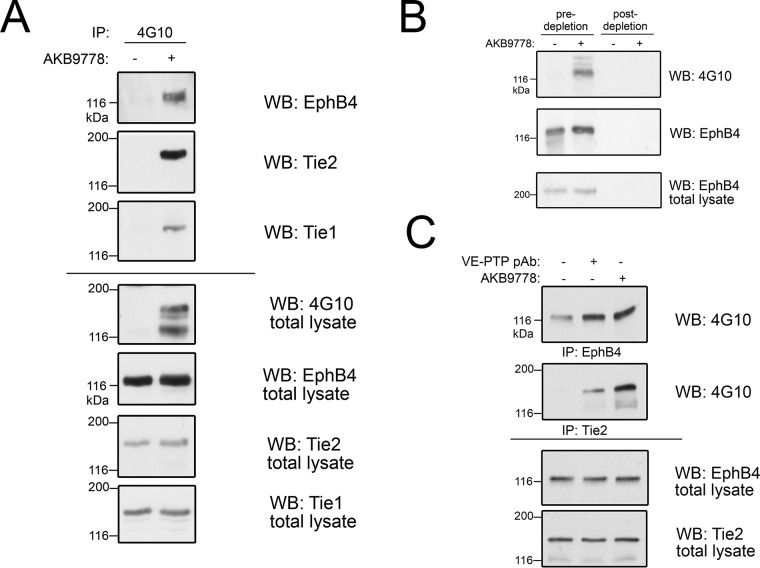
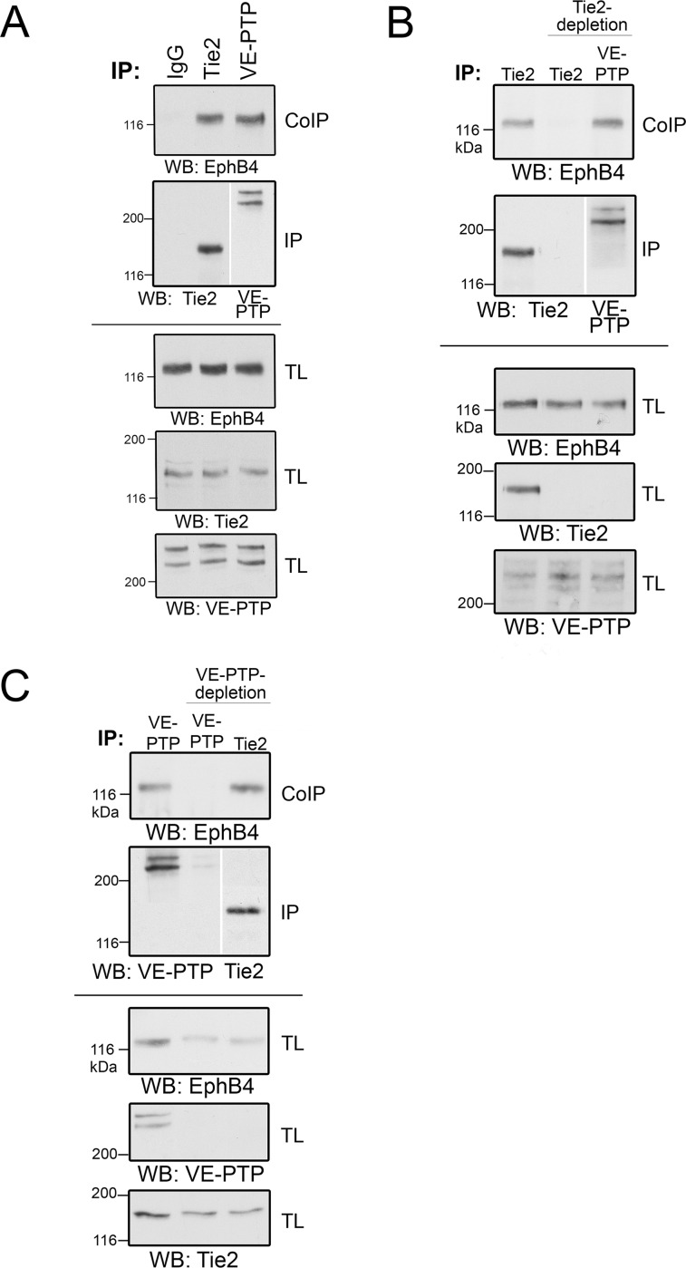
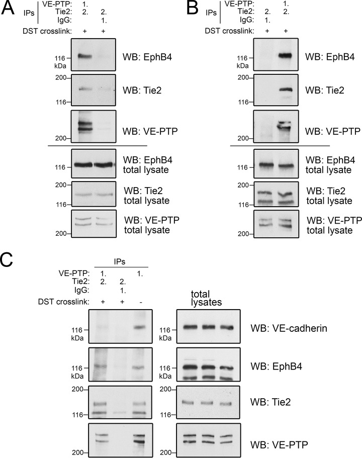
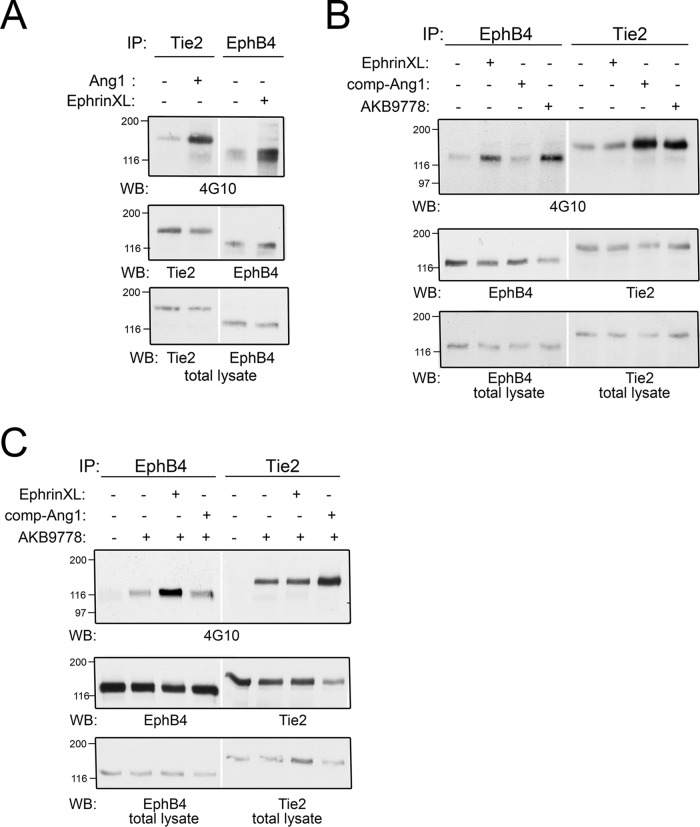
Vascular endothelial protein tyrosine phosphatase (VE-PTP, PTPRB) is a receptor type phosphatase that is crucial for the regulation of endothelial junctions and blood vessel development. We and others have shown recently that VE-PTP regulates vascular integrity by dephosphorylating substrates that are key players in endothelial junction stability, such as the angiopoietin receptor TIE2, the endothelial adherens junction protein VE-cadherin and the vascular endothelial growth factor receptor VEGFR2. Here, we have systematically searched for novel substrates of VE-PTP in endothelial cells by utilizing two approaches. First, we studied changes in the endothelial phosphoproteome on exposing cells to a highly VE-PTP-specific phosphatase inhibitor followed by affinity isolation and mass-spectrometric analysis of phosphorylated proteins by phosphotyrosine-specific antibodies. Second, we used a substrate trapping mutant of VE-PTP to pull down phosphorylated substrates in combination with SILAC-based quantitative mass spectrometry measurements. We identified a set of substrate candidates of VE-PTP, of which a remarkably large fraction (29%) is related to cell junctions. Several of those were found in both screens and displayed very high connectivity in predicted functional interaction networks. The receptor protein tyrosine kinase EPHB4 was the most prominently phosphorylated protein on VE-PTP inhibition among those VE-PTP targets that were identified by both proteomic approaches. Further analysis revealed that EPHB4 forms a ternary complex with VE-PTP and TIE2 in endothelial cells. VE-PTP controls the phosphorylation of each of these two tyrosine kinase receptors. Despite their simultaneous presence in a ternary complex, stimulating each of the receptors with their own specific ligand did not cross-activate the respective partner receptor. Our systematic approach has led to the identification of novel substrates of VE-PTP, of which many are relevant for the control of cellular junctions further promoting the importance of VE-PTP as a key player of junctional signaling.

Keywords: cell adhesion; cell-cell interactions; immunoaffinity; phosphorylation; protein complex analysis; protein phosphatases; substrate identification; substrate trapping; tyrosine kinases.


Figures

None
Graphical abstract
Fig. 1.
Fig. 1.
Identification of potential VE-PTP substrates by anti-phosphotyrosine affinity purification after VE-PTP inhibition. A, Schematic representation of label free quantification of immunoprecipitated pY-containing and associated proteins on VE-PTP inhibitor treatment. Phosphotyrosine-containing proteins in untreated or AKB-9778-treated cells were precipitated by 4G10-antibody, separated by SDS-PAGE, in gel - digested and analyzed by label free quantitative LC-MS/MS. B, Functional interaction network of significantly enriched proteins from the immunoprecipitation experiment. Only proteins that are part of the network are displayed (40 out of 54). The size of the protein nodes reflects quantitative differences in enrichment based on label free quantification of proteins. Red label: Proteins associated with GO process signal transduction; green label: proteins associated with GO compartment cell junction.
Fig. 2.
Fig. 2.
Identification of potential VE-PTP substrates by substrate trapping. A, Domain structure and catalytic domains of wildtype VE-PTP (FN III: extracellular fibronectin type III-like repeat; TM: transmembrane domain) and schematic representation of the engineered GST-VE-PTP fusion proteins. B, Mouse bEnd.5 endothelioma cells, pretreated with 1 mm pervanadate for 30 min, were subjected to substrate trapping pull downs with GST (GST only), GST-VE-PTP wt, GST-VE-PTP C/S, GST-VE-PTP D/A, GST-VE-PTP C/S D/A or GST-VE-PTP D/A Q/A, followed by immunoblotting for phospho-tyrosine (upper, middle panel) and GST (lower panel). C, Synopsis of the SILAC-based substrate trapping strategy. bEnd.3 endothelioma cells labeled with either Lys4/Arg6 (medium condition) or Lys8/Arg10 (heavy condition) were treated with 100 μm pervanadate for 30 min to achieve maximal protein-tyrosine phosphorylation. The heavy lysate was incubated with GST-VE-PTP C/S D/A double mutant and the medium lysate was incubated with GST-VE-PTP wt. Enzyme-substrate complexes were purified by GST-pulldowns, combined and analyzed by GeLC-MS/MS. D, Functional interaction network of significantly enriched proteins from the substrate trapping experiment. Only those proteins that are part of the network are displayed. VE-PTP (PTPRB) was added to the network as additional node, as well as 5 nearest neighbors. The size of the protein nodes reflects quantitative differences in enrichment. Proteins in this network associated with signal transduction are labeled in red and proteins associated with cell junction in green.
Fig. 3.
Fig. 3.
Comparison of Co-immunoprecipitations and Trapping SILAC approach. A, Size proportional Venn diagram to show the overlap of enriched proteins from both experimental approaches (SILAC substrate trapping and AKB-9778 inhibitor treatment/4G10-mediated CoIP). B, Combined functional interaction network of all significantly regulated proteins from both exerimental approaches. Node sizes are based on the degree of connectivity within the network, which allows for the visualization of likely network hubs. Highlighting arbitrarily proteins with 15 or more connections to neighboring protein nodes (red circles) reveals the impact of VE-PTP on signal transduction by transmembrane receptors at cell junctions and on elements regulating the actin cytoskeleton.
Fig. 4.
Fig. 4.
EPHB4 phosphorylation is enhanced on VE-PTP inhibition. A, bEnd.5 cells were treated with (+) or without (−) VE-PTP inhibitor AKB9778, phosphorylated proteins were then immunoprecipitated with 4G10 antibody and immunoblotted for EPHB4, TIE2 and Tie1 (3 top panels). Total cell lysates were botted with indicated antibodies (bottom four panels). B, bEnd.5 cells were treated without (−) or with (+) VE-PTP inhibitor and pY-containing proteins were immunoprecipitated either after control-depletion or after depletion of cell lysates for EPHB4 (as indicated above). Immunoprecipitates were blotted for anti pY (4G10) or for EPHB4. Bottom panel: Immunoblot of total cell lysate for EPHB4. C, HUVEC were either untreated (−) or pre-treated with polyclonal Ab against the extracellular part of VE-PTP (to induce endocytosis) or with AKB9778, followed by immunoprecipitation for either TIE2 or EPHB4 and immunoblotting with anti-pY (4G10). Bottom panels: Immunoblots of total cell lysates.
Fig. 5.
Fig. 5.
EPHB4 is a direct interactor of both VE-PTP and TIE2. A, bEnd5 cell lysates were immunopreciptated with control antibodies (IgG) or with antibodies against TIE2 or VE-PTP, followed by immunoblotting for EPHB4 (top panel) or TIE2 or VE-PTP (second panel). Bottom panels: Immunoblots of total cell lysates for the indicated antigens. B, bEnd5 cell lysates were either mock depleted (left lane) or depleted for TIE2 (two lanes on the right), followed by immunoprecipitation of either TIE2 or VE-PTP (as indicated above) and immunoblotting for EPHB4, TIE2 or VE-PTP (as indicated below). Note that EPHB4 co-precipitates with VE-PTP in the absence of TIE2. Bottom panels: Immunoblots of total lysates for the indicated antigens. C, Similar as for B, except that VE-PTP was depleted instead of TIE2. Note that EPHB4 co-precipitates with TIE2 in the absence of VE-PTP.
Fig. 6.
Fig. 6.
Biochemical characterization of the VE-PTP-TIE2-EPHB4 complex. Sequential immunoprecipitations from bEnd.5 cell lysates (A) or bEnd.3 cell lysates (B) with either first (1.) anti VE-PTP antibodies followed by (2.) anti-TIE2 antibodies after elution (left lane in A, right lane in B) or first (1.) with control antibodies (IgG), followed by (2.) anti-TIE2 antibodies (right lane in A, left lane in B). Cells have been pre-treated with cleavable DST crosslinker (+) to stabilize complexes. Immunoprecipitates were immunoblotted for the antigens indicated on the right. Bottom panels. Immunoblots of total cell lysates for the indicated antigens. C, Sequential immunoprecipitations of bEnd.5 cell lysates were done as in A. followed by immunoblotting for the indicated antigens. Cells had been either pre-treated with DST crosslinker (+), to stabilize complexes or were left untreated (−). Panels on the right depict immunoblots of total cell lysates. Note that VE-cadherin was not found associated with the ternary VE-PTP/EPHB4/TIE2 complex.
Fig. 7.
Fig. 7.
Activation of EPHB4 or TIE2 does not lead to receptor transactivation. A, bEnd5 cells were either stimulated with Angiopoietin1 (Ang1) or by ephrinB2-Fc crosslinking (EphrinXL) followed by immunoprecipitation of either TIE2 or EPHB4 and subsequent immunoblotting for the antigens indicated below. Bottom panel: immunoblot of total cell lysate for the indicated antigens. B, and C, Principally similar as in A. Note that transactivation - such as activation of TIE2 by ephrin crosslinking or activation of EPHB4 by stimulation with Ang1 - was not detectable neither in comparison (B), nor in combination (C) with VE-PTP inhibitor.

References

    1. Tiganis T., and Bennett A. M. (2007) Protein tyrosine phosphatase function: the substrate perspective. Biochem. J. 402, 1–15 - PMC - PubMed
    1. Tonks N. K. (2006) Protein tyrosine phosphatases: from genes, to function, to disease. Nat. Rev. Mol. Cell Biol. 7, 833–846 - PubMed
    1. Yao Z., Darowski K., St-Denis N., Wong V., Offensperger F., Villedieu A., Amin S., Malty R., Aoki H., Guo H., Xu Y., Iorio C., Kotlyar M., Emili A., Jurisica I., Neel B. G., Babu M., Gingras A. C., and Stagljar I. (2017) A Global Analysis of the Receptor Tyrosine Kinase-Protein Phosphatase Interactome. Mol. Cell. 65, 347–360 - PMC - PubMed
    1. Fachinger G., Deutsch U., and Risau W. (1999) Functional interaction of vascular endothelial-protein-tyrosine phosphatase with the angiopoietin receptor Tie-2. Oncogene 18, 5948–5953 - PubMed
    1. Nawroth R., Poell G., Ranft A., Samulowitz U., Fachinger G., Golding M., Shima D. T., Deutsch U., and Vestweber D. (2002) VE-PTP and VE-cadherin ectodomains interact to facilitate regulation of phosphorylation and cell contacts. EMBO J. 21, 4885–4895 - PMC - PubMed

Publication types

MeSH terms