Review
. 2020 Jan 2;12(1):a038802.
doi: 10.1101/cshperspect.a038802.

Fundamentals of Cellular Calcium Signaling: A Primer

Affiliations
Review

Fundamentals of Cellular Calcium Signaling: A Primer

Martin D Bootman et al. Cold Spring Harb Perspect Biol. .

Abstract

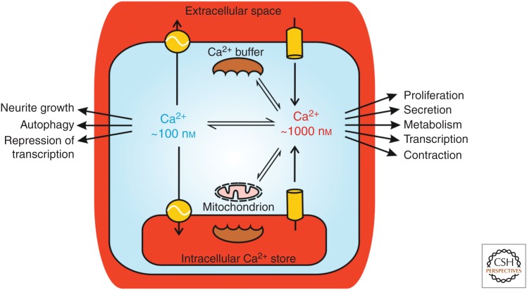
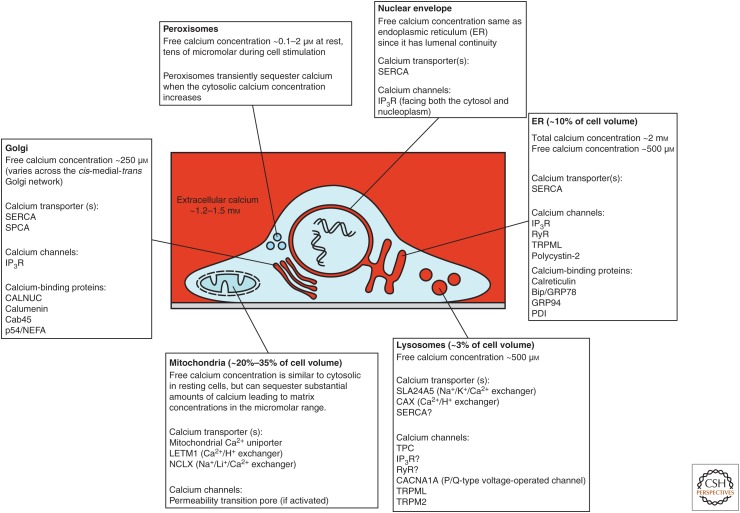
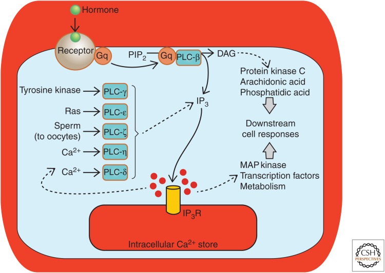
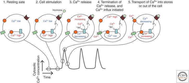
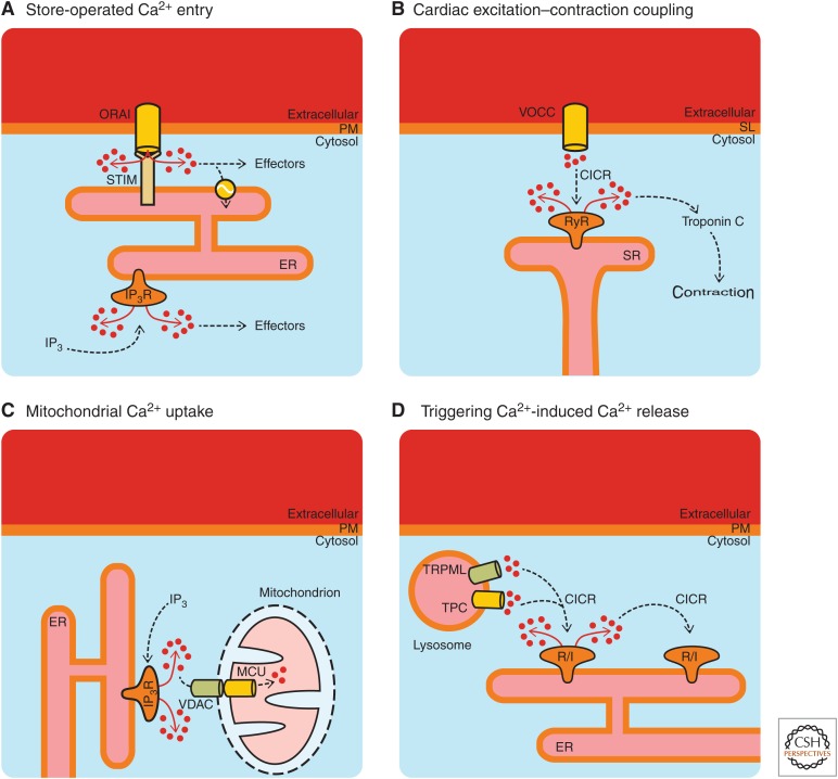
Ionized calcium (Ca2+) is the most versatile cellular messenger. All cells use Ca2+ signals to regulate their activities in response to extrinsic and intrinsic stimuli. Alterations in cellular Ca2+ signaling and/or Ca2+ homeostasis can subvert physiological processes into driving pathological outcomes. Imaging of living cells over the past decades has demonstrated that Ca2+ signals encode information in their frequency, kinetics, amplitude, and spatial extent. These parameters alter depending on the type and intensity of stimulation, and cellular context. Moreover, it is evident that different cell types produce widely varying Ca2+ signals, with properties that suit their physiological functions. This primer discusses basic principles and mechanisms underlying cellular Ca2+ signaling and Ca2+ homeostasis. Consequently, we have cited some historical articles in addition to more recent findings. A brief summary of the core features of cellular Ca2+ signaling is provided, with particular focus on Ca2+ stores and Ca2+ transport across cellular membranes, as well as mechanisms by which Ca2+ signals activate downstream effector systems.


Figures

Figure 1.
Figure 1.
Cytosolic Ca2+ signals arise via Ca2+ release from intracellular organelles and/or Ca2+ influx across the plasma membrane. During physiological Ca2+ signaling, the averaged cytosolic Ca2+ concentration typically increases from ∼100 nm to ∼1 µm, depending on the stimulus and factors such as buffering of Ca2+ by mitochondria and Ca2+-binding proteins. Cellular processes are specifically switched on or off when the cytosolic Ca2+ concentration is altered. The main intracellular Ca2+ store is the endoplasmic reticulum (ER), but also the nuclear envelope, Golgi, and acidic compartments such as lysosomes function as Ca2+ stores (see text).
Figure 2.
Figure 2.
Ca2+-sequestering organelles. Cellular Ca2+ signaling involves combinations of organelles depending on the tissue type and stimulus. Although there is considerable overlap in some of the organelles’ characteristics, they also have discrete properties that imbue each of the organelles with the ability to generate distinctive Ca2+ signals. Mitochondria and peroxisomes are not constitutive Ca2+ stores, but have the capacity to sequester Ca2+ during cytosolic Ca2+ increases. For more details, see Wang et al. (2019), Chen et al. (2019), Lloyd-Evans and Waller-Evans (2019), Wacquier et al. (2019), and Vangeel and Voets (2019). A significant Ca2+ store not shown in the figures is the sarcoplasmic reticulum (SR), which plays a critical Ca2+ signaling role in muscle cells. For details about SR function and Ca2+ homeostasis see Wang et al. (2019) and Gilbert et al. (2019). SERCA, sarcoendoplasmic reticulum Ca2+-ATPase; SPCA, Golgi/secretory pathway Ca2+ ATPase; TPC, two-pore channel; IP3R, inositol 1,4,5-trisphosphate receptor; RyR, ryanodine receptor.
Figure 3.
Figure 3.
IP3-mediated Ca2+ release is a common outcome from a number of cellular signaling processes and evokes a range of downstream outcomes. A generic pathway leading from hormone-receptor activation, activation of a heterotrimeric G-protein (Gq) and phospholipase C β (PLC-β) is depicted centrally. However, the other PLC isoforms provide alternative mechanisms for activating Ca2+ signaling via IP3Rs. (From Bootman et al. 2009; adapted, with permission, from Company of Biologists © 2009.)
Figure 4.
Figure 4.
Generation of Ca2+ oscillations via positive and negative feedback on IP3Rs. The sequence of cartoons 1–5 depict the status of cytosolic Ca2+, IP3R activity, and Ca2+ store content during the various phases of a Ca2+ oscillation. Phase 1 represents the cell at rest, before application of an extracellular stimulus. Phase 2 shows the cell subsequent to agonist–receptor interaction, when there is IP3 production but no Ca2+ release via IP3Rs. Phase 3 indicates the initial opening of IP3Rs and an increasing cytosolic Ca2+ concentration, which exerts a positive (+Ve) feedback on the IP3R, thereby promoting further Ca2+ release. At phase 4, the cytosolic Ca2+ concentration has increased to the point where it inhibits IP3R activity via negative (−Ve) feedback and hence Ca2+ release terminates. At this point, the Ca2+ store is depleted and Ca2+ influx is activated. Phase 5 indicates the recovery of the cell as Ca2+ is transported from the cytosol toward the intracellular stores and the extracellular environment and the negative feedback from cytosolic Ca2+ is relaxed.
Figure 5.
Figure 5.
Examples of Ca2+ signaling via membrane contact sites. (A)–(D) Well-known situations where the close apposition of membranes/organelles is essential for initiation or communication of Ca2+ signals. VOCC, voltage-operated Ca2+ channel; SL, sarcolemma; VDAC, voltage-dependent anion channel; MCU, mitochondrial Ca2+ uniporter; TPC, two-pore channel; TRPML, mucolipin subfamily of transient receptor potential channels; CICR, Ca2+-induced Ca2+ release; R/I, ryanodine receptor or IP3 receptor.

References

    1. Ahuja M, Young Chung W, Lin W-Y, McNally BA, Muallem S. 2019. Ca2+ signaling in exocrine cells. Cold Spring Harb Perspect Biol 10.1101/cshperspect.a035279 - DOI - PMC - PubMed
    1. Ambudkar IS, de Souza LB, Ong HL. 2017. TRPC1, Orai1, and STIM1 in SOCE: Friends in tight spaces. Cell Calcium 63: 33–39. 10.1016/j.ceca.2016.12.009 - DOI - PMC - PubMed
    1. Atakpa P, Thillaiappan NB, Mataragka S, Prole DL, Taylor CW. 2018. IP3 receptors preferentially associate with ER-lysosome contact sites and selectively deliver Ca2+ to lysosomes. Cell Rep 25: 3180–3193.e7. 10.1016/j.celrep.2018.11.064 - DOI - PMC - PubMed
    1. Barak P, Parekh AB. 2019. Signaling through Ca2+ microdomains from store-operated CRAC channels. Cold Spring Harb Perspect Biol 10.1101/cshperspect.a035097 - DOI - PMC - PubMed
    1. Berridge MJ. 1997. The AM and FM of calcium signalling. Nature 386: 759–760. 10.1038/386759a0 - DOI - PubMed