. 2019 Oct 18;87(11):e00540-19.
doi: 10.1128/IAI.00540-19. Print 2019 Nov.

Functional Characterization of an Interferon Gamma Receptor-Like Protein on Entamoeba histolytica

Affiliations

Functional Characterization of an Interferon Gamma Receptor-Like Protein on Entamoeba histolytica

Julieta Pulido-Ortega et al. Infect Immun. .

Abstract

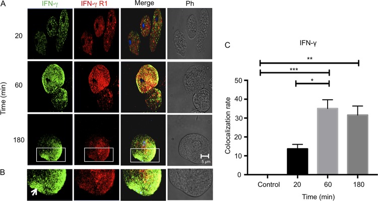
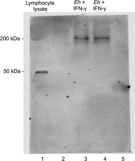
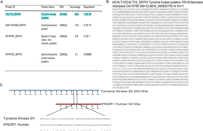
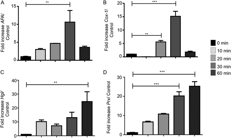
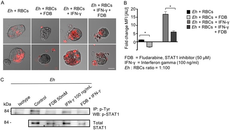
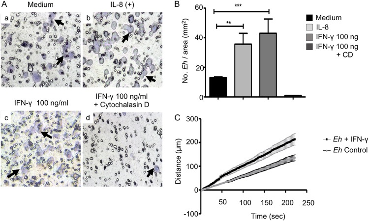
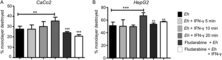
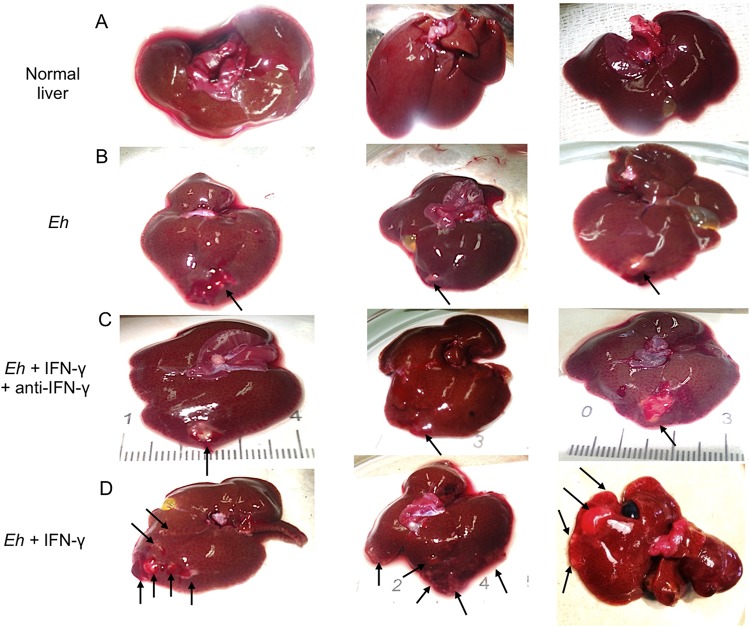
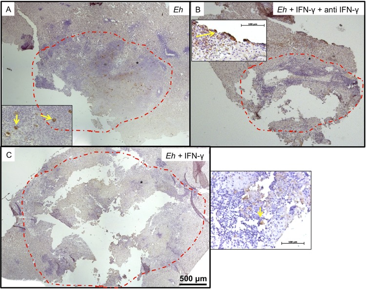
Entamoeba histolytica is an anaerobic parasitic protozoan and the causative agent of amoebiasis. E. histolytica expresses proteins that are structurally homologous to human proteins and uses them as virulence factors. We have previously shown that E. histolytica binds exogenous interferon gamma (IFN-γ) on its surface, and in this study, we explored whether exogenous IFN-γ could modulate parasite virulence. We identified an IFN-γ receptor-like protein on the surface of E. histolytica trophozoites by using anti-IFN-γ receptor 1 (IFN-γR1) antibody and performing immunofluorescence, Western blot, protein sequencing, and in silico analyses. Coupling of human IFN-γ to the IFN-γ receptor-like protein on live E. histolytica trophozoites significantly upregulated the expression of E. histolytica cysteine protease A1 (EhCP-A1), EhCP-A2, EhCP-A4, EhCP-A5, amebapore A (APA), cyclooxygenase 1 (Cox-1), Gal-lectin (Hgl), and peroxiredoxin (Prx) in a time-dependent fashion. IFN-γ signaling via the IFN-γ receptor-like protein enhanced E. histolytica's erythrophagocytosis of human red blood cells, which was abrogated by the STAT1 inhibitor fludarabine. Exogenous IFN-γ enhanced chemotaxis of E. histolytica, its killing of Caco-2 colonic and Hep G2 liver cells, and amebic liver abscess formation in hamsters. These results demonstrate that E. histolytica expresses a surface IFN-γ receptor-like protein that is functional and may play a role in disease pathogenesis and/or immune evasion.

Keywords: Entamoeba histolytica; IFN-γ; amoebiasis; cysteine proteases; cytopathic effect; erythrophagocytosis.


Figures

FIG 1
FIG 1
Localization of IFN-γ on the surface of E. histolytica trophozoites. (A) Immunodetection of the IFN-γ protein on the surface of E. histolytica trophozoites incubated with anti-human IFN-γ antibody (1:100 dilution), followed by a secondary antibody conjugated to Alexa 488 (1:1,000), imaged by confocal microscopy. Note that the intense green staining is absent in the control (without IFN-γ) at time zero. Ph contrast, phase-contrast images. (B) Western blot detection of the 17-kDa protein corresponding to IFN-γ on E. histolytica membrane (M) and cytoplasmic (C) fractions at 60 and 180 min. The 220-kDa lectin was used as an internal control to confirm membrane localization and subcellular fractionation. Scale bar represents 10 μm.
FIG 2
FIG 2
Colocalization of IFN-γ and IFN-γ receptor-like protein on E. histolytica trophozoites. E. histolytica trophozoites were incubated with IFN-γ for 20, 60, or 180 min and analyzed by confocal microscopy. After the interaction, trophozoites were incubated with a polyclonal anti-IFN-γ antibody (1:100 dilution) and a monoclonal anti-IFN-γR1 antibody (1:100 dilution), followed by Alexa Fluor 594- and Alexa Fluor 488-labeled secondary antibodies, respectively. (A) Micrographs show that IFN-γ bound to the surface of E. histolytica trophozoites; an increase in positive signal was obtained with longer exposure time. The label for IFN-γ receptor-like protein was constant after 3 h of interaction and was displaced to the uroid side of trophozoites. Increased colocalization at 1 h of interaction was observed. (B) Images illustrating morphology that demonstrate capping (arrow) on an E. histolytica trophozoite after interaction with IFN-γ for 3 h. Ph, phase-contrast images. (C) Analysis was performed according to the Costes methodology, which includes the elimination of autofluorescence with the Coloc2 plugin of Fiji software (ImageJ). The graph was made with GraphPad Prism 7. *, P < 0.05; **, P < 0.01; ***, P < 0.001.
FIG 3
FIG 3
Western blot of E. histolytica membrane fractions using anti-human IFN-γR1 antibody. E. histolytica proteins were analyzed by 12% SDS-PAGE and immunoblotting. E. histolytica trophozoites were incubated with or without 100 ng/ml IFN-γ. Note strong immunoreactivity with the anti-IFN-γR1 antibody at 200 kDa in E. histolytica membrane fractions (lanes 3 and 4) and at 50 kDa in lymphocyte lysate used as controls (lane 1). Lanes 2 and 5 are blanks.
FIG 4
FIG 4
Analysis of the anti-human IFN-γR1 antibody detected a 200-kDa amebic protein. (A) Analysis of the 200-kDa-protein band by mass spectrometry (MS) identified four E. histolytica proteins by peptide analysis. The highlighted protein at the top of the list is reported as a putative amebic surface antigen and as a putative tyrosine kinase of E. histolytica. (B) Sequence of the reported E. histolytica 200-kDa surface antigen (putative tyrosine kinase). (C) Alignment of IFN-γR1 human amino acid sequence and E. histolytica 200-kDa surface antigen (putative tyrosine kinase). The similar amino acid motifs, with 20% identity between the two proteins, are flanked by slanted lines. Ten of the amino acids in this motif are identical, and the other 20 have similar physicochemical properties.
FIG 5
FIG 5
IFN-γ upregulated cysteine protease expression in E. histolytica trophozoites. (A to D) E. histolytica trophozoites treated or not with IFN-γ for 10, 20, 30, or 60 min showed increased expression of cysteine protease genes associated with E. histolytica pathogenicity. Note maximal upregulation of EhCP-A1, EhCP-A2, EhCP-A4, and EhCP-A5 expression after 20 min of IFN-γ stimulation. *, P < 0.05; **, P < 0.01; ***, P < 0.001.
FIG 6
FIG 6
IFN-γ upregulated other virulence genes in E. histolytica in a temporal fashion. (A to D) Interaction of E. histolytica trophozoites with IFN-γ upregulated APA, Cox-1, Hgl, and Prx (peroxiredoxin), with maximal expression occurring after 30 min. *, P < 0.05; **, P < 0.01; ***, P < 0.001.
FIG 7
FIG 7
IFN-γ enhanced erythrophagocytosis in E. histolytica trophozoites. (A) Images of erythrophagocytosis by nontreated E. histolytica trophozoites (Eh) or by trophozoites in the presence of fludarabine or IFN-γ or both. Note that IFN-γ-stimulated erythrophagocytosis was substantially inhibited with the STAT1 inhibitor fludarabine. Red fluorescence shows ingested erythrocytes. Scale bar represents 10 μm. (B) Histogram showing quantification of the fluorescence of erythrocytes phagocytosed in the different assays. MFI, mean fluorescence intensity; AU, arbitrary units. *, P < 0.05. (C) Basal and IFN-γ-induced phosphorylation of STAT1 in E. histolytica is inhibited by the phospho-STAT1 inhibitor fludarabine. IP, immunoprecipitation; WB, Western blotting.
FIG 8
FIG 8
Chemotaxis of E. histolytica trophozoites toward human IFN-γ. (A) Microscopy images of E. histolytica trophozoites that were deposited in the upper chambers of Transwell units and allowed to migrate to the lower chambers in the absence (a) or presence (c) of IFN-γ or in the presence of IL-8 as a positive migration control (b) or of IFN-γ in the presence of 10 μg/ml of cytochalasin D (d). Arrows indicate E. histolytica trophozoites. (B) Numbers of E. histolytica trophozoites/mm2. Data represent values from four independent experiments. CD, cytochalasin D. (C) E. histolytica trophozoite velocities tracked in the presence or absence of IFN-γ. The graph represents migration velocities recorded by real-time video microscopy. **, P < 0.01; ***, P < 0.001.
FIG 9
FIG 9
IFN-γ enhanced E. histolytica’s cytopathic effects on Caco-2 and HepG2 cells. (A, B) Caco-2 cells (A) and HepG2 cell monolayers (B) were exposed to E. histolytica trophozoites pretreated with IFN-γ at different times with or without the STAT1 inhibitor fludarabine (50 μM). Following interaction with E. histolytica, the remaining cells were stained with methylene blue and the released color measured at 655 nm. Data represent values from 4 independent experiments. **, P < 0.01; ***, P < 0.001.
FIG 10
FIG 10
Amebic liver abscesses (ALA) 4 days postinoculation. Macroscopic development of ALA in male hamsters inoculated with E. histolytica trophozoites with or without stimulation with IFN-γ for 20 min. (A) Control uninfected livers of hamsters. (B) Single lesions (arrows) are seen in the liver lobes inoculated with E. histolytica. (C) The lesions (arrows) in the livers of hamsters treated with IFN-γ and anti-IFN-γ antibody appear similar to the ALA formed by control E. histolytica trophozoites, shown in panel B. (D) Morphology of ALA when E. histolytica trophozoites were pretreated with IFN-γ, demonstrating several granulomas distributed in the left and right lobes of the livers (arrows).
FIG 11
FIG 11
(A) Normal development of ALA. Necrotic tissue is seen in the center of the lesion (*), and the edges show areas of inflammatory infiltrate (arrowheads) that are in contact with healthy areas of the liver (+). (B) ALA formed by E. histolytica trophozoites in the presence of IFN-γ–anti-IFN antibody complex: the distribution of necrotic area and inflammatory infiltrates is similar to those of ALA induced by E. histolytica trophozoites. (C) ALA formed by E. histolytica trophozoites preincubated with IFN-γ (100 ng/ml). Note increased ALA development with multiple zones of new inflammatory infiltrate (yellow arrows) at the periphery of the lesions. The black dashed-line circles indicate the zones of ALA development. Hematoxylin-and-eosin staining was used. (D) Quantification of ALA development. One-way ANOVA and Tukey’s post hoc test were used. *, P < 0.05; **, P < 0.01.
FIG 12
FIG 12
Immunohistochemical detection of E. histolytica trophozoites in hamster ALA. (A) ALA formed by control E. histolytica trophozoites. (B) ALA formed by E. histolytica trophozoites treated with IFN-γ–anti-IFN-γ complex. (C) ALA formed by E. histolytica trophozoites treated with IFN-γ. The red dashed-line circles indicate the zones of ALA development. In insets, the yellow arrows show E. histolytica trophozoites in liver tissues. The asterisk indicates the location zone of E. histolytica trophozoites.

References

    1. Bruckner DA. 1992. Amebiasis. Clin Microbiol Rev 5:356–369. doi: 10.1128/cmr.5.4.356. - DOI - PMC - PubMed
    1. Chávez-Munguía B, Talamás-Rohana P, Castañón G, Salazar-Villatoro L, Hernández-Ramírez V, Martínez-Palomo A. 2012. Differences in cap formation between invasive Entamoeba histolytica and non-invasive Entamoeba dispar. Parasitol Res 111:215–221. doi: 10.1007/s00436-012-2820-2. - DOI - PubMed
    1. Garcia-Nieto RM, Rico-Mata R, Arias-Negrete S, Avila EE. 2008. Degradation of human secretory IgA1 and IgA2 by Entamoeba histolytica surface-associated proteolytic activity. Parasitol Int 57:417–423. doi: 10.1016/j.parint.2008.04.013. - DOI - PubMed
    1. Talamás-Rohana P, Schlie-Guzmán MA, Hernández-Ramírez VI, Rosales-Encina JL. 1995. T-cell suppression and selective in vivo activation of TH2 subpopulation by the Entamoeba histolytica 220-kilodalton lectin. Infect Immun 63:3953–3958. - PMC - PubMed
    1. Wang W, Chadee K. 1995. Entamoeba histolytica suppresses gamma interferon-induced macrophage class II major histocompatibility complex Ia molecule and I-A beta mRNA expression by a prostaglandin E2-dependent mechanism. Infect Immun 63:1089–1094. - PMC - PubMed

Publication types