. 2019 Sep 3;116(36):17659-17665.
doi: 10.1073/pnas.1902273116. Epub 2019 Aug 19.

Biomineralization by particle attachment in early animals

Affiliations

Biomineralization by particle attachment in early animals

Pupa U P A Gilbert et al. Proc Natl Acad Sci U S A. .

Abstract

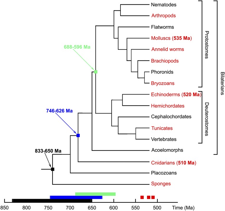
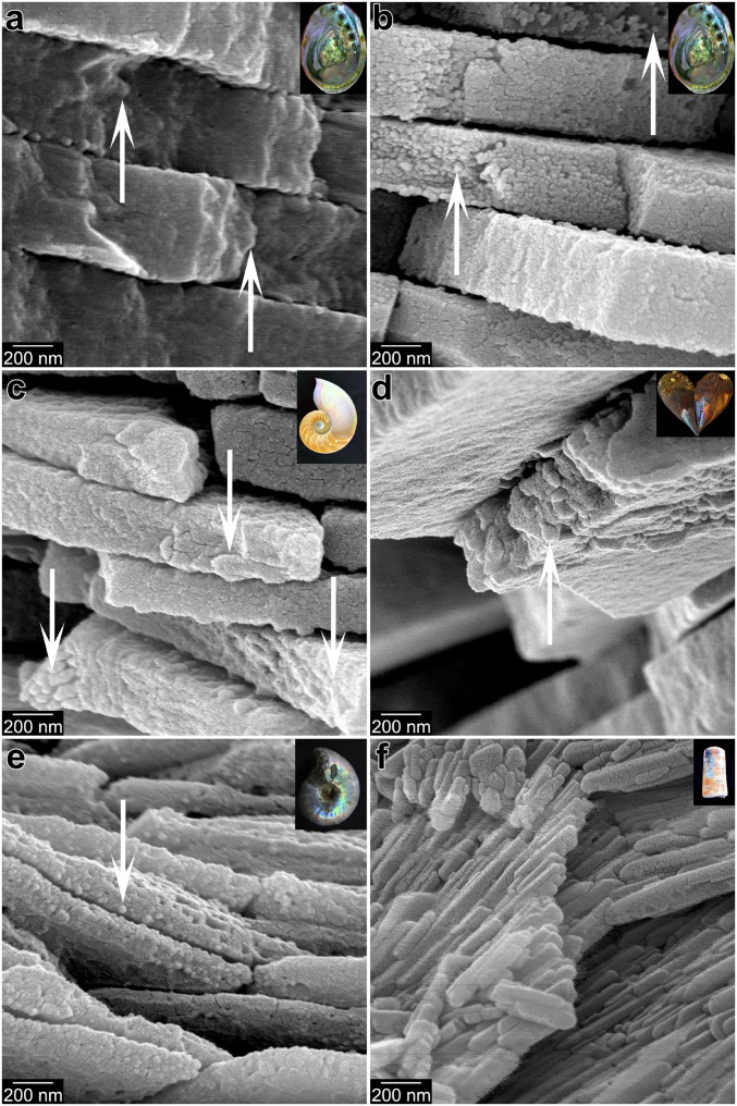
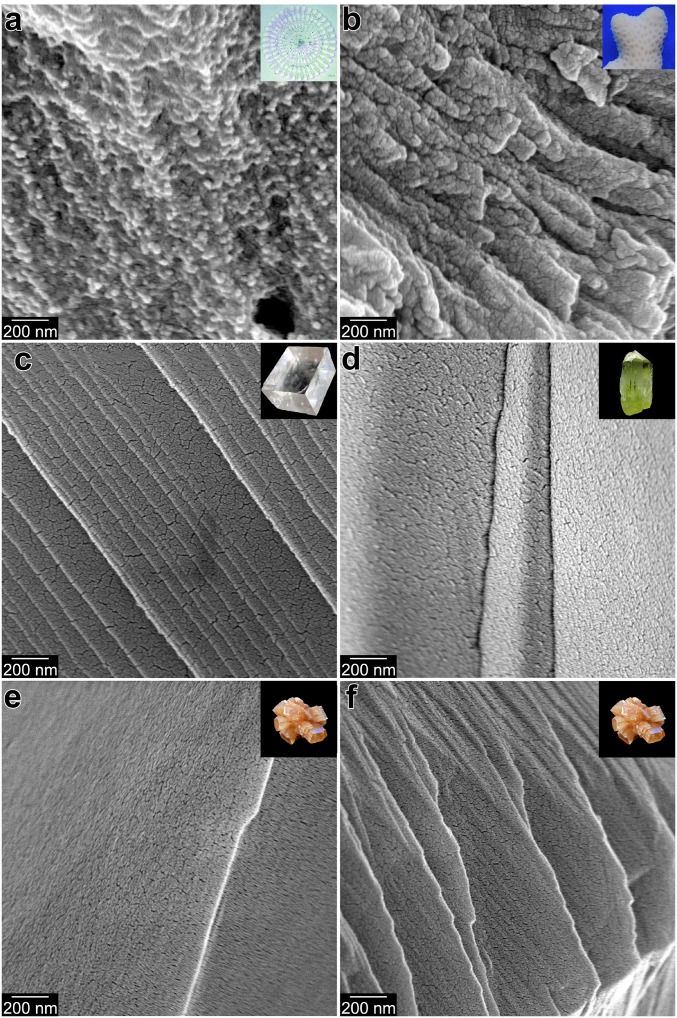
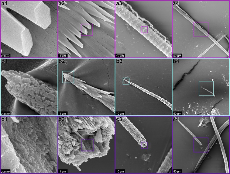
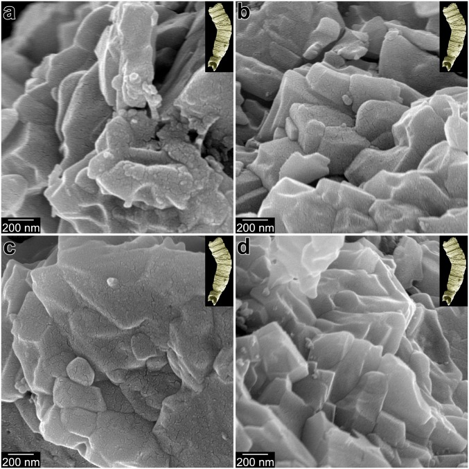
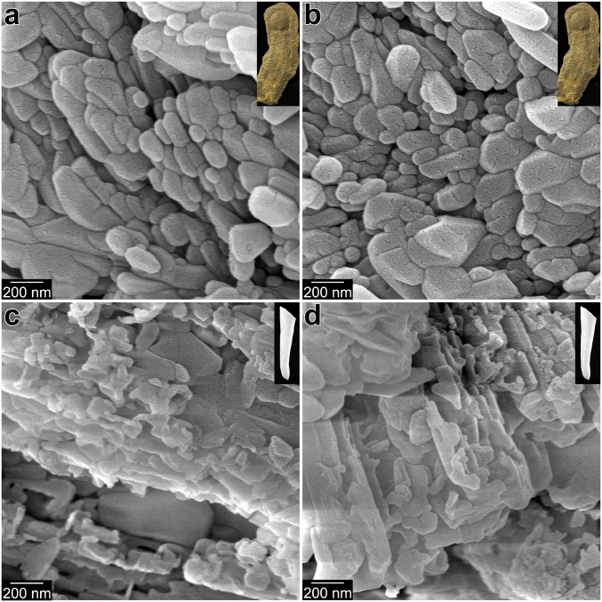
Crystallization by particle attachment (CPA) of amorphous precursors has been demonstrated in modern biomineralized skeletons across a broad phylogenetic range of animals. Precisely the same precursors, hydrated (ACC-H2O) and anhydrous calcium carbonate (ACC), have been observed spectromicroscopically in echinoderms, mollusks, and cnidarians, phyla drawn from the 3 major clades of eumetazoans. Scanning electron microscopy (SEM) here also shows evidence of CPA in tunicate chordates. This is surprising, as species in these clades have no common ancestor that formed a mineralized skeleton and appear to have evolved carbonate biomineralization independently millions of years after their late Neoproterozoic divergence. Here we correlate the occurrence of CPA from ACC precursor particles with nanoparticulate fabric and then use the latter to investigate the antiquity of the former. SEM images of early biominerals from Ediacaran and Cambrian shelly fossils show that these early calcifiers used attachment of ACC particles to form their biominerals. The convergent evolution of biomineral CPA may have been dictated by the same thermodynamics and kinetics as we observe today.

Keywords: biomineralization; calcium carbonate; particle attachment; skeleton.


Conflict of interest statement

Conflict of interest statement: P.U.P.A.G., C.-Y.S., and Tali Mass are coauthors on 2 research articles published in 2017.

Figures

Fig. 1.
Fig. 1.
A simplified phylogenetic tree of animals showing the distribution of CaCO3 biomineralization. The major clades in which at least some members form CaCO3 skeletons are highlighted in red. The phylogeny is after ref. . Dates in black, blue, or green are molecular clock estimates for early animal divergences (48, 49), and all nodes are positioned in time according to the molecular clock estimates from refs. , . Red dates in parentheses are the ages of the oldest fossils referable to the 3 principal phyla known to use ACC precursors in skeleton formation (mollusks, echinoderms, and cnidarians). The mollusk date comes from ref. , the cnidaria date is from ref. , and the echinoderm date is from ref. . Beyond any molecular clock uncertainties (48), the 3 phyla started biomineralizing long after diverging from one another.
Fig. 2.
Fig. 2.
Modern and fossil nacre from 3 molluscan classes, exhibiting irregularly shaped nanoparticles. Arrows indicate fractured tablets. (A) Cryofractured modern nacre from the gastropod Haliotis rufescens. (B) Nacre as in A, but bleached and etched to better reveal its nanoparticulate texture. (C) Nacre from the modern cephalopod Nautilus pompilius, cryofractured, bleached, etched. (D) Nacre from the Miocene (∼13 Ma) bivalve Atrina harrisii nacre, which is still 100% aragonite (28). (E) Nacre from a Cretaceous (∼100 Ma) ammonite Desmoceras sp. (F) Nacre from the Upper Ordovician (∼450 Ma) cephalopod Isorthoceras sociale nacre, which is secondarily phosphatized. Lower-magnification, cocentered images from the same locations as these are presented in SI Appendix, Fig. S1, where they are correspondingly labeled. Insets show photographs of similar samples.
Fig. 3.
Fig. 3.
Modern biominerals and nonbiogenic mineral crystals, all cryofractured. The biominerals exhibit irregularly shaped nanoparticulate fracture figures, whereas the nonbiogenic minerals show smooth, flat fracture figures. (A) Modern Strongylocentrotus purpuratus sea urchin spine, cryofractured and etched to reveal its nanoparticulate texture. (B) Modern Stylophora pistillata coral skeleton. (C) Nonbiogenic calcite. (D) Nonbiogenic apatite. Nonbiogenic aragonite, with typical featureless appearance (E) and rare feature-rich surface (F). The cracked appearance on all smooth, flat surfaces in CF originates from the 20-nm Pt coating. Lower-magnification, cocentered, colabeled images from these locations are presented in SI Appendix, Fig. S2. Insets show photographs of similar samples.
Fig. 4.
Fig. 4.
Modern vaterite spicule from the ascidian tunicate Herdmania momus. (A) Hexagonal pyramidal outer spines, exhibiting a smooth cryofracture figure. (B) The forming end of another vaterite spicule, in which both the outer spines and the inner core appear nanoparticulate. (C) The inner core of a third vaterite spicule, exhibiting a nanoparticulate cryofracture figure. Boxes in panels 2, 3, 4 indicate the area magnified in panels 1, 2, 3.
Fig. 5.
Fig. 5.
Irregularly shaped nanoparticles in the fractured walls of phosphatized Ediacaran (∼550 Ma) Cloudina skeletons. Lower-magnification, cocentered, colabeled images from these locations are presented in SI Appendix, Fig. S5.
Fig. 6.
Fig. 6.
Cambrian phosphatized small shelly fossils after fracturing. (A and B) Halkieriid sclerite. (C and D) Hyolith conch. All 4 cross-sections show irregularly shaped, noneuhedral nanoparticles. Lower-magnification, cocentered, colabeled images from these locations and others are presented in SI Appendix, Fig. S7. Additional data in SI Appendix, Fig. S8.

References

    1. De Yoreo J. J., et al. , Crystallization by particle attachment in synthetic, biogenic, and geologic environments. Science 349, aaa6760 (2015). - PubMed
    1. Penn R. L., Banfield J. F., Oriented attachment and growth, twinning, polytypism, and formation of metastable phases: Insights from nanocrystalline TiO2. Am. Mineral. 83, 1077–1082 (1998).
    1. Banfield J. F., Welch S. A., Zhang H., Ebert T. T., Penn R. L., Aggregation-based crystal growth and microstructure development in natural iron oxyhydroxide biomineralization products. Science 289, 751–754 (2000). - PubMed
    1. Niederberger M., Cölfen H., Oriented attachment and mesocrystals: Non-classical crystallization mechanisms based on nanoparticle assembly. Phys. Chem. Chem. Phys. 8, 3271–3287 (2006). - PubMed
    1. Killian C. E., et al. , Mechanism of calcite co-orientation in the sea urchin tooth. J. Am. Chem. Soc. 131, 18404–18409 (2009). - PubMed

Publication types

Substances