. 2019 Nov;3(11):870-879.
doi: 10.1038/s41551-019-0437-9. Epub 2019 Aug 19.

Computationally guided personalized targeted ablation of persistent atrial fibrillation

Affiliations

Computationally guided personalized targeted ablation of persistent atrial fibrillation

Patrick M Boyle et al. Nat Biomed Eng. 2019 Nov.

Abstract

Atrial fibrillation (AF)-the most common arrhythmia-significantly increases the risk of stroke and heart failure. Although catheter ablation can restore normal heart rhythms, patients with persistent AF who develop atrial fibrosis often undergo multiple failed ablations, and thus increased procedural risks. Here, we present personalized computational modelling for the reliable predetermination of ablation targets, which are then used to guide the ablation procedure in patients with persistent AF and atrial fibrosis. First, we show that a computational model of the atria of patients identifies fibrotic tissue that, if ablated, will not sustain AF. Then, we report the results of integrating the target ablation sites in a clinical mapping system and testing its feasibility in ten patients with persistent AF. The computational prediction of ablation targets avoids lengthy electrical mapping and could improve the accuracy and efficacy of targeted AF ablation in patients while eliminating the need for repeat procedures.


Figures

Fig. 1 ∣
Fig. 1 ∣. OPTIMA Approach Flowchart.
Top row presents an overview of the approach. Late gadolinium enhancement magnetic resonance imaging (LGE-MRI) scans for each persistent atrial fibrillation (AF) patient are processed to reconstruct a personalized 3D model of the patient’s fibrotic atria. Personalized simulations are then conducted to determine all the arrhythmias that could arise in the fibrotic substrate, and the custom-tailored OPTIMA ablation targets are determined. In the final panel of this row, tact indicates activation time. Bottom row presents the detailed steps undertaken in finding the OPTIMA ablation targets based on the arrhythmias that arise in the atrial fibrotic substrate following rapid pacing from 40 uniformly-distributed bi-atrial sites. From the analysis of the pacing-induced arrhythmias, an initial set of lesions is determined, consisting of locations of the persistent reentrant drivers and ablation lines connecting these sites to the closest non-conductive anatomical barriers (pulmonary vein ostia, superior/inferior vena cava openings, or mitral/tricuspid valve annuli). Virtual ablation of these targets is next performed, where the lesions are rendered non-conductive in the model. The rapid pacing protocol is then repeated to determine whether new arrhythmias arise in the post-ablation substrate. The latter two steps are repeated until AF can no longer be induced in the patient’s atrial model. Finally, the personalized ablation treatment plan is exported to a format compatible with the CARTO™ system.
Fig. 2 ∣
Fig. 2 ∣. Schematic summarizing the process of importing OPTIMA ablation targets into the clinical electroanatomic navigation system (i.e., CARTO™).
The starting point for this process is the set of outputs from the OPTIMA approach (i.e., ablation targets on bi-atrial volumetric mesh reconstructed from LGE-MRI). Red regions indicate ablation targets from analysis; yellow regions indicate connecting lines. Targets located in the LA are mapped via co-registration onto a geometric reconstruction of the LA endocardial surface extracted from the same patient’s MRA scan, which includes anatomical features needed for peri-procedural registration (e.g., prominent ridge between left PVs and lateral LA). Notably, the LA geometry from MRA is a surface mesh (i.e., a 2D manifold in 3D space) and not a volumetric mesh. Since RA segmentation from the MRA scan is not possible, we instead derive a surface mesh of the RA endocardium, including any ablation targets, from the LGE-MRI-based model. The RA surface mesh with ablation targets is then down-sampled since high-resolution meshes cannot be loaded into the clinical mapping system. Finally, the LA-MRA and down-sampled RA surface meshes with the OPTIMA ablation plan are aligned and scale-matched via affine transformation, resulting in a geometric model ready to be converted into a Visualization Toolkit (VTK) file suitable for importing into the CARTO™ system. PV: pulmonary vein; MRA: magnetic resonance angiography; LA/RA: left/right atria; LGE-MRI: late gadolinium enhancement magnetic resonance imaging.
Fig. 3 ∣
Fig. 3 ∣
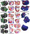
Examples of the process to determine the OPTIMA ablation targets for three patients. Patient 5, is a 68-year-old man with one prior failed ablation (see also Movie S2). Patient 7 is a 49-year-old man with one prior failed ablation (see also Movie S3). Patient 9 is a 72-year-old man with two prior failed ablations (see also Movie S4). a, Posterior (top) and anterior (bottom) views of the three patient-specific atrial models as reconstructed from segmented LGE-MRI scans, including the distribution of fibrotic tissue. b, Two examples per model of arrhythmia activation sequences induced by the rapid pacing protocol, and the corresponding persistent reentrant drivers (RDs, pink) obtained from the analysis of the induced arrhythmias. For patient 9, no RD-perpetuated AF was observed following the initial pacing protocol. However, peri-mitral macroreentrant tachycardia was found, and transient episodes of non-sustained reentry were documented in two locations on the posterior left atrium. All activation maps share the same time scale except the lower panel activation map for Patient 7; tact indicates activation time. c, Two examples per model of activation sequences associated with arrhythmia emerging anew in the models following virtual ablation (lesions shown in orange), and the corresponding emergent RDs. For patient 9, following virtual ablation to render the initiation of peri-mitral reentry impossible, stable emergent RDs were observed in the same two locations where non-sustained reentries were observed following the initial rapid pacing. D, Custom-tailored OPTIMA ablation treatment plans, including targets corresponding to all RDs (pre-ablation and emergent) and lesion lines connecting the latter to non-conductive tissue boundaries. LA/RA: left/right atria; LAA/RAA: left/right atrial appendage.
Fig. 4 ∣
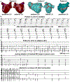
Fig. 4 ∣. Data from the OPTIMA-driven ablation procedures in the three patients from Fig.2.
a, Sites of ablation delivery (catheter tip locations marked by red dots) in the left atrium (LA) as rendered in CARTO intracardiac mapping system at the end of the clinical ablation procedure in three patients. Dashed ellipses indicate locations ablated based on locations of persistent RDs as identified by OPTIMA. As patient 7 also had targets in the right atrium (RA), Fig. S3 shows the annotated RA CARTO map from the same procedure. b, Bipolar electrogram recordings (five per panel) from a decapolar catheter placed in the coronary sinus during the procedure; recordings are from proximal (top) to distal (bottom) leads. Intracardiac electrograms recorded during the ablation procedure in patient 5. Ablation of the marked anterior LA reentrant driver (RD) target (asterisk in a, patient 5) resulted in a transient change from atrial fibrillation (AF) (top row of five electrograms) to an organized atrial tachycardia/flutter (AT/Afl) (bottom row of five electrograms). c, Intracardiac electrograms for patient 9. The top panel shows 5 electrograms for patient 9 prior to ablation (AF). Stable induced AF (top row of the middle panel) terminated abruptly (middle row of the middle panel) upon ablation of the marked posterior LA target (** in Figure 4a, patient 9) and AF could not be re-induced despite aggressive rapid pacing (bottom row of the bottom panel).

References

    1. Andrade J, Khairy P, Dobrev D & Nattel S The clinical profile and pathophysiology of atrial fibrillation: relationships among clinical features, epidemiology, and mechanisms. Circ Res 114, 1453–1468 (2014). - PubMed
    1. Go AS et al. Prevalence of diagnosed atrial fibrillation in adults: national implications for rhythm management and stroke prevention: the AnTicoagulation and Risk Factors in Atrial Fibrillation (ATRIA) Study. JAMA 285, 2370–2375 (2001). - PubMed
    1. Dorian P et al. The impairment of health-related quality of life in patients with intermittent atrial fibrillation: implications for the assessment of investigational therapy. J Am Coll Cardiol 36, 1303–1309 (2000). - PubMed
    1. Kalantarian S, Stern TA, Mansour M & Ruskin JN Cognitive impairment associated with atrial fibrillation: a meta-analysis. Ann Intern Med 158, 338–346 (2013). - PMC - PubMed
    1. Stewart S, Murphy NF, Walker A, McGuire A & McMurray JJ Cost of an emerging epidemic: an economic analysis of atrial fibrillation in the UK. Heart 90, 286–292 (2004). - PMC - PubMed

Publication types