Review
. 2019 Sep;15(9):853-864.
doi: 10.1038/s41589-019-0350-2. Epub 2019 Aug 19.

Emerging structural insights into glycosyltransferase-mediated synthesis of glycans

Affiliations
Review

Emerging structural insights into glycosyltransferase-mediated synthesis of glycans

Kelley W Moremen et al. Nat Chem Biol. 2019 Sep.

Abstract

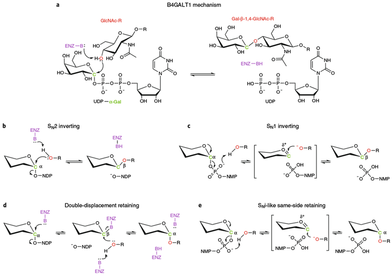
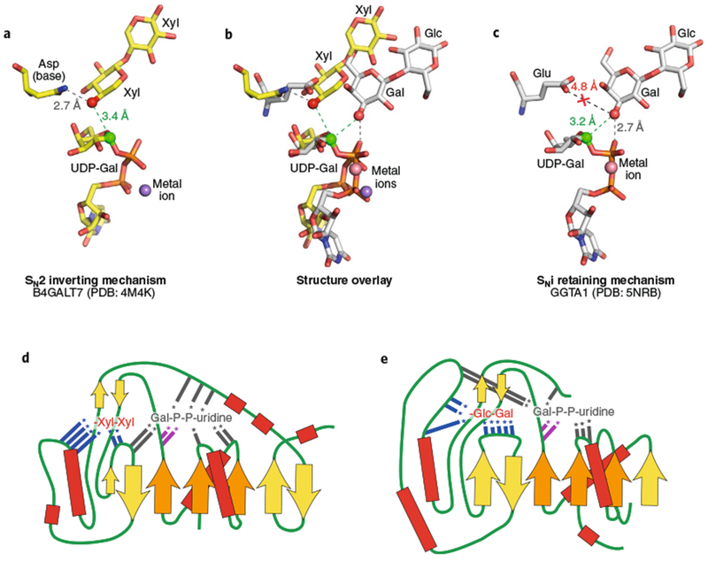
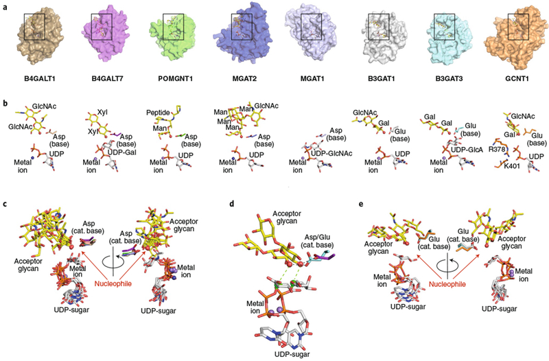
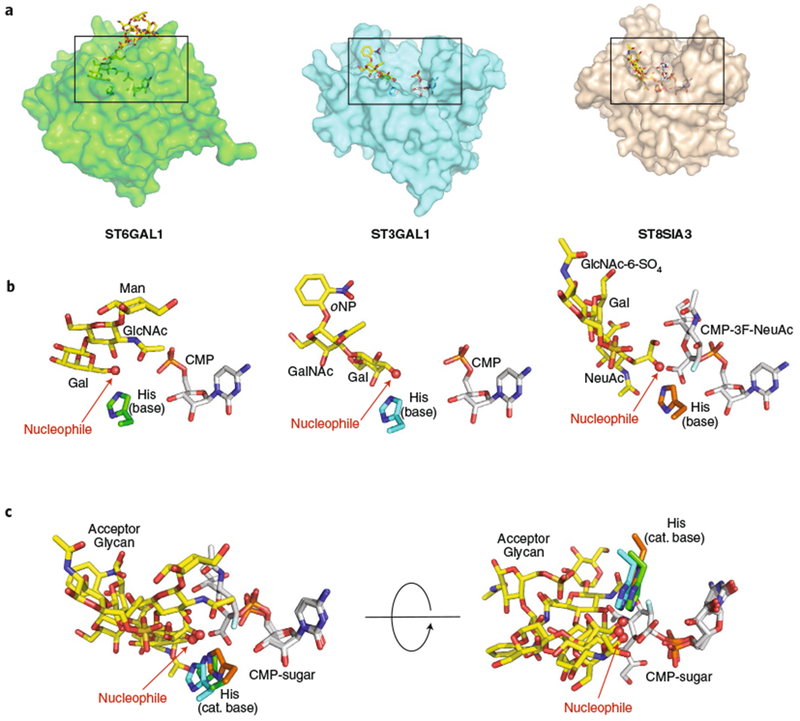
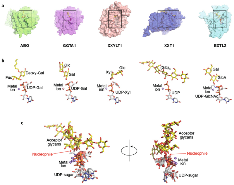
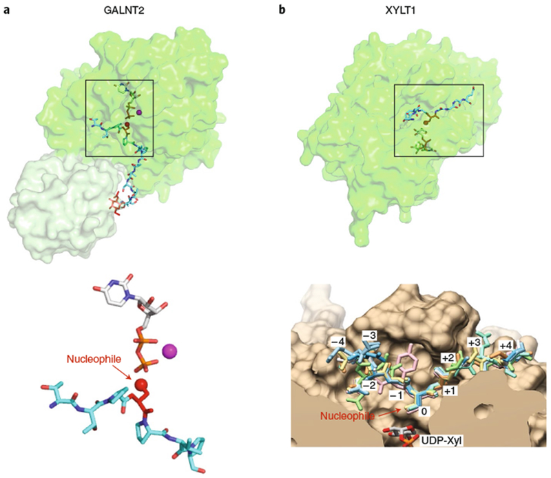
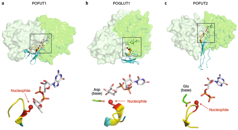
Glycans linked to proteins and lipids play key roles in biology; thus, accurate replication of cellular glycans is crucial for maintaining function following cell division. The fact that glycans are not copied from genomic templates suggests that fidelity is provided by the catalytic templates of glycosyltransferases that accurately add sugars to specific locations on growing oligosaccharides. To form new glycosidic bonds, glycosyltransferases bind acceptor substrates and orient a specific hydroxyl group, frequently one of many, for attack of the donor sugar anomeric carbon. Several recent crystal structures of glycosyltransferases with bound acceptor substrates reveal that these enzymes have common core structures that function as scaffolds upon which variable loops are inserted to confer substrate specificity and correctly orient the nucleophilic hydroxyl group. The varied approaches for acceptor binding site assembly suggest an ongoing evolution of these loop regions provides templates for assembly of the diverse glycan structures observed in biology.


Figures

Fig. 1 |
Fig. 1 |. GT reaction mechanisms.
a, B4GALT1 reaction. b, SN2 inverting mechanism. c, SN1 inverting mechanism. d, Double-displacement retaining mechanism. e, SNi-like same-side retaining mechanism. All mechanisms in b-e are drawn using a generic hexose in α-linkage to a nucleoside diphosphate. The β-phosphate is drawn out when it participates in the reaction (panels c and e). Enz-B and Enz-BH (purple) indicate the catalytic base that deprotonates the acceptor nucleophile. NMP and NDP are nucleoside monophosphate and nucleoside diphosphate, respectively. Red O indicates nucleophilic oxygen of acceptor substrate. Green C indicates anomeric carbon of donor sugar. R indicates acceptor substrate. The linkage of anomeric carbon in the hexose is indicated in both substrates and products.
Fig. 2 |
Fig. 2 |. Comparison of representative inverting and retaining catalytic mechanisms and enzyme topologies for substrate interactions.
Bound ligands for B4GALT7 (a, PDB: 4M4K) and GGTA1 (c, PDB: 5NRD) are displayed along with putative catalytic bases. a, The inverting SN2 catalytic mechanism for B4GALT7 is catalyzed by deprotonation of the Xyl acceptor hydroxyl (red sphere) by the catalytic base (Asp211 in the wild type enzyme mutated to Asn211 in PDB ID 4M4K) and attack of the anomeric carbon (green sphere) in line with the departing nucleotide leaving group. c, GGTA1 has a retaining SNi-like mechanism in which the nucleophile position (red sphere) is shifted relative to the inverting enzymes. The equivalent catalytic base residue (Glu317) is too far away for deprotonation, but the acceptor Gal hydroxyl (red sphere) is positioned adjacent to the β-phosphate oxygen for deprotonation (gray dotted line) and in proximity to attack the anomeric carbon (green sphere). b, Overlay of the two structures based on the Gal C1-phosphate oxygen bond illustrates the differences in geometry of nucleophilic attack on the anomeric carbons. d,e, Donor and acceptor binding sites for B4GALT7 (d) and GGTA1 (e) are displayed as topology diagrams illustrating the positions of residues that interact with donors and acceptors in loops extending from the core β-sheets of the Rossmann fold. β-Sheets are indicated as filled arrows (orange for conserved Rossmann-fold elements and yellow for nonconserved sheets among GT-A fold enzymes) and helices as red boxes connected by loop regions (green lines). Positions of interacting residues are indicated by colored lines with asterisks (gray, interacting with donors; blue, interacting with acceptors; magenta, DxD motif). Acceptors for each enzyme (B4GALT7: Xyl-β-1,4-Xyl and GGTA1: Gal-β-1,4-Glc) are indicated in abbreviated form, but with inverted orientation from standard nomenclature to indicate the proximity of the terminal sugar acting as nucleophile in the glycosyltransfer reaction.
Fig. 3 |
Fig. 3 |. Structural gallery of eukaryotic GT-A fold inverting enzymes.
a, Eight eukaryotic GT-A fold inverting enzyme structures were aligned using Coot (transparent surface) with bound donor analogs (white sticks), acceptor analogs (yellow sticks), divalent cations (spheres), and catalytic bases (sticks). Structures include B4GALT1 (PDB: 1TW5), B4GALT7 (PDB: 4M4K), POMGNT1 (PDB: 5GGI), MGAT2 (overlay of PDB IDs 5VCM and 5VCS), MGAT1 (PDB: 1FOA), B3GAT1 (PDB: 1V84), B3GAT3 (PDB: 1FGG), and GCNT1 (overlay of PDB IDs 2GAM and 3OTK,). Boxes represent the regions of the respective structures shown in b. b, Enlarged views of the bound ligands are displayed in the same orientation and coloring as in a, with individual molecule components labeled. For GCNT1, the Lys and Arg residues that interact with the phosphodiester are indicated (sticks). c, Bound ligand components for enzymes that employ Asp as the catalytic base, shown in two different rotations. e, Bound ligand components for enzymes that employ Glu as the catalytic base, shown in two different rotations. d, Overlay of two representative enzymes (B4GALT7 employing an Asp catalytic base and B3GAT3 employing a Glu catalytic base) illustrating the differences in position for the nucleophiles and sugar donors as a consequence of the differences of length of the catalytic base side chains. Acceptors, donors (C1 as green sphere) metal ions and the nucleophilic hydroxyl (red sphere) are labeled, and the positions of nucleophilic attack (green dotted line) and catalytic base (black dotted line) action are indicated.
Fig. 4 |
Fig. 4 |. Structural gallery of eukaryotic GT29 sialyltransferases as acceptor-bound complexes.
a, Three eukaryotic GT29 sialyltransferase structures (GT-A fold (variant 2)) were aligned using Coot (transparent surface) with bound donor analogs (white sticks), acceptor analogs (yellow sticks), and catalytic bases (sticks). Structures include ST6GAL1 (PDB: 4JS2), ST3GAL1 (PDB: 2WNB), and ST8SIA3 (PDB: 5BO9). Boxes represent the regions of the respective structures shown in b. b, Enlarged views of the bound ligands are displayed in the same orientation and coloring as in a, with individual donors, acceptors, catalytic bases labeled as in Fig. 3. For ST6GAL1, the large Gal2GlcNAc2Man3GlcNAc2-Asn ligand has been simplified to the terminal Gal-β-1,4-GlcNAc-β-1,2-Man acceptor. c, Overlay of the three bound ligand complexes in two different orientations.
Fig. 5 |
Fig. 5 |. Structural gallery of eukaryotic GT-A fold retaining enzymes.
a, Five eukaryotic GT-A fold retaining enzyme structures were aligned using Coot (transparent surface) with bound donor analogs (white sticks), acceptor analogs (yellow sticks), and divalent cations (spheres). Structures are ABO (PDB: 2RJ7), GGTA1 (PDB: 5NRD), XXYLT1 (PDB: 4WNH), XXT1 (PDB: 6BSW) and EXTL2 (overlay of PDB IDs 1ON8 and 1OMZ). Boxes represent the regions of the respective structures shown in b. b, Enlarged views of the bound ligands are displayed in the same orientation and coloring as in a, with donors, acceptors, metal ions, catalytic bases labeled as in Fig. 3. c, Overlay of the aligned ligand components, shown in two different rotations.
Fig. 6 |
Fig. 6 |. Glycosyltransferases recognizing linear peptides.
a, GALNT2 (PDB: 5AJP). Enzyme represented in transparent surface representation, with the catalytic domain in green and lectin domain in light green. Donor nucleotide, white sticks; acceptor peptide, cyan sticks; GalNAc on acceptor peptide, red sticks; manganese, magenta sphere. Lower panel: enlarged view of the bound ligands are displayed in the same orientation and coloring as that shown on top with individual molecule components labeled. Acceptor amino acid is shown in red sticks, with the acceptor oxygen as a red sphere. b, XYLT1 (overlay of PDB IDs 6EJ7 and 6EJ8). The catalytic domain of the enzyme is represented in transparent surface representation in green. Donor nucleotide, white sticks; acceptor peptide, cyan sticks. Lower panel: enlarged cut-through view of the active site in tan with donor UDP-Xyl at the bottom, showing superposition of eight acceptor peptides in sticks (reproduced with permission from ref. ). Boxes in the upper panels represent the regions of the respective structures shown in the lower panels.
Fig. 7 |
Fig. 7 |. Domain specific GTs (POFUT1, POFUT2, and POGLUT1) with substrates.
a, POFUT1 (overlay of PDB IDs 5KXH and 5KY3). b, POGLUT1 (overlay of PDB IDs 5L0R and 5L0U). c, POFUT2 (PDB: 5FOE). Upper panels: all three GT-B fold enzymes are displayed in transparent surface representations with A domains in light green and B domains in darker green. Folded domains (EGF repeats for POFUT1 and POGLUT2; TSR for POFUT2) are in cyan cartoon with respective consensus sequences in yellow. Acceptor amino acid is shown in red sticks, with the acceptor oxygen as a red sphere. Donor nucleotides, white sticks. Note that the POGLUT1 structure has the donor analog UDP-CH2-Glc. Lower panels: enlarged views of the bound ligands are displayed in the same orientation and coloring as the upper panels, with individual molecule components labeled. Boxes in the upper panels represent the regions of the respective structures shown in the lower panels.

References

    1. Cummings RD & Pierce JM The challenge and promise of glycomics. Chem. Biol 21, 1–15 (2014). - PMC - PubMed
    1. Varki A Biological roles of glycans. Glycobiology 27, 3–49 (2017). - PMC - PubMed
    1. Moremen KW, Tiemeyer M & Nairn AV Vertebrate protein glycosylation: diversity, synthesis and function. Nat. Rev. Mol. Cell Biol 13, 448–462 (2012). - PMC - PubMed
    1. Moremen KW et al. Expression system for structural and functional studies of human glycosylation enzymes. Nat. Chem. Biol 14, 156–162 (2018). - PMC - PubMed
    1. Lairson LL, Henrissat B, Davies GJ & Withers SG Glycosyltransferases: structures, functions, and mechanisms. Annu. Rev. Biochem 77, 521–555 (2008). - PubMed

Publication types

MeSH terms