

The Effect of Glycemic Control on Endothelial and Cardiac Dysfunction Induced by Red Blood Cells in Type 2 Diabetes
- PMID: 31427970
- PMCID: PMC6688094
- DOI: 10.3389/fphar.2019.00861
The Effect of Glycemic Control on Endothelial and Cardiac Dysfunction Induced by Red Blood Cells in Type 2 Diabetes
Abstract
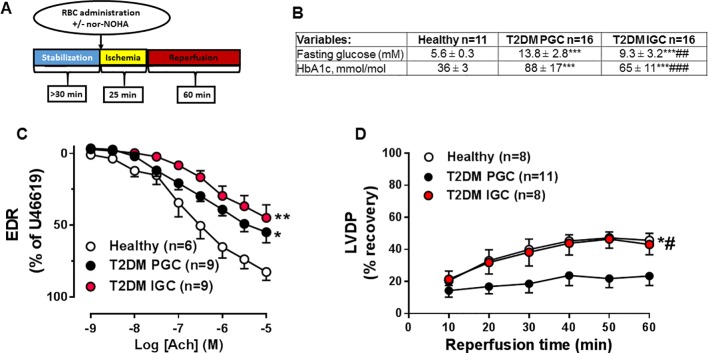
Red blood cells (RBCs) from patients with type 2 diabetes mellitus (T2DM) induce endothelial dysfunction and impair cardiac function following ischemia via increase in RBC arginase and oxidative stress. Here, we aimed to elucidate whether the effect of RBC-mediated cardiac impairment following ischemia and endothelial dysfunction in T2DM is dependent on glycemic control. Patients with T2DM at poor glycemic control (T2DM PGC) and at improvement in glycemic control (T2DM IGC) and healthy subjects were recruited. Isolated RBCs from subjects were incubated with aortic rings from healthy wild-type rats with subsequent evaluation of endothelium-dependent relaxation (EDR) using wire myograph. Moreover, RBCs were administered to isolated wild-type rat hearts with subsequent evaluation of left ventricular developed pressure (LVDP) during reperfusion using Langendorff setup. In separate experiments, RBCs were preincubated with an arginase inhibitor before perfusion. Blood glucose and glycated hemoglobin were 33 and 26%, respectively, lower in T2DM IGC compared with those in T2DM PGC. RBCs from T2DM PGC and T2DM IGC impaired EDR to a similar magnitude compared with RBCs from healthy subjects. LVDP was significantly impaired in hearts given RBCs from T2DM PGC as compared with those from healthy subjects. The impairment of LVDP induced by T2DM PGC was attenuated by RBCs from T2DM IGC. Arginase inhibition improved LVDP to a similar extent between T2DM PGC and IGC groups. These observations indicate that glycemic control abrogate the impairment in postischemic recovery but not endothelial dysfunction induced by RBCs from T2DM. Moreover, inhibition of RBC arginase improves cardiac function irrespective of glycemic control.
Keywords: diabetes; endothelial dysfunction; glycemic control; ischemia; red blood cells; reperfusion.
Figures

References
-
- Costantino S., Paneni F., Battista R., Castello L., Capretti G., Chiandotto S., et al. (2017). Impact of glycemic variability on chromatin remodeling, oxidative stress, and endothelial dysfunction in patients with type 2 diabetes and with target hba1c levels. Diabetes 66 (9), 2472–2482. 10.2337/db17-0294 - DOI - PubMed
-
- Kosiborod M., Rathore S. S., Inzucchi S. E., Masoudi F. A., Wang Y., Havranek E. P., et al. (2005). Admission glucose and mortality in elderly patients hospitalized with acute myocardial infarction: implications for patients with and without recognized diabetes. Circulation 111 (23), 3078–3086. 10.1161/CIRCULATIONAHA.104.517839 - DOI - PubMed
LinkOut - more resources
Full Text Sources