

Frontal Sinus Fractures: Management and Complications
- PMID: 31428249
- PMCID: PMC6697471
- DOI: 10.1055/s-0038-1675560
Frontal Sinus Fractures: Management and Complications
Abstract
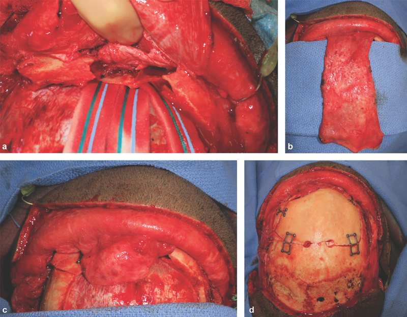
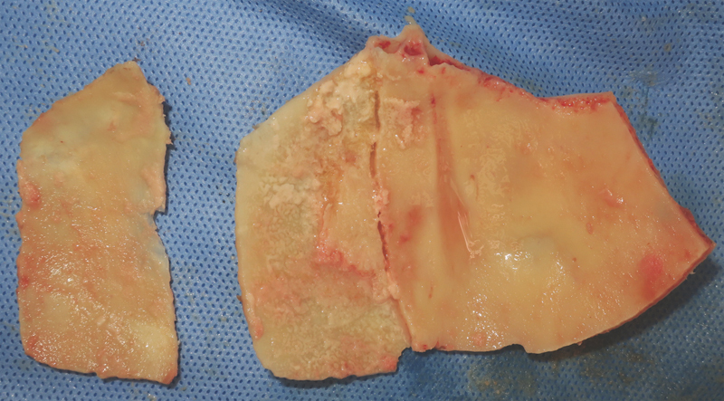
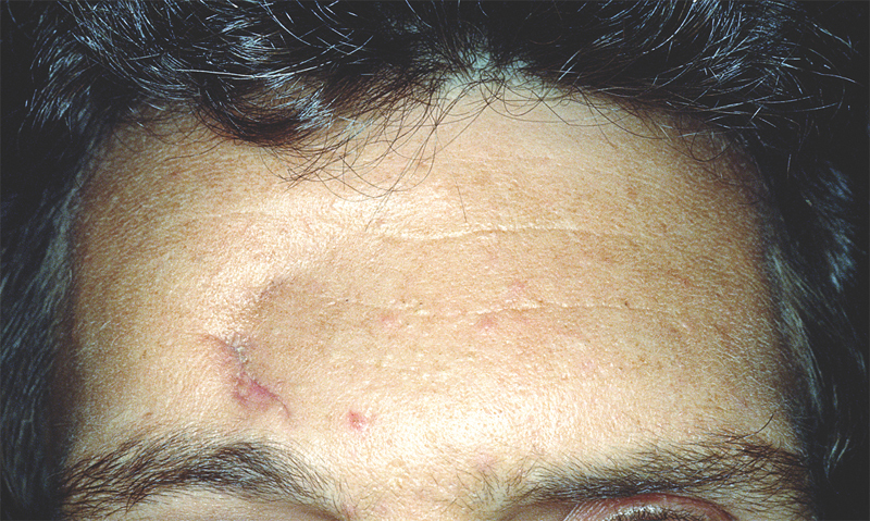
Frontal sinus fractures are relatively rare maxillofacial injuries (only 5-15% of all facial fractures). The appropriate management of frontal sinus fracture and associated pathology is controversial. Diagnosis and treatment of frontal sinus fractures has improved with the advances of high-resolution computed tomography technology. Treatment of frontal sinus fractures depends on several factors, including contour deformity of anterior table; the presence of CSF leak or air-fluid level in the sinus, likelihood of nasofrontal duct obstruction, and degree of displacement of posterior table. Nasofrontal duct patency should be checked if fracture pattern is highly suspicious of ductal injury. Cranialization is performed in cases of severely comminuted posterior wall fracture. Long-term complication of frontal sinus fracture can occur up to 10 years after initial injury or intervention; so, judicious long-term follow-up is warranted. This article presents the management and complications of frontal sinus fractures.
Keywords: calvarial bone graft; complications; frontal sinus fracture; management.
Conflict of interest statement
Figures
References
-
- Gerbino G, Roccia F, Benech A, Caldarelli C. Analysis of 158 frontal sinus fractures: current surgical management and complications. J Craniomaxillofac Surg. 2000;28(03):133–139. - PubMed
-
- Nahum A M. The biomechanics of maxillofacial trauma. Clin Plast Surg. 1975;2(01):59–64. - PubMed
-
- McLaughlin R B, Jr, Rehl R M, Jr, Lanza D C. Clinically relevant frontal sinus anatomy and physiology. Otolaryngol Clin North Am. 2001;34(01):1–22. - PubMed
-
- Hollinshead W H. New York: Harper and Row; 1968. Anatomy of Surgeons, Vol. 1: Head and Neck; p. 283.
-
- Donald P J. The tenacity of the frontal sinus mucosa. Otolaryngol Head Neck Surg (1979) 1979;87(05):557–566. - PubMed
LinkOut - more resources
Full Text Sources