Case Reports
. 2019 Jul 25:2019:4375839.
doi: 10.1155/2019/4375839. eCollection 2019.

A Rare Case of Steroid Cell Tumor, Not Otherwise Specified (NOS), of the Ovary in a Young Woman

Affiliations
Case Reports

A Rare Case of Steroid Cell Tumor, Not Otherwise Specified (NOS), of the Ovary in a Young Woman

Eek Chaw Tan et al. Case Rep Obstet Gynecol. .

Abstract

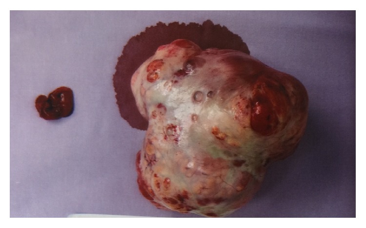
Steroid cell tumour is a rare sex cord-stromal tumor of the ovary. It may produce steroids and is associated with testosterone secretion which causes symptoms like hair loss, hirsutism, and oligomenorrhea/amenorrhea due to hormonal activity and virilizing properties of tumor. In this article, we reported a 27-year-old woman who presented with hirsutism, hoarseness of voice, scalp hair fall, and amenorrhea for 8 years. Clinical and diagnostic evaluation revealed a left adnexal mass and elevated serum levels of testosterone and she was diagnosed as having a Sertoli Leydig cell tumour of ovary. She underwent left salpingooophorectomy and both histopathological examination and immunohistochemistry confirmed the diagnosis. Her serum testosterone levels normalized 3 days after the surgery and her menses resumed spontaneously a few months after the operation. In addition, we reviewed the literature on the epidemiology, clinical presentations, imaging and histological findings, and the treatment options on this disease.


Conflict of interest statement

The authors declare that they have no conflicts of interest.

Figures

Figure 1
Figure 1
The ultrasound pelvis showed a multicystic avascular structure in the right adnexa measuring 4.5 x 4.3 x 3.3cm. Another multicystic avascular structure was seen in the left adnexa measuring 5.4 x 4.4 x 4.1cm. These could possibly represent polycystic ovaries containing multiple cysts.
Figure 2
Figure 2
MRI abdomen and pelvis showed the presence of a large heterogeneous predominantly solid mass arising from the left ovary with cystic areas within. The solid components showed restricted diffusion and avid contrast enhancement. The mass was measured 8.4 x 6.1 x 8.9 cm. The solid components were hyperintense to muscle on T2w imaging and iso/hypointense on T1w imaging.
Figure 3
Figure 3
The specimen showed the left ovarian tumour and fallopian tube of the patient. The gross specimen consisted of a lobulated mass 100 x 100 x 30 mm with a defect measuring 20 mm in size externally, altogether weighing 177 grams. The mass had morcellated appearance. On cut section had solid and multicystic appearance. The solid areas had a golden yellow appearance punctuated by foci of haemorrhage. A small fleshy tan solid-cystic area was present at one end measuring 20 x 15 x 15 mm. In cytological examination, the sections of the main tumour showed a predominantly diffuse proliferation of polygonal cells with ample, pale, vacuolated cytoplasm and round central nuclei. Small areas of a more nested appearance were also seen. Foci showing cells with more eosinophilic cytoplasm were also evident, but no Reinke crystals were evident. The nuclei were central and round, with no significant atypia. Immunohistochemical stains for calretinin and alpha-inhibin were diffusely and strongly positive. CD 10 is negative. An area showing multiple cystic structures lined by bland cuboidal to columnar epithelium with focal ciliation in a fibromatous stroma was seen. No significant epithelial proliferation or atypia was seen.

References

    1. Young R. H., Clement P. B., Scully R. E. Sex-cord, stromal, steroid cell and germ cell tumours of the ovary. In: Mills S.-E., Carter D., Greenson J.-K., Oberman H.-A., Reuter V., Stoler M.-H., editors. Sternbergs Diagnostic Surgical Pathology. 4. Philadelphia, Pa, USA: Lippincott Williams & Wilkins; 2004. pp. 2579–2615.
    1. Hayes M. C., Scully R. E. Ovarian steroid cell tumors (not otherwise specified): a clinicopathological analysis of 63 cases. The American Journal of Surgical Pathology. 1987;11(11):835–845. doi: 10.1097/00000478-198711000-00002. - DOI - PubMed
    1. Kim Y. T., Kim S. W., Yoon B. S., et al. An ovarian steroid cell tumor causing virilization and massive ascites. Yonsei Medical Journal. 2007;48(1):142–146. doi: 10.3349/ymj.2007.48.1.142. - DOI - PMC - PubMed
    1. Revathy M., Kanchana M. P. Incidence of virilisation in sex cord stromal tumours of ovary, a 5-year experience in a tertiary care gynaecological centre. Journal of Evolution of Medical and Dental Sciences. 2018;7(7):886–891. doi: 10.14260/jemds/2018/202. - DOI
    1. Seles F. M., Revathy M., Kanchana M. P. Steroid cell tumour of the ovary: a case report with review of literature. International Journal of Reproduction, Contraception, Obstetrics and Gynecology. 2018;7:3425–3428.

Publication types

LinkOut - more resources