. 2019 Aug 2:9:680.
doi: 10.3389/fonc.2019.00680. eCollection 2019.

Fact or Fiction, It Is Time for a Verdict on Vasculogenic Mimicry?

Affiliations

Fact or Fiction, It Is Time for a Verdict on Vasculogenic Mimicry?

Andrés Valdivia et al. Front Oncol. .

Abstract

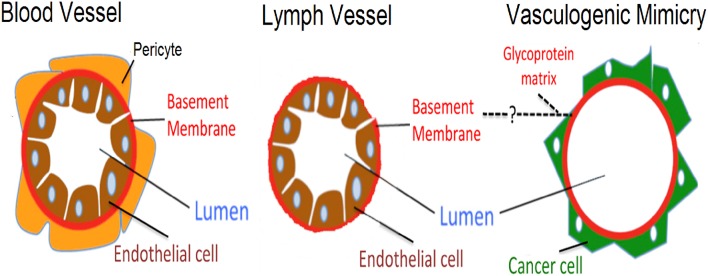
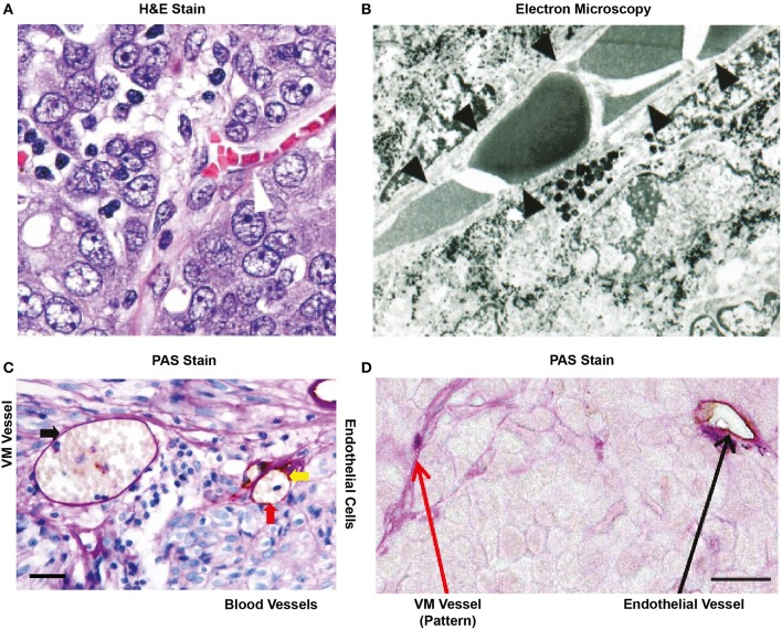
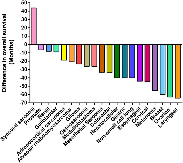
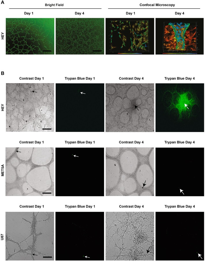
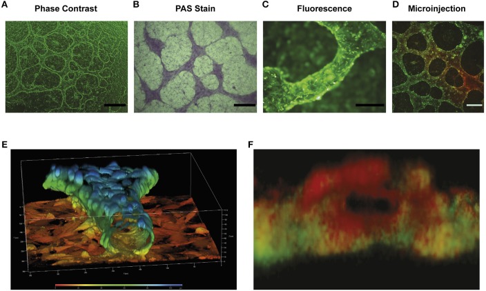
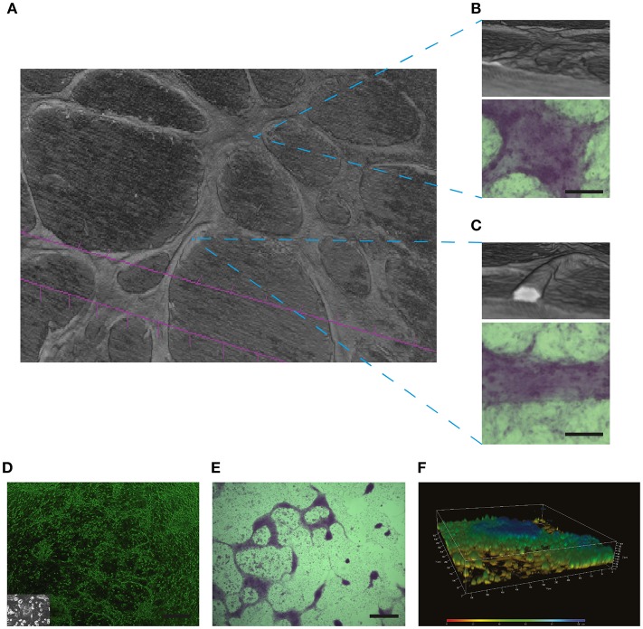
The term vasculogenic mimicry (VM) refers to the capacity of certain cancer cells to form fluid-conducting structures within a tumor in an endothelial cell (EC)-free manner. Ever since its first report by Maniotis in 1999, the existence of VM has been an extremely contentious issue. The overwhelming consensus of the literature suggests that VM is frequently observed in highly aggressive tumors and correlates to lower patient survival. While the presence of VM in vivo in animal and patient tumors are claimed upon the strong positive staining for glycoproteins (Periodic Acid Schiff, PAS), it is by no means universally accepted. More controversial still is the existence of an in vitro model of VM that principally divides the scientific community. Original reports demonstrated that channels or tubes occur in cancer cell monolayers in vitro when cultured in matrigel and that these structures may support fluid movement. However, several years later many papers emerged stating that connections formed between cancer cells grown on matrigel represented VM. We speculate that this became accepted by the cancer research community and now the vast majority of the scientific literature reports both presence and mechanisms of VM based on intercellular connections, not the presence of fluid conducting tubes. In this opinion paper, we call upon evidence from an exhaustive review of the literature and original data to argue that the majority of in vitro studies presented as VM do not correspond to this phenomenon. Furthermore, we raise doubts on the validity of concluding the presence of VM in patient samples and animal models based solely on the presence of PAS+ staining. We outline the requirement for new biomarkers of VM and present criteria by which VM should be defined in vitro and in vivo.

Keywords: angiogenesis; endothelial; in vivo–in vitro; model; vasculogenic mimicry (VM).


Figures

Figure 1
Figure 1
Schematic representation highlighting differences between blood vessels, lymph vessels, and VM structures. Differences are shown between an endothelial lined blood vessel (left side), a lymph vessel (center), and the hypothesized VM structure (right side). Blood vessels present an external layer of pericytes which overlay a basement membrane, with endothelial cells lining the luminal side. In lymph vessels, a similar architecture is present, however pericytes are absent and the basement membrane is thinner. Finally, the proposed structure present in VM has cancer cells sitting on the non-luminal side of a glycoprotein-rich (PAS+) matrix, with the total absence of endothelial cells.
Figure 2
Figure 2
Historical representation of in vivo VM from the literature. (A) H&E stain of an ovarian tumor showing that red blood cells are contained in a structure lined by cancer cells (arrow head), not endothelial cells. Image taken with kind permission from the publication by Sood et al. (12). (B) Electron microscopy of a uveal melanoma showing that red blood cells are contained within a structure lined by cancer cells (as confirmed by the presence of melanin in these cells). Taken with kind permission from the publication by Maniotis et al. (13). (C) Cross section of a glioblastoma after staining for PAS and CD31. The red arrow indicates an endothelial lined blood vessel with PAS+ luminal stain and positive for the endothelial marker CD31 (yellow arrow). RBCs (red arrow) are shown within the structure. In the same cross-section, a PAS+ yet, and CD31 negative structure is also present that contains red blood cells (black arrow). This may represent VM. Size bar represents 100 μm. Taken with kind permission from the publication by Sun et al. (14). (D) Small lung cell carcinoma cross section showing the presence of a PAS+/CD31+ blood vessel (black arrow) and the presence of PAS+ “patterned structures” that have been speculated to represent VM (red arrow). Size bar represents 100 μm. Image taken with kind permission from the publication by Williamson et al. (15) http://creativecommons.org/licenses/by/4.0/. All appropriate permissions have been obtained from the copyright holders.
Figure 3
Figure 3
Presence of VM is associated with poor patient prognosis. Waterfall plot presenting the difference in survival time (years) of patients presenting structures claimed to represent VM in their tumors. The zero value represents patient survival in the absence of VM. In reports to date VM has a negative impact on the overall patient survival in all but one cohort (synovial sarcoma).
Figure 4
Figure 4
Structures present in matrigel culture may not represent VM. The upper panel (A) demonstrates light (size bar represents 200 μm) and confocal microscopy (size bar represents 500 μm) images of day 1 and day 4 cell cultures of the HEY cancer cell line. The presence of a tubular hollow structure can clearly be seen at day 4 in culture. (B) demonstrates dye microinjections of the HEY cell lines at day 1 and 4, showing movement of fluid and thus functional tubes (upper panel). Despite the formation of what is often reported in the literature to refer to tubular structures (VM), no movement of dye was observed in the MET5A or the U87 cell lines at day 1 and 4 (middle and lower panel respectably). In fact, the dye either remained in an individual cell or dispersed from culture. Arrow heads display injection sites, all size bars represent 250 μm.
Figure 5
Figure 5
Characterization of in vitro model of VM: (A) is a representative light microscope image of the tubular structures formed by the HEY ovarian cell line after 4 days in matrigel culture. Elevated tubular structures are observed above a cell monolayer. Size bar represents 500 μm. (B) PAS stain showing that the elevated tubular structures possess a higher concentration of glycoproteins that cell monolayer present below. Size bar represents 500 μm. (C) Epifluorescence image of the tubular structures of the formed in the HEY cell line transfected with GFP (HEY-GFP). Note the difference in height of the tubular structure compared with cell monolayer below and that the tubular structure (~100 μm in diameter) is composed of numerous cancer cells. Size bar represents 200 μm. (D) A dye micro injection of 4-day old HEY-GFP culture, showing that these structures are capable of moving a colorant dye within their interior. Size bar represents 200 μm. (E) Confocal 3D reconstruction of the tubular structures found in the HEY-GFP cell lines using ZEN 2012 program. The color scale represents the height in culture over the cell monolayer. Note how the cells form a tubular hollow structure with a central lumen. The size bar demonstrates that the structure is ~80 μm in diameter. (F) A 3D reconstruction of a cross section of the tubular structure showing the PAS positive (red) show up preferentially on the luminal side of the tubular structure, while the green cancer cells are confined to the outer sides (as schematically represented in Figure 1). Size bar represents 100 μm.
Figure 6
Figure 6
PAS positive structures may not necessarily represent the presence of VM: In (A) Micro-CT 3D reconstruction of the SKOV3 ovarian cancer cell line after 4 days culture in Matrigel demonstrating the presence of elevated structures over a cell monlayer. (B) A flatter yet elevated section of the culture did not demonstrate a lumen when reconstructed by Micro-CT, yet this structure stained positive for PAS (lower panel). Size bar represents 100 μm. (C) Reconstruction by Micro-CT demonstrates the presence of a lumen containing structure, as demonstrated by the interior white space. The PAS positive stain is shown in the lower panel. Size bar represents 100 μm. In (D–F) a characterization is shown of structures formed by primary cultures of glioblastoma cells. These samples where obtained with Ethical Committee approval and written patient consent from the Clinical Hospital of the University of Chile, Santiago, Chile. Cell culture was as described previously in Racordon et al. (6). (D) Light microscopy imagery of primary cultured cells grown on matrigel, with an image of the cells grown in plastic in the inlay. Size bar represents 500 μm. (E) Primary cultured glioblastoma cells presented elevated structures over the cell monolayer that stained for PAS. Size bar represents 500 μm. (F) Confocal 3D reconstruction using ZEN 2012 demonstrates that the PAS positive structures observed in (E) are elevated over the cell monolayer but do not possess a lumen.

References

    1. Pinto MP, Sotomayor P, Carrasco-Avino G, Corvalan AH, Owen GI. Escaping antiangiogenic therapy: strategies employed by cancer cells. Int J Mol Sci. (2016) 17:1–20. 10.3390/ijms17091489 - DOI - PMC - PubMed
    1. Barnett FH, Rosenfeld M, Wood M, Kiosses WB, Usui Y, Marchetti V, et al. . Macrophages form functional vascular mimicry channels in vivo. Sci Rep. (2016) 6:36659. 10.1038/srep36659 - DOI - PMC - PubMed
    1. Kertschanska S, Štulcová B, Kaufmann P, Štulc J. Distensible transtrophoblastic channels in the rat placenta. Placenta. (2000) 21:670–7. 10.1053/plac.2000.0558 - DOI - PubMed
    1. Kučera T, Strilić B, Regener K, Schubert M, Laudet V, Lammert E. Ancestral vascular lumen formation via basal cell surfaces. PLoS ONE. (2009) 4:e4132. 10.1371/journal.pone.0004132 - DOI - PMC - PubMed
    1. Langheinrich MC, Schellerer V, Perrakis A, Lohmüller C, Schildberg C, Naschberger E, et al. . Molecular mechanisms of lymphatic metastasis in solid tumors of the gastrointestinal tract. Int J Clin Exp Pathol. (2012) 5:614–23. - PMC - PubMed