. 2019 Aug 13:2:311.
doi: 10.1038/s42003-019-0556-6. eCollection 2019.

Hemoglobin S and C affect biomechanical membrane properties of P. falciparum-infected erythrocytes

Affiliations

Hemoglobin S and C affect biomechanical membrane properties of P. falciparum-infected erythrocytes

Benjamin Fröhlich et al. Commun Biol. .

Abstract

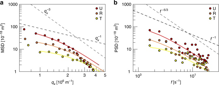
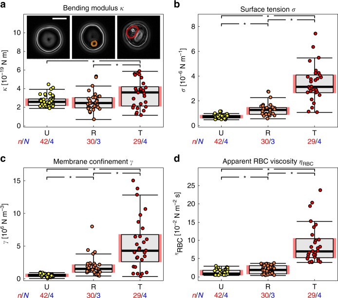
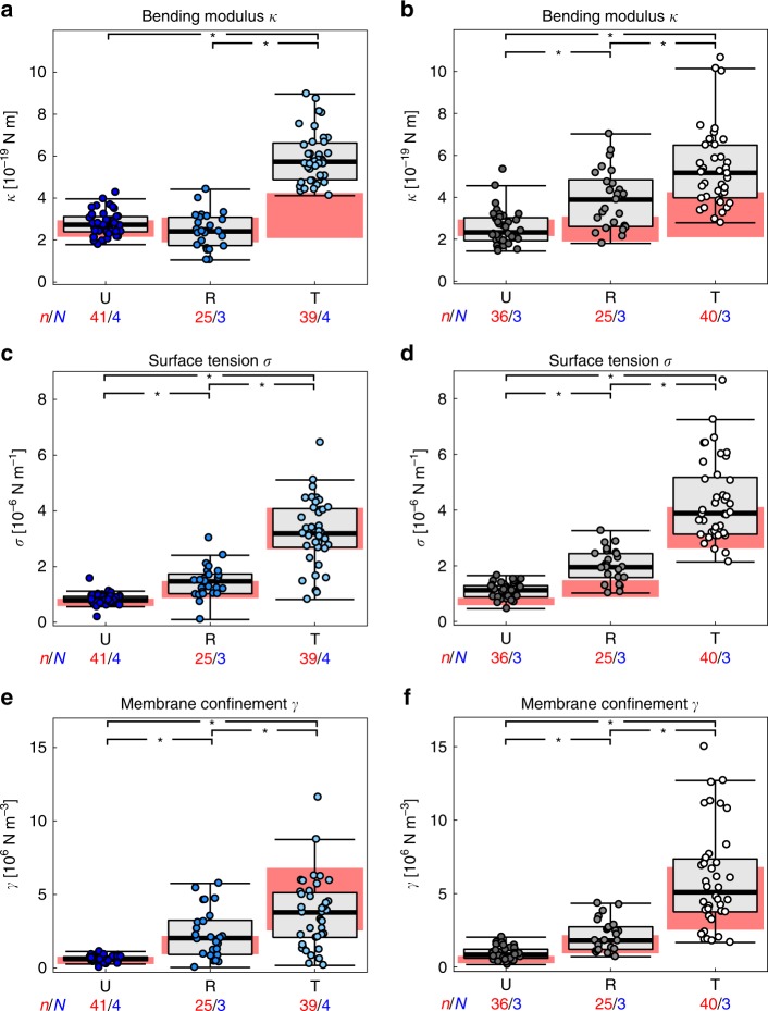
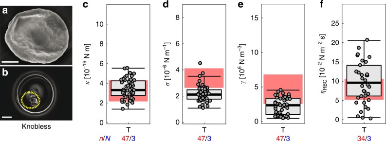
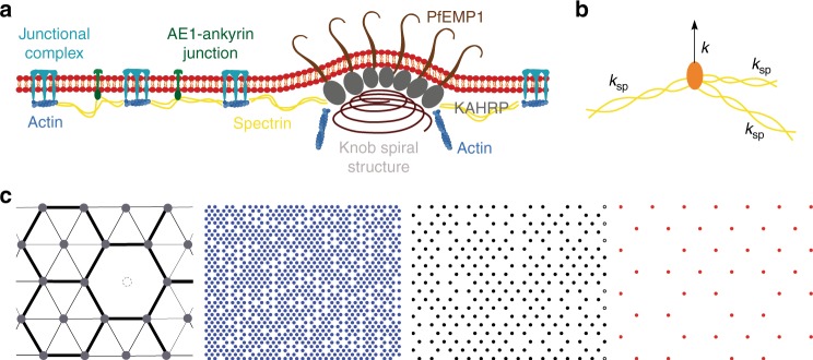
During intraerythrocytic development, the human malaria parasite Plasmodium falciparum alters the mechanical deformability of its host cell. The underpinning biological processes involve gain in parasite mass, changes in the membrane protein compositions, reorganization of the cytoskeletons and its coupling to the plasma membrane, and formation of membrane protrusions, termed knobs. The hemoglobinopathies S and C are known to partially protect carriers from severe malaria, possibly through additional changes in the erythrocyte biomechanics, but a detailed quantification of cell mechanics is still missing. Here, we combined flicker spectroscopy and a mathematical model and demonstrated that knob formation strongly suppresses membrane fluctuations by increasing membrane-cytoskeleton coupling. We found that the confinement increased with hemoglobin S but decreases with hemoglobin C in spite of comparable knob densities and diameters. We further found that the membrane bending modulus strongly depends on the hemoglobinopathetic variant, suggesting increased amounts of irreversibly oxidized hemichromes bound to membranes.

Keywords: Membrane biophysics; Parasitic infection.


Conflict of interest statement

Competing interestsThe authors declare no competing interests.

Figures

Fig. 1
Fig. 1
Membrane fluctuation spectra obtained from uninfected and P. falciparum-infected erythrocytes at the ring and trophozoite stage. a Mean squared displacement (MSD) as a function of the wavenumber qx. b The corresponding power spectrum density (PSD) as a function of frequency f. U (red); uninfected, R (orange); ring phase, and T (yellow); trophozoite. Representative spectra from single cell measurements are shown. The two different power law exponents predicted by the theoretical model for the low and high values of the wavenumber qx and the frequency f respectively, are presented to guide the eye
Fig. 2
Fig. 2
Changes in biomechanical parameters in uninfected and P. falciparum-infected HbAA erythrocytes. a Bending modulus κ, b surface tension σ, c membrane confinement γ, d and apparent red blood cell viscosity ηRBC. U (red); uninfected, R (orange); ring phase, and T (yellow); trophozoite. Individual data points represent a single determination and n the total number of data points obtained using blood of N different donors. Box plots (gray) were laid over the data points. The 25–75 percentile ranges are highlighted in red and replotted in the following figures. The gradient images of representative cells at the corresponding infection stages are presented as insets of panel a. The cytoplasmic area occupied by the parasite is highlighted by the dotted line. *p < 0.05 according to Welch t-test. Scale bar, 5 µm
Fig. 3
Fig. 3
Changes in biomechanical parameters in uninfected and P. falciparum-infected HbAS and HbAC erythrocytes. a Bending modulus κ of U (dark blue); uninfected, R (blue); ring phase, and T (light blue); trophozoite HbAS erythrocytes. b Bending modulus κ of U (dark gray); uninfected, R (gray); ring phase, and T (white); trophozoite HbAC erythrocytes. Corresponding data for the c and d surface tension σ and e and f membrane confinement γ. Individual data points represent a single determination and n the total number of data points obtained using blood of N different donors. Box plots (gray) were laid over the data points. The 25–75 percentile ranges of the corresponding HbAA results are highlighted in red for visual comparison. *p < 0.05 according to Welch t-test
Fig. 4
Fig. 4
Biomechanical properties of HbAA erythrocytes infected with a knobless, FCR3-derived mutant at the trophozoite stage. a Representative SEM image and b representative gradient map calculated from a phase contrast image. c Bending modulus κ, d surface tension σ e membrane confinement γ, and f apparent red blood cell viscosity ηRBC. Individual data points represent a single determination and n the total number of data points obtained using blood of N different donors. Box plots (gray) were laid over the data points. The 25–75 percentile ranges of the corresponding HbAA results are highlighted in red for visual comparison. *p < 0.05 according to Welch t-test. Scale bar, 2 µm
Fig. 5
Fig. 5
Graphic representation of key parameters of the numerical model. a Simplified model of the membrane skeleton, including the junctional complexes and AE-1-ankyrin junctions (left, uninfected) and additional anchoring structures via the knob components Plasmodium falciparum erythrocyte membrane protein 1 (PfEMP1) and knob-associated histidine rich protein (KAHRP) (right, trophozoite). b The structure of a singular anchoring point is depicted, with spectrin filaments indicated in yellow and connections to the lipid bilayer indicated in orange. The whole complex can be modeled by a vertical spring with a spring constant k. c The model treats the membrane–spectrin connections as springs that are distributed in a hexagonal array. Inhomogeneities are introduced by removing some midpoints of hexagons as indicated in the left panel. The other three arrays show examples of spring distributions for densities of 1096, 257, and 44 springs per µm2 (from left to right)
Fig. 6
Fig. 6
Simulated effect of the spring density and the spring constant on the membrane confinement. Simulated mean square displacement as a function of the wavenumber q calculated a for different connector densities ρ (see Fig. 5c) and b for different spring constants k. The discrete data points represent the results of numerical calculations and the continuous lines are the corresponding fit of the continuum theory. Membrane confinement γ as a function of c the density ρ of connectors d the spring constant k. The arrows indicate values calculated for uninfected red blood cells
Fig. 7
Fig. 7
Influence of knobs size and density on the membrane confinement. Spring positions of two realizations for a 1 µm × 1 µm membrane patch. Knobs are represented by a clusters of 13 springs and b 49 springs. The background represents a membrane with a thinned out anchor density of 102 µm−2. c The relative membrane confinement γ/γ0 as normalized to an array without knobs is shown as a function of the cumulative number of knob-specific springs N. Each data point represents the mean value obtained from ten individual simulations. Three different knob sizes as defined by spring number were considered. The spring numbers are indicated

References

    1. Fung, Y., Skalak, R. Biomechanics: mechanical properties of living tissues. (Springer Science & Business Media, Berlin, Germany, 2013).
    1. Tomaiuolo G. Biomechanical properties of red blood cells in health and disease towards microfluidics. Biomicrofluidics. 2014;8:051501. doi: 10.1063/1.4895755. - DOI - PMC - PubMed
    1. Lux S. E. Anatomy of the red cell membrane skeleton: unanswered questions. Blood. 2015;127(2):187–199. doi: 10.1182/blood-2014-12-512772. - DOI - PubMed
    1. Machnicka B, et al. Spectrins: a structural platform for stabilization and activation of membrane channels, receptors and transporters. Biochim. Biophys. Acta (BBA)-Biomembr. 2014;1838:620–634. doi: 10.1016/j.bbamem.2013.05.002. - DOI - PubMed
    1. Lavazec C. Molecular mechanisms of deformability of Plasmodium-infected erythrocytes. Curr. Opin. Microbiol. 2017;40:138–144. doi: 10.1016/j.mib.2017.11.011. - DOI - PubMed

Publication types

MeSH terms