Review
. 2019 Aug 19;10(1):83.
doi: 10.1186/s13244-019-0761-3.

Magnetic resonance imaging of the papillary muscles of the left ventricle: normal anatomy, variants, and abnormalities

Affiliations
Review

Magnetic resonance imaging of the papillary muscles of the left ventricle: normal anatomy, variants, and abnormalities

Prabhakar Rajiah et al. Insights Imaging. .

Abstract

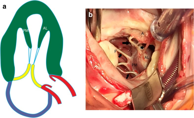
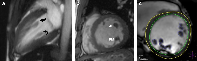
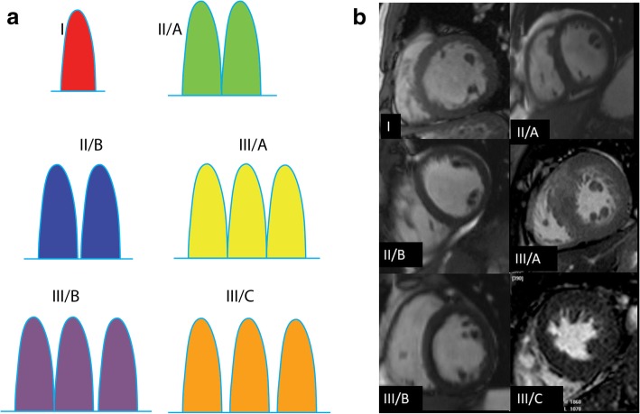
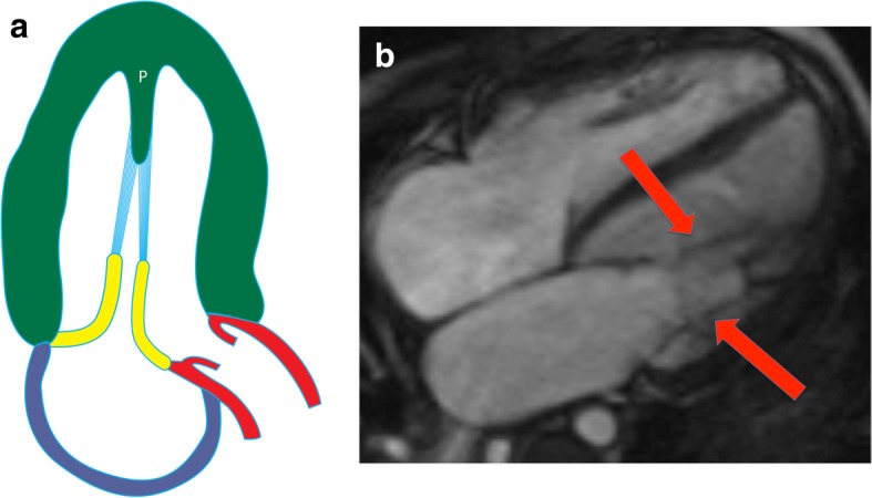
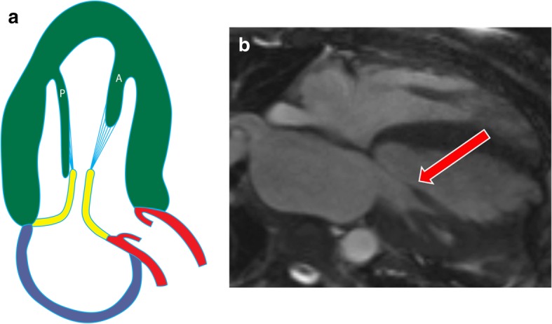
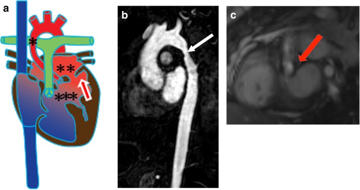
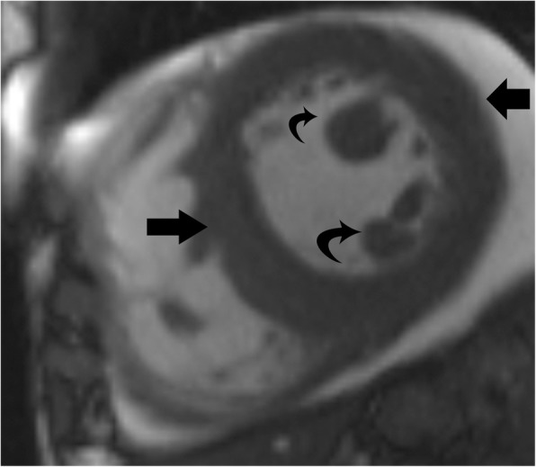
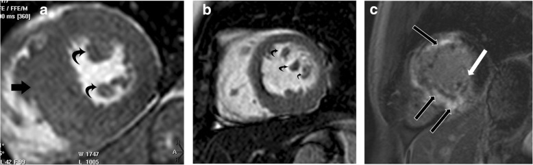
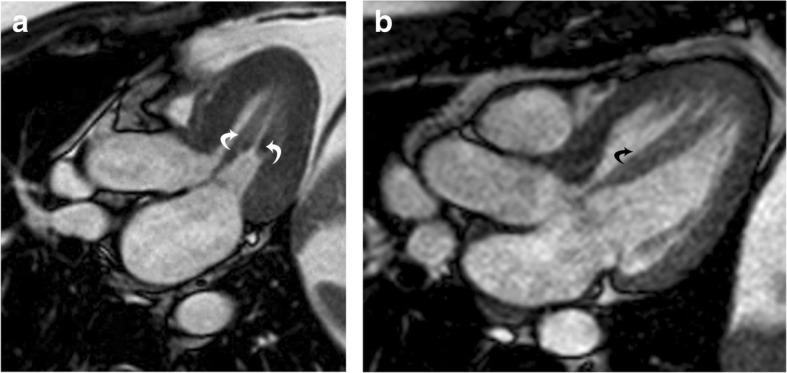
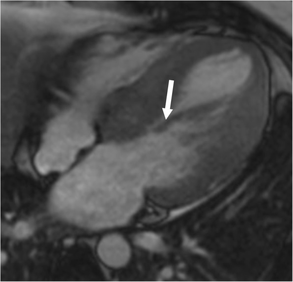
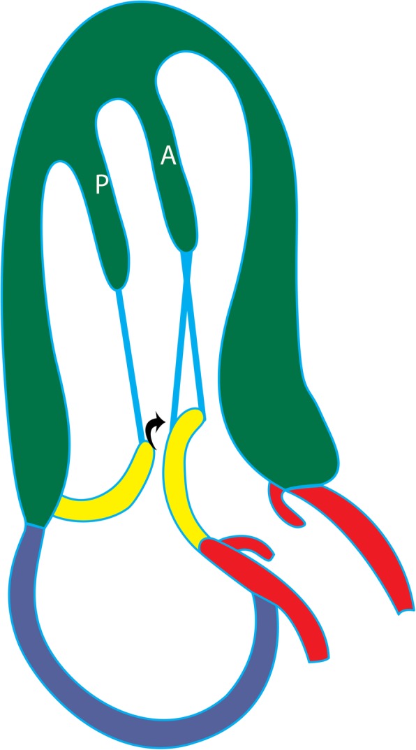
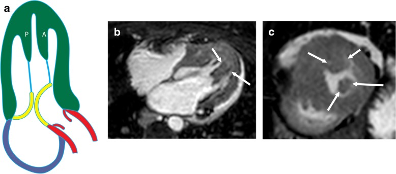
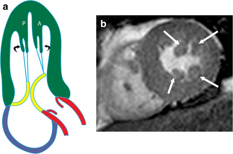
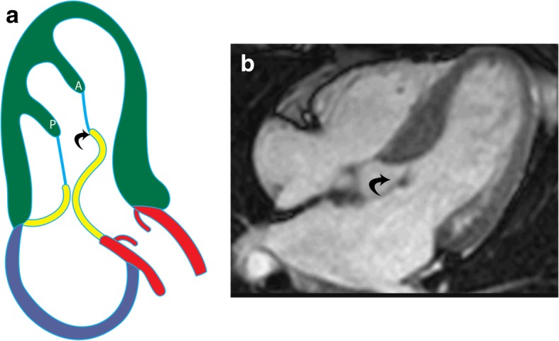
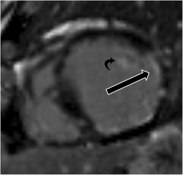
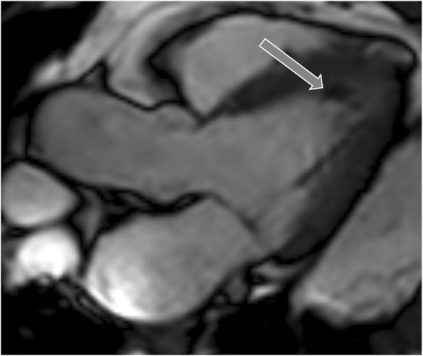
Left ventricular papillary muscles are small myocardial structures that play an important role in the functioning of mitral valve and left ventricle. Typically, there are two groups of papillary muscles, namely the anterolateral and the posteromedial groups. Cardiovascular magnetic resonance (CMR) is a valuable imaging modality in the evaluation of papillary muscles, providing both morphological and functional information. There is a remarkably wide variation in the morphology of papillary muscles. These variations can be asymptomatic or associated with symptoms related to LV outflow tract obstruction, often associated with hypertrophic cardiomyopathy. Abnormalities of the papillary muscles range from congenital disorders to neoplasms. Parachute mitral valve is the most common congenital abnormality of papillary muscles, in which all the chordae insert into a single papillary muscle. Papillary muscles can become dysfunctional, most commonly due to ischemia. Papillary muscle rupture is a major complication of acute myocardial infarction that results in mitral regurgitation and associated with high mortality rates. The most common papillary neoplasm is metastasis, but primary benign and malignant neoplasms can also be seen. In this article, we discuss the role of CMR in the evaluation of papillary muscle anatomy, function, and abnormalities.

Keywords: Anatomy; CMR; Hypertrophic cardiomyopathy; Papillary muscles; Variants.


Conflict of interest statement

The authors declare that they have no competing interests.

Figures

Fig. 1
Fig. 1
Normal anatomy of papillary muscles. a Illustration showing the anterolateral (AL) and posteromedial (PM) in a vertical long-axis projection. The papillary muscles originate from the free wall of the LV attached to trabecula carnea. The papillary muscles give rise to multiple chordae tendinae (blue), which attach to the mitral valve (yellow). b Surgical image showing the head of the anterolateral (AL) and posteromedial (PM) muscles attached to their chordae tendinae (arrows), which then attach to the anterior (A) and posterior (P) mitral leaflets
Fig. 2
Fig. 2
MRI appearances of normal papillary muscles. a Two-chamber vertical long-axis SSFP MRI through the left ventricle shows the anterolateral (straight arrow) and posteromedial (curved arrow) papillary muscles. b Short-axis SSFP MRI image shows the anterolateral (AL) and posteromedial (PM) papillary muscles. c Short-axis SSFP MRI image shows the technique for measuring papillary muscle mass. The papillary muscles have been contoured (blue) in end-diastolic image to derive papillary muscle mass. The endocardial (green) and epicardial (yellow) contours are also seen
Fig. 3
Fig. 3
Papillary muscle variations. a Illustration of six papillary muscle variations. Type I is a single muscle, type II/A is two heads with a common origin, type II/B is two heads separated at the basal portion, type III/A is three heads with a common origin, type III/B is three heads with two sharing a common origin, and type III/C is three heads with no common origin. b Multiple short-axis SSFP MRI images demonstrating each of the papillary muscle variations
Fig. 4
Fig. 4
Parachute mitral valve. a Illustration of a parachute mitral valve. The chordae tendinae (blue) originate from a single papillary muscle (P). b Four-chamber SSFP image of parachute mitral valve, with all of the chordae (arrows) arising from a single papillary muscle
Fig. 5
Fig. 5
Parachute-like asymmetric mitral valve. a Illustration showing the posteromedial papillary muscle (P) is longer and attaches more proximally to the mitral valve, compared to the anterolateral (A) papillary muscle. b Four-chamber cine SSFP image shows asymmetric elongation of a single papillary muscle (anterolateral muscle) (arrow) attaching to the mitral valve leaflets, resulting in an eccentric mitral orifice
Fig. 6
Fig. 6
Shone complex. a Illustration of Shone complex showing aortic coarctation (*), supravalvular mitral ring (**), subaortic stenosis (***) and a parachute mitral valve (arrow). b Sagittal MRA of the aorta shows coarctation (arrow). c Short-axis cine SSFP image at the level of LVOT depicting a subaortic stenosis (arrow) with flow acceleration seen at the subaortic level
Fig. 7
Fig. 7
Papillary muscle hypertrophy. Short-axis cine-SSFP image in a patient with systemic hypertension shows hypertrophy of the papillary muscles (curved arrow), which is proportionate to the concentric LV hypertrophy (straight arrows)
Fig. 8
Fig. 8
Hypertrophic cardiomyopathy. a Short-axis view SSFP image shows severe hypertrophy of the ventricular septum (straight arrow) and hypertrophy of the papillary muscles (curved arrows). b Short-axis SSFP image in another patient shows 3 papillary muscles which are hypertrophied (curved arrows). c Short-axis view delayed-enhancement image shows extensive mid-myocardial delayed enhancement (black arrows) due to interstitial fibrosis in hypertrophic cardiomyopathy. The posteromedial papillary muscle also shows patchy areas of delayed enhancement (white arrow)
Fig. 9
Fig. 9
Solitary papillary muscle hypertrophy. a Three-chamber SSFP image shows significant hypertrophy of the papillary muscles (curved arrows), without significant myocardial hypertrophy, which is causing narrowing of the mid-ventricular cavity in systole. b Three-chamber SSFP image in another patient shows hypertrophy of the anterolateral papillary muscle (curved arrow) with normal thickness of the LV myocardium
Fig. 10
Fig. 10
Anomalous papillary muscle insertion. Anomalous direct insertion of the anterolateral papillary muscle to the anterior mitral leaflet (arrow) without intervening chorda tendinae
Fig. 11
Fig. 11
Anomalous papillary muscle insertion. Illustration of anomalous insertion of the anterolateral papillary muscle (A) into the mid-portion of anterior mitral leaflet (arrow), which results in leaflet slack and narrowing of the LVOT
Fig. 12
Fig. 12
Anteroapical displacement. a Illustration showing apical displacement of the papillary muscles (A and P) resulting in leaflet slack and left ventricular outflow tract obstruction during systole. b Four-chamber cine 3D-SSFP image shows anteroapical displacement of hypertrophied bifid papillary muscles (arrow) in a patient with HCM. c Short-axis SSFP MRI image at the apical level shows the presence of double bifid papillary muscles (arrows), indicating anteroapical displacement
Fig. 13
Fig. 13
a Illustration shows double bifid papillary muscles (curved arrows), which causes systolic LVOT obstruction due to leaflet slack. b Short-axis MRI image shows double bifid papillary muscles (arrows)
Fig. 14
Fig. 14
Elongated mitral leaflet. a Illustration of elongated mitral valve leaflet (curved arrow), resulting in systolic obstruction of the left ventricular outflow tract, without evidence of other abnormalities related to hypertrophic cardiomyopathy. b Four-chamber cine SSFP image shows an elongated anterior mitral leaflet (curved arrow), which causes LVOT obstruction
Fig. 15
Fig. 15
Papillary muscle infarction. Delayed enhancement image shows full thickness infarction of the anterolateral papillary muscle (curved arrow). There is also partial thickness scarring of the lateral ventricular wall due to myocardial infarction
Fig. 16
Fig. 16
Calcification. Three-chamber LVOT view demonstrates hypointense signal at the apical portion of the anterolateral papillary muscle (arrow) related to calcification
Fig. 17
Fig. 17
Thrombus. a Two-chamber vertical long axis delayed enhancement MRI image shows the thrombus (straight arrow) showing no contrast enhancement. Infarct is seen in the apical region (curved arrow). b Short-axis delayed enhancement image shows non-enhancing thrombus in the apical region (straight arrow) and apical infarct (curved arrow)
Fig. 18
Fig. 18
Accessory papillary muscle mimicking thrombus. a Short-axis SSFP shows hypointense region in the ventricular apex (arrow), thought to be thrombus on echocardiography. b Delayed-enhanced images with an inversion time > 600 ms show that the structure is not hypointense (arrow), consistent with an accessory papillary muscle rather than thrombus
Fig. 19
Fig. 19
Myxoma. a Two-chamber T2-weighted dark blood image shows a hyperintense mass involving the anterolateral papillary muscle (arrow). b The mass enhances after contrast-enhancement (arrow) making it isointense to the blood pool. This was proven to be a myxoma
Fig. 20
Fig. 20
Fibroelastoma. a Three-chamber T2-weighted dark blood MR image shows a hyperintense mass attached to the anterolateral papillary muscle (arrow). b Short-axis delayed enhancement image shows delayed contrast enhancement (arrow). This was shown to be a fibroelastoma
Fig. 21
Fig. 21
Rhabdomyoma. Four-chamber cine SSFP image shows a subtle mass isointense to myocardium attached to the papillary muscle (arrow). This was shown to be a rhabdomyoma. The patient also had a large subependymal nodule in the brain due to subependymal giant cell astrocytoma, related to the patient’s underlying tuberous sclerosis
Fig. 22
Fig. 22
Metastasis. a Four-chamber SSFP image demonstrates a large hypointense mass of the anterolateral papillary muscle (arrows). b Short-axis delayed-enhancement image shows heterogeneous enhancement of the mass (black arrow). Attached to the mass is a markedly hypointense focus (white arrows) consistent with adherent thrombus. The patient had a history of metastatic thyroid cancer
Fig. 23
Fig. 23
Leukemia. Short-axis delayed-enhanced image in a patient with leukemia demonstrates diffuse infiltration involving the papillary muscles (curved arrow) and myocardium (straight arrows)

References

    1. McCarthy KP, Ring L, Rana BS. Anatomy of the mitral valve: Understanding the mitral valve complex in mitral regurgitation. Eur J Echocardiogr. 2010;11(10):i3–i9. doi: 10.1093/ejechocard/jeq153. - DOI - PubMed
    1. Ranganathan N, Burch G. Gross morphology and arterial supply of the papillary muscles of the left ventricle of man. Am Heart J. 1969;77(4):506–516. doi: 10.1016/0002-8703(69)90160-4. - DOI - PubMed
    1. Axel L. Papillary muscles do not attach directly to the solid heart wall. Circulation. 2004;109(25):3145–3148. doi: 10.1161/01.CIR.0000134276.06719.F3. - DOI - PubMed
    1. Roberts WC. Left ventricular papillary muscles causing them to be abnormal. Circulation. 1972;46(1):138–154. doi: 10.1161/01.CIR.46.1.138. - DOI - PubMed
    1. Voci P, Bilotta F, Caretta Q, Mercanti C, Marino B. Papillary muscle perfusion pattern. A hypothesis for ischemic papillary muscle dysfunction. Circulation. 1995;91(6):1714–1718. doi: 10.1161/01.CIR.91.6.1714. - DOI - PubMed

LinkOut - more resources