Review
. 2019 Jan-Dec:18:2325958219867315.
doi: 10.1177/2325958219867315.

HIV-Related Cerebral Toxoplasmosis Revisited: Current Concepts and Controversies of an Old Disease

Affiliations
Review

HIV-Related Cerebral Toxoplasmosis Revisited: Current Concepts and Controversies of an Old Disease

José Ernesto Vidal. J Int Assoc Provid AIDS Care. 2019 Jan-Dec.

Abstract

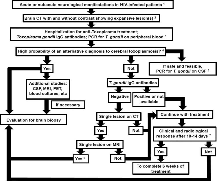
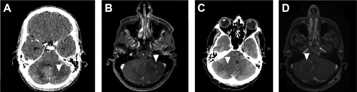
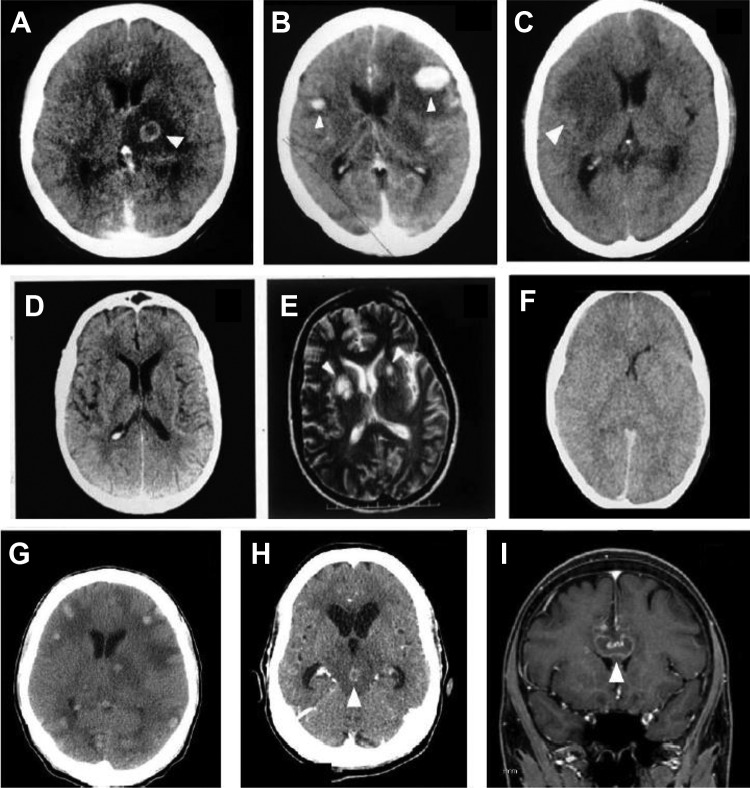
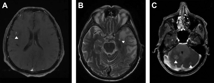
Cerebral toxoplasmosis is the most common cause of expansive brain lesions in people living with HIV/AIDS (PLWHA) and continues to cause high morbidity and mortality. The most frequent characteristics are focal subacute neurological deficits and ring-enhancing brain lesions in the basal ganglia, but the spectrum of clinical and neuroradiological manifestations is broad. Early initiation of antitoxoplasma therapy is an important feature of the diagnostic approach of expansive brain lesions in PLWHA. Pyrimethamine-based regimens and trimethoprim-sulfamethoxazole (TMP-SMX) seem to present similar efficacy, but TMP-SMX shows potential practical advantages. The immune reconstitution inflammatory syndrome is uncommon in cerebral toxoplasmosis, and we now have more effective, safe, and friendly combined antiretroviral therapy (cART) options. As a consequence of these 2 variables, the initiation of cART can be performed within 2 weeks after initiation of antitoxoplasma therapy. Herein, we will review historical and current concepts of epidemiology, diagnosis, and treatment of HIV-related cerebral toxoplasmosis.

Keywords: acquired immunodeficiency syndrome; central nervous system; cerebral toxoplasmosis; toxoplasmic encephalitis; toxoplasmosis.


Conflict of interest statement

Declaration of Conflicting Interests: The author(s) declared no potential conflicts of interest with respect to the research, authorship, and/or publication of this article.

Figures

Figure 1.
Figure 1.
Proposed algorithm for the management of suspicion cases of cerebral toxoplasmosis in people living with HIV/AIDS (PLWHA). This algorithm is meant as guidance, and individual clinical situation may render deviation from this algorithm preferable; expert opinion remains vital. Up to 4 weeks of suggestive clinical manifestations of expansive brain lesions, such as headache, motor focal deficit, or altered mental status. Most common patterns in brain computed tomography (CT) scan are ring-enhancing lesions with perilesional edema, nodular-enhancing lesions with perilesional edema, and nonenhancing lesions with expansive effect. Magnetic resonance imaging (MRI) should be obtained in patients with equivocal or negative CT scans. If available, MRI is the imaging modality of choice for evaluating PLWHA with expansive brain lesions. Antitoxoplasma therapy is a tool for the diagnosis of expansive brain lesions in PLWHA. Therefore, close clinical follow-up is imperative. The absence of Toxoplasma gondii immunoglobulin G (IgG) antibodies and the negative polymerase chain reaction (PCR) in blood samples do not rule out the possibility of cerebral toxoplasmosis. This step is very important to suspect early an alternative diagnosis to cerebral toxoplasmosis. The main issues to be evaluated are: local neuroepidemiology; degree of immunosuppression; and individual clinical, laboratorial, and neuroradiological features. We have carefully evaluated whether there is a risk of cerebral herniation that may contraindicate lumbar puncture. A negative polymerase chain reaction (PCR) for T gondii in cerebrospinal fluid (CSF) does not rule out the possibility of cerebral toxoplasmosis. The absence of T gondii IgG antibodies and the presence of a single lesion on MRI suggest an alternative diagnosis to cerebral toxoplasmosis and a brain biopsy is usually indicated. However, if the patient demonstrates clinical improvement, a new brain imaging can be performed with 1 to 2 weeks of antitoxoplasma therapy, and if there is radiological improvement, biopsy will not be necessary. Clinical improvement usually precedes radiological improvement, but always consider the impact of corticosteroids regardless of the cause of the disease.
Figure 2.
Figure 2.
Contrast-enhanced computed tomography (CT) scan and magnetic resonance imaging (MRI) of an HIV-infected patient with cerebral toxoplasmosis (A and B). At admission, a hypodense lesion without contrast-enhancing in the left cerebellar hemisphere (A). After 3 days, an MRI showed several ring- or heterogeneous-enhancing cerebellar lesions associated with perilesional edema. Computed tomography scan and MRI of an HIV-infected patient with cerebral toxoplasmosis (C and D). At admission, a hypodense lesion without contrast enhancing in the right cerebellar hemisphere associated with perilesional edema and deviation of the fourth ventricle (C). After 5 days, an MRI showed a ring-enhancing cerebellar lesion associated with perilesional edema and lesser deviation of the fourth ventricle (D).
Figure 3.
Figure 3.
Brain computed tomography (CT) images showing the spectrum of neuroradiological findings of cerebral toxoplasmosis in people living with HIV/AIDS. Hypodense lesion with ring-enhancing and perilesional edema (A); nodular-enhancing and perilesional edema (B); expansive hypodense lesion without contrast enhancing and with mass effect (C); contrast-enhanced CT scan without abnormalities (D) and corresponding T2-weighted magnetic resonance imaging (MRI) showing multiple basal ganglia lesions, with high-intensity signals (E); contrast-enhanced CT scan showing diffuse edema (F). Non-contrast-enhanced CT scan of an HIV-infected patient showing several spontaneous hyperdense lesions associated with perilesional edema. Histopathology study confirmed the diagnosis of hemorrhagic toxoplasmosis (G). Contrast-enhanced CT scan of an HIV-infected patient showing a ring-enhancing lesion in the mesencephalon associated with perilesional edema and hydrocephalus. This lesion showed complete resolution after 4 weeks of antitoxoplasma therapy (H). Sagittal contrast-enhanced T1-weighted MRI shows a single ring-enhancing lesion crossing the corpus callosum (I). This patient underwent brain biopsy with the suspicion of primary central nervous system (CNS) lymphoma, but the histopathology study confirmed the diagnosis of cerebral toxoplasmosis. The arrows show the abnormalities.
Figure 4.
Figure 4.
Magnetic resonance imaging (MRI) of HIV-infected patients with cerebral toxoplasmosis. T1-weighted imaging showed ring-enhancing brain lesion with a small, enhancing asymmetric nodule along the wall of the lesion (the “eccentric target sign; A). T2-weighted imaging showed a lesion with concentric alternating zones of hypo- and hyperintensities (the “concentric target sign”; B). T1-weighted imaging showed ring-enhancing brain lesion with a small, enhancing central nodule (the “target sign”; C). The arrows show the abnormalities.
Figure 5.
Figure 5.
Magnetic resonance imaging (MRI) and spectroscopy of an HIV-infected patient with cerebral toxoplasmosis (A-C). Gadolinium T1-weighted imaging showing a heterogeneous ring-enhancing brain lesion in right temporoparietal region (B). Spectroscopy image showing increased lipid peak and diminution of other metabolic activity corresponding to the known lesion (C). Magnetic resonance imaging (MRI) and 18F-fluorodeoxyglucose positron emission tomography–computed tomography (18F-FDG PET-CT) image of an HIV-infected patient with histopathologically confirmed primary central nervous system lymphoma (D-F). Gadolinium T1-weighted imaging showed irregular and nodular-enhancing brain lesion in left nucleocapsular region (E). 18F-FDG PET-CT image showing foci of increased metabolic activity corresponding to the known lesion (F). The arrows show the abnormalities.
Figure 6.
Figure 6.
Magnetic resonance imaging (MRI) of an HIV-infected patient with cerebral toxoplasmosis (A-C). At admission, single lesion was observed in the left parietal lobe (A). After 2 weeks of antitoxoplasma therapy without corticosteroids, partial reduction in both size and perilesional edema was observed (B). After 6 weeks of antitoxoplasma therapy, marked decrease in lesion size and perilesional edema was seen (C). Contrast-enhanced computed tomography (CT) imaging of an HIV-infected patient with cerebral toxoplasmosis (D-F). At admission, extensive single lesion in the left basal ganglia causing brain herniation was seen (D). After 4 weeks of antitoxoplasma therapy with corticosteroids, marked reduction was observed in both size and perilesional edema (E). After 8 weeks of antitoxoplasma therapy, another CT scan showed residual alterations only (F). The arrows show the abnormalities.

References

    1. Dunay IR, Gajurel K, Dhakal R, Liesenfeld O, Montoya JG. Treatment of toxoplasmosis: historical perspective, animal models, and current clinical practice. Clin Microbiol Rev. 2018;31(4):pii:e00057–17. - PMC - PubMed
    1. Lyons RE, McLeod R, Roberts CW. Toxoplasma gondii tachyzoite-bradyzoite interconversion. Trends Parasitol. 2002;18(5):198–201. - PubMed
    1. Luft BJ, Hafner R, Korzun AH. et al. Toxoplasmic encephalitis in patients with the acquired immunodeficiency syndrome. Members of the ACTG 077p/ANRS 009 Study Team. N Engl J Med. 1993;329(14):995–1000. - PubMed
    1. Vilaseca J, Arnau JM, Bacardi R, Mieras C, Serrano A, Navarro C. Kaposi’s sarcoma and Toxoplasma gondii brain abscess in a Spanish homosexual. Lancet. 1982;1(8271):572. - PubMed
    1. Centers for Disease Control (CDC). Opportunistic infections and Kaposi’s sarcoma among Haitians in the United States. MMWR Morb Mortal Wkly Rep. 1982;31(26):353–354, 360-361. - PubMed

MeSH terms

Substances