. 2019 Aug 19;20(16):4044.
doi: 10.3390/ijms20164044.

Stem Cells from a Female Rat Model of Type 2 Diabetes/Obesity and Stress Urinary Incontinence Are Damaged by In Vitro Exposure to its Dyslipidemic Serum, Predicting Inadequate Repair Capacity In Vivo

Affiliations

Stem Cells from a Female Rat Model of Type 2 Diabetes/Obesity and Stress Urinary Incontinence Are Damaged by In Vitro Exposure to its Dyslipidemic Serum, Predicting Inadequate Repair Capacity In Vivo

Istvan Kovanecz et al. Int J Mol Sci. .

Abstract

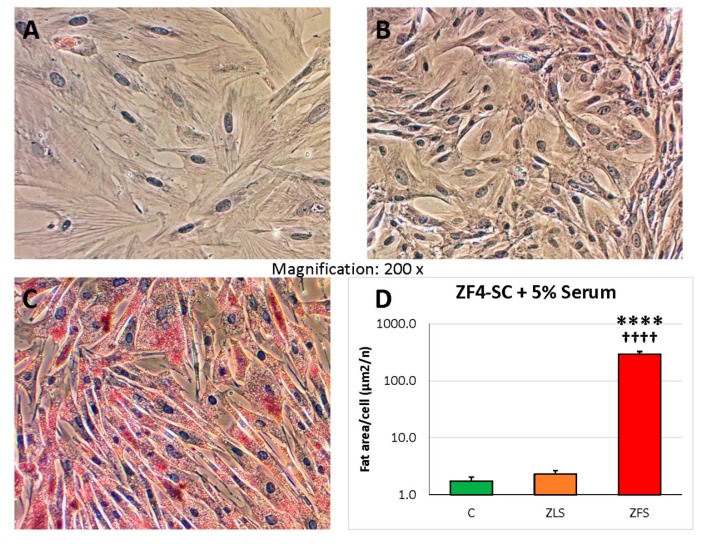
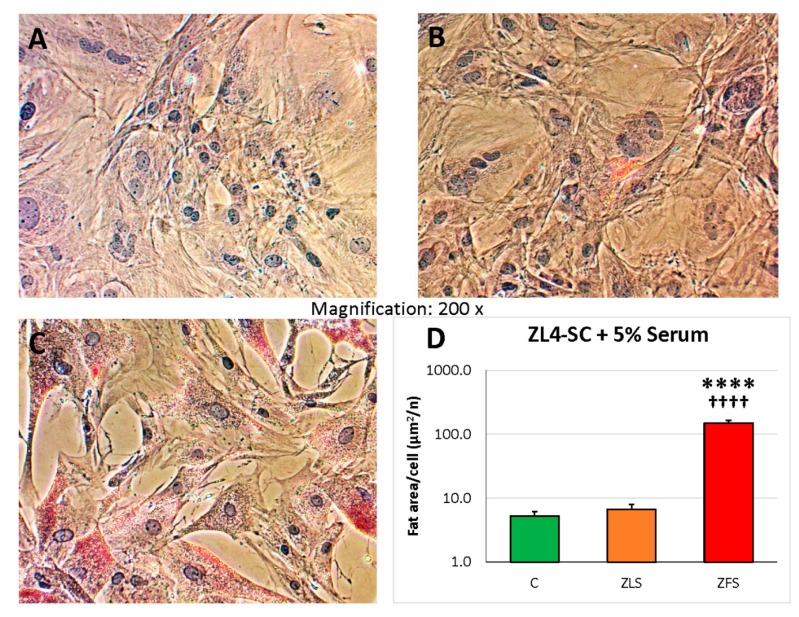
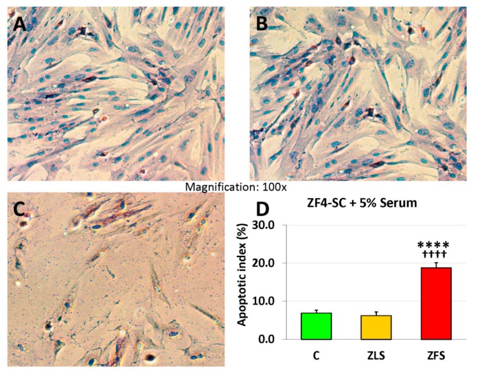
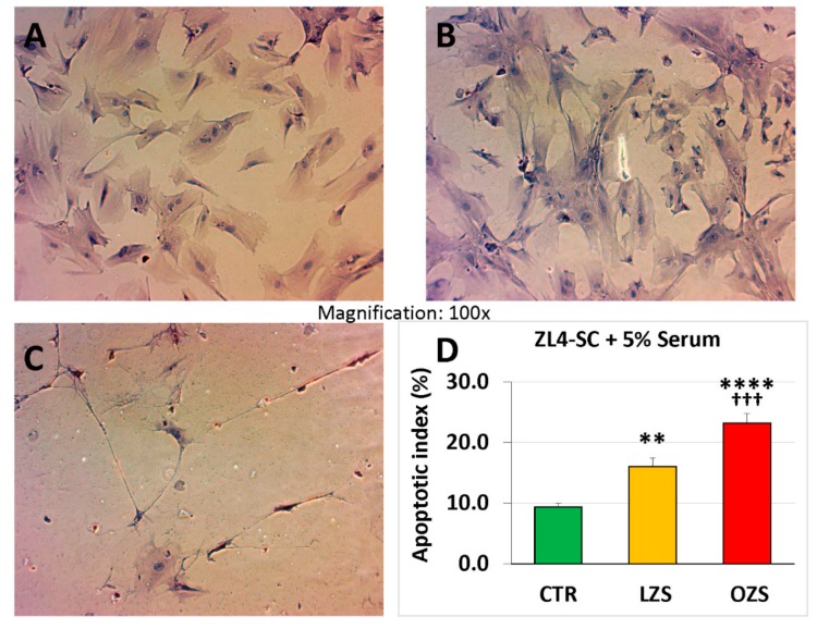
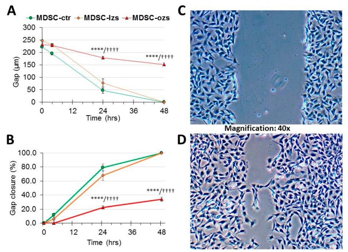
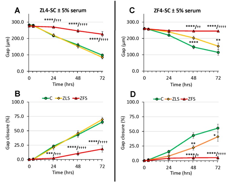
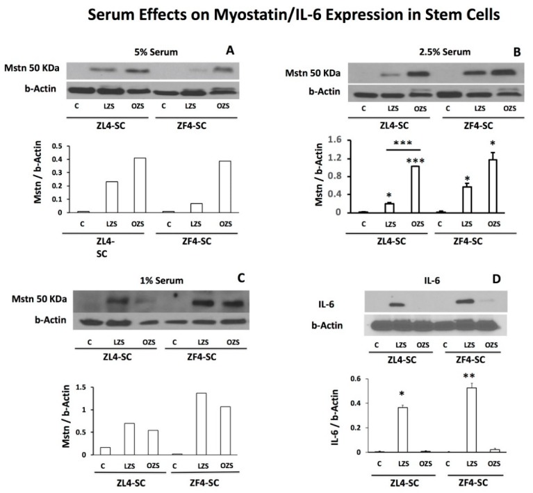
Female stress urinary incontinence (FSUI) is prevalent in women with type 2 diabetes/obesity (T2D/O), and treatment is not optimal. Autograph stem cell therapy surprisingly has poor efficacy. In the male rat model of T2D/O, it was demonstrated that epigenetic changes, triggered by long-term exposure to the dyslipidemic milieu, led to abnormal global transcriptional signatures (GTS) of genes and microRNAs (miR), and impaired the repair capacity of muscle-derived stem cells (MDSC). This was mimicked in vitro by treatment of MDSC with dyslipidemic serum or lipid factors. The current study aimed to predict whether these changes also occur in stem cells from female 12 weeks old T2D/O rats, a model of FSUI. MDSCs from T2D/O (ZF4-SC) and normal female rats (ZL4-SC) were treated in vitro with either dyslipidemic serum (ZFS) from late T2D/O 24 weeks old female Zucker fatty (ZF) rats, or normal serum (ZLS) from 24 weeks old female Zucker lean (ZL) rats, for 4 days and subjected to assays for fat deposition, apoptosis, scratch closing, myostatin, interleukin-6, and miR-GTS. The dyslipidemic ZFS affected both female stem cells more severely than in the male MDSC, with some gender-specific differences in miR-GTS. The changes in miR-GTS and myostatin/interleukin-6 balance may predict in vivo noxious effects of the T2D/O milieu that might impair autograft stem cell (SC) therapy for FSUI, but this requires future studies.

Keywords: apoptosis; dyslipidemia; fat infiltration; interleukin-6; microRNA; muscle-derived stem cells; myostatin; wound closure.


Conflict of interest statement

The authors declare no conflict of interest.

Figures

Figure 1
Figure 1
Incubation of ZF4-SC with ZF (Zucker fatty) serum (ZFS) induced intracellular infiltration by fat globules, whereas the one caused by ZL (Zucker lean) serum (ZLS) was very low and similar to the one in control with no addition. ZF4-SC were incubated for 4 days with no addition (A), or with added 5% ZLS from 20 weeks old ZL rats (B) or 5% ZFS from age-matched rats (C), and stained at 4–5 days with Oil Red O for fat infiltration. Pictures were taken at 200X, but QIA was applied to multiple fields at 100× (D) showing the bar graph of red area (fat) per cell, in a semi-logarithmic scale, C: no serum addition; **** p ≤ 0.0001 (C vs. ZFS); ++++ p ≤ 0.0001 (ZFS vs. ZLS).
Figure 2
Figure 2
The fat infiltration of ZL4-SC caused by ZFS was similar to the one experienced by the ZF4-SC in Figure 1, with negligible infiltration by ZLS, like in no rat serum addition, indicating a similar response of both MDSC to ZFS. ZL4-SC were incubated as the ZF4-SC in Figure 1 with no addition (A), or added ZLS (B), or ZFS (C), then stained with Oil Red O, and pictures were taken as in Figure 1. (D) The bar graph is equivalent to the one in Figure 1D, with **** p ≤ 0.0001 (C vs. ZFS); ++++ p ≤ 0.0001 (ZFS vs. ZLS).
Figure 3
Figure 3
Incubation of ZF4-SC with ZFS-induced apoptosis, but the one caused by ZLS was negligible, similar to when no rat serum was added. ZF4-SC were incubated with no addition (A), or added ZLS (B), or ZFS (C), as in Figure 1, then subjected to the TUNEL reaction, and pictures were taken at 100X. The bar graph in (D) is equivalent to the one in Figure 1D, but on a linear scale with **** p ≤ 0.0001 (C vs. ZFS); ++++ p ≤ 0.0001 (ZFS vs. ZLS).
Figure 4
Figure 4
ZL4-SC were more sensitive to apoptosis caused by ZFS than the ZF4-SC shown in Figure 3, but they were also mildly affected by ZLS. ZL4-SC were incubated, as in Figure 1, with no addition (A) or added ZLS (B) or ZFS (C), and then subjected to TUNEL reaction, and pictures were taken at 100×. (D) The bar graph is equivalent to the one in Figure 1 but on a linear scale. C: no serum addition; **** p ≤ 0.0001 (C vs. ZFS); ** p ≤ 0.01 (C vs. ZLS); +++ p ≤ 0.001 (ZFS vs. ZLS).
Figure 5
Figure 5
Gap closure of in vitro scratch injury by male ED-MDSC at 48 h was partially abrogated by the male lean Zucker serum, and it did not occur by the addition of the male obese Zucker serum, thus showing MDSC damage specifically by the latter. In this case, pertaining to the male ED-MDSC isolated in our studies of refs #18 and #19, and using their male lean Zucker serum (lzs), or obese Zucker serum (ozs), or no addition control (ctr), these preceding abbreviations are used, differing from the ones for the female rats used elsewhere (including Figure 6), to emphasize that both the stem cells and the sera were from male animals and not from their female counterparts. A gap was created in confluent monolayers of ED-MDSC, and either ozs or lzs was immediately added to 5% or not. Pictures were immediately taken at 0 h, and then at 3, 24, and 48 h, measuring both the gap width (µm) and its closure (%). Panel (C) is a micrograph at 0 h in the presence of added ZFS, and Panel (D) is the final one at 48 h with still a residual gap. Panel (A) is a time plot of the gap width, and Panel (B) is for gap closure. **** p ≤ 0.0001 (ctr vs. ozs), and ++++ p ≤ 0.0001 (lzs vs. ozs).
Figure 6
Figure 6
The efficacy of gap closure by ZF4-SC after gap injury was lower than the one for ED-MDSC, and it was more damaged by ZFS, but ZL4-SC were less affected than ZF4-SC. Experiments and measures were as in Figure 5, but with the female stem cells, and the female abbreviations. Panels (A) and (C): ZL4-SC; Panels (B) and (D): ZF4-SC; **** p ≤ 0.0001 (C vs. ZFS), *** p ≤ 0.005 (C vs. ZLS), ** p ≤ 0.01 (C vs. ZLS), and + p ≤ 0.05 (ZLS vs. ZFS), +++ p ≤ 0.005 (ZLS vs. ZFS), and ++++ p ≤ 0.0001 (ZLS vs. ZFS).
Figure 7
Figure 7
Myostatin in ZF4-SC and ZL4-SC was increased by ZFS (OZS) added to 5% and 2.5%, but little by ZLS (LZS), and the effects were less specific at 1%. This was associated with a potential counteraction by ZFS (OZS) of IL-6 expression. The respective stem cells were incubated in vitro for 4 days with either no addition, or with ZFS (OZS) or ZLS (LZS) added to 5% (A), 2.5% (B,D), or 1% (C), and the cell protein homogenates were assayed by western blot. The 50 kDa band was quantitated and corrected by the housekeeping gene beta-actin. Separate western blots were done for interleukin-6 (IL-6). Statistics were not applied on panels A and C, but based on this comparison, the effects of 2.5% incubations were repeated for statistical analysis as shown in panels B and D. *** p ≤ 0.001 (ZFS (OZS) vs C and vs ZLS (LZS)), ** p ≤ 0.01 (ZFS (OZS) vs ZLS (LZS)), and *p ≤ 0.05 (ZFS (OZS) vs. ZLS (LZS)).

References

    1. Aoki Y., Brown H.W., Brubaker L., Cornu J.N., Daly J.O., Cartwright R. Correction: Urinary incontinence in women. Nat. Rev. Dis. Primers. 2017;3:17097. doi: 10.1038/nrdp.2017.97. - DOI - PubMed
    1. Capobianco G., Madonia M., Morelli S., Dessole F., De Vita D., Cherchi P.L., Dessole S. Management of female stress urinary incontinence: A care pathway and update. Maturitas. 2018;109:32–38. doi: 10.1016/j.maturitas.2017.12.008. - DOI - PubMed
    1. Lukacz E.S., Santiago-Lastra Y., Albo M.E., Brubaker L. Urinary Incontinence in Women. JAMA. 2017;318:1592–1604. doi: 10.1001/jama.2017.12137. - DOI - PubMed
    1. Bennington J., Williams J.K., Andersson K.-E. New concepts in regenerative medicine approaches to the treatment of female stress urinary incontinence. Curr. Opin. Urol. 2019;29:380–384. doi: 10.1097/MOU.0000000000000617. - DOI - PubMed
    1. Thomaz R.P., Colla C., Darski C., Paiva L.L. Influence of pelvic floor muscle fatigue on stress urinary incontinence: A systematic review. Int. Urogynecol. J. 2018;29:197–204. doi: 10.1007/s00192-017-3538-6. - DOI - PubMed