Epigallocatechin gallate (EGCG) suppresses growth and tumorigenicity in breast cancer cells by downregulation of miR-25
- PMID: 31431131
- PMCID: PMC6738446
- DOI: 10.1080/21655979.2019.1657327
Epigallocatechin gallate (EGCG) suppresses growth and tumorigenicity in breast cancer cells by downregulation of miR-25
Abstract
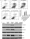
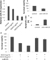
The aim of the present study was to investigate the anticancer effects and potential mechanisms of polyphenol epigallocatechin-3-gallate (EGCG) on breast cancer MCF-7 cells in vitro and in vivo. Our results showed that EGCG significantly inhibited MCF-7 cell viability in a time- and dose-dependent manner. Flow cytometry analysis indicated that EGCG induced apoptosis and disrupted cell cycle progression at G2/M phase. Moreover, EGCG inhibited miR-25 expression and increased PARP, pro-caspase-3 and pro-caspase-9 at protein levels. Restoration of miR-25 inhibited EGCG-induced cell apoptosis. Furthermore, EGCG suppressed tumor growth in vivo by downregulating the expression of miR-25 and proteins associated with apoptosis, which was further confirmed by a reduction of Ki-67 and increase of pro-apoptotic PARP expression as determined by immunohistochemistry staining. These findings indicate that EGCG possesses chemopreventive potential in breast cancer which may serve as a promising anticancer agent for clinical applications.
Keywords: Polyphenol epigallocatechin-3-gallate; apoptosis; breast cancer; proliferation.
Figures


Similar articles
-
Suppressing glucose metabolism with epigallocatechin-3-gallate (EGCG) reduces breast cancer cell growth in preclinical models.Food Funct. 2018 Nov 14;9(11):5682-5696. doi: 10.1039/c8fo01397g. Food Funct. 2018. PMID: 30310905 Free PMC article.
-
Molecular mechanism of epigallocatechin-3-gallate in human esophageal squamous cell carcinoma in vitro and in vivo.Oncol Rep. 2015 Jan;33(1):297-303. doi: 10.3892/or.2014.3555. Epub 2014 Oct 20. Oncol Rep. 2015. PMID: 25333353
-
Epigallocatechin-3-gallate suppresses the expression of HSP70 and HSP90 and exhibits anti-tumor activity in vitro and in vivo.BMC Cancer. 2010 Jun 10;10:276. doi: 10.1186/1471-2407-10-276. BMC Cancer. 2010. PMID: 20537126 Free PMC article.
-
The Role of EGCG in Breast Cancer Prevention and Therapy.Mini Rev Med Chem. 2021;21(7):883-898. doi: 10.2174/1389557520999201211194445. Mini Rev Med Chem. 2021. PMID: 33319659 Review.
-
Protective Effects of Epigallocatechin Gallate (EGCG) on Endometrial, Breast, and Ovarian Cancers.Biomolecules. 2020 Oct 25;10(11):1481. doi: 10.3390/biom10111481. Biomolecules. 2020. PMID: 33113766 Free PMC article. Review.
Cited by
-
Role of epigallocatechin-3- gallate in the regulation of known and novel microRNAs in breast carcinoma cells.Front Genet. 2022 Oct 6;13:995046. doi: 10.3389/fgene.2022.995046. eCollection 2022. Front Genet. 2022. PMID: 36276982 Free PMC article.
-
Epigallocatechin-3-O-gallate promotes extracellular matrix and inhibits inflammation in IL-1β stimulated chondrocytes by the PTEN/miRNA-29b pathway.Pharm Biol. 2022 Dec;60(1):589-599. doi: 10.1080/13880209.2022.2039722. Pharm Biol. 2022. PMID: 35260041 Free PMC article.
-
Inhibition of Cancer Development by Natural Plant Polyphenols: Molecular Mechanisms.Int J Mol Sci. 2023 Jun 26;24(13):10663. doi: 10.3390/ijms241310663. Int J Mol Sci. 2023. PMID: 37445850 Free PMC article. Review.
-
An update of Nrf2 activators and inhibitors in cancer prevention/promotion.Cell Commun Signal. 2022 Jun 30;20(1):100. doi: 10.1186/s12964-022-00906-3. Cell Commun Signal. 2022. PMID: 35773670 Free PMC article. Review.
-
Green tea epigallocatechin gallate suppresses 3T3-L1 cell growth via microRNA-143/MAPK7 pathways.Exp Biol Med (Maywood). 2022 Sep;247(18):1670-1679. doi: 10.1177/15353702221108925. Epub 2022 Jul 27. Exp Biol Med (Maywood). 2022. PMID: 35894140 Free PMC article.
References
-
- Baselga J, Norton L.. Focus on breast cancer. Cancer Cell. 2002;1:319–322. - PubMed
-
- Coughlin SS, Ekwueme DU. Breast cancer as a global health concern. Cancer Epidemiol. 2009;33:315–318. - PubMed
-
- Mukherjee S, Ghosh S, Das DK, et al. Gold-conjugated green tea nanoparticles for enhanced anti-tumor activities and hepatoprotection–synthesis, characterization and in vitro evaluation. J Nutr Biochem. 2015;26:1283–1297. - PubMed
-
- Lu G, Liao J, Yang G, et al. Inhibition of adenoma progression to adenocarcinoma in a 4-(methylnitrosamino)-1-(3-pyridyl)-1-butanone-induced lung tumorigenesis model in A/J mice by tea polyphenols and caffeine. Cancer Res. 2006;66:11494–11501. - PubMed
MeSH terms
Substances
LinkOut - more resources
Full Text Sources
Medical
Research Materials
