

Engineering approaches to studying cancer cell migration in three-dimensional environments
- PMID: 31431175
- PMCID: PMC6627017
- DOI: 10.1098/rstb.2018.0219
Engineering approaches to studying cancer cell migration in three-dimensional environments
Abstract
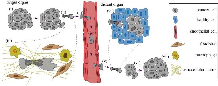
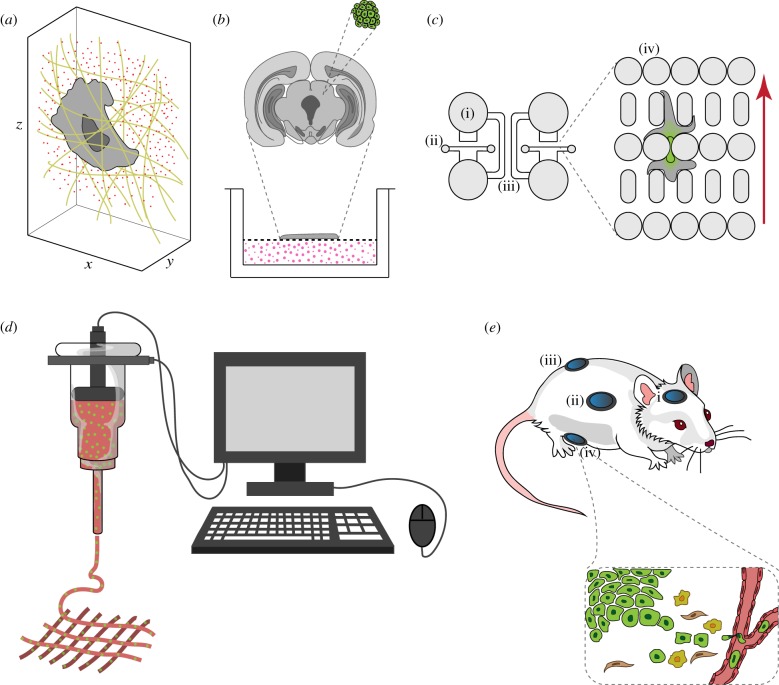
Cancer is one of the most devastating diseases of our time, with 17 million new cancer cases and 9.5 million cancer deaths in 2018 worldwide. The mortality associated with cancer results primarily from metastasis, i.e. the spreading of cancer cells from the primary tumour to other organs. The invasion and migration of cells through basement membranes, tight interstitial spaces and endothelial cell layers are key steps in the metastatic cascade. Recent studies demonstrated that cell migration through three-dimensional environments that mimic the in vivo conditions significantly differs from their migration on two-dimensional surfaces. Here, we review recent technological advances made in the field of cancer research that provide more 'true to the source' experimental platforms and measurements for the study of cancer cell invasion and migration in three-dimensional environments. These include microfabrication, three-dimensional bioprinting and intravital imaging tools, along with force and stiffness measurements of cells and their environments. These techniques will enable new studies that better reflect the physiological environment found in vivo, thereby producing more robust results. The knowledge achieved through these studies will aid in the development of new treatment options with the potential to ultimately lighten the devastating cost cancer inflicts on patients and their families. This article is part of a discussion meeting issue 'Forces in cancer: interdisciplinary approaches in tumour mechanobiology'.
Keywords: decellularized tissue; force generation; intravital imaging; microfluidics; nuclear mechanics; three-dimensional bioprinting.
Conflict of interest statement
We have no competing interests.
Figures
Similar articles
-
Characterizing Cell Migration Within Three-dimensional In Vitro Wound Environments.J Vis Exp. 2017 Aug 16;(126):56099. doi: 10.3791/56099. J Vis Exp. 2017. PMID: 28872146 Free PMC article.
-
Design of a microfluidic device to quantify dynamic intra-nuclear deformation during cell migration through confining environments.Integr Biol (Camb). 2015 Dec;7(12):1534-46. doi: 10.1039/c5ib00200a. Epub 2015 Nov 9. Integr Biol (Camb). 2015. PMID: 26549481 Free PMC article.
-
Cell migration through three-dimensional confining pores: speed accelerations by deformation and recoil of the nucleus.Philos Trans R Soc Lond B Biol Sci. 2019 Aug 19;374(1779):20180225. doi: 10.1098/rstb.2018.0225. Epub 2019 Jul 1. Philos Trans R Soc Lond B Biol Sci. 2019. PMID: 31431171 Free PMC article.
-
The matrix environmental and cell mechanical properties regulate cell migration and contribute to the invasive phenotype of cancer cells.Rep Prog Phys. 2019 Jun;82(6):064602. doi: 10.1088/1361-6633/ab1628. Epub 2019 Apr 4. Rep Prog Phys. 2019. PMID: 30947151 Review.
-
Bioengineering paradigms for cell migration in confined microenvironments.Curr Opin Cell Biol. 2014 Oct;30:41-50. doi: 10.1016/j.ceb.2014.06.001. Epub 2014 Jun 26. Curr Opin Cell Biol. 2014. PMID: 24973724 Free PMC article. Review.
Cited by
-
The forces of cancer.Philos Trans R Soc Lond B Biol Sci. 2019 Aug 19;374(1779):20190103. doi: 10.1098/rstb.2019.0103. Epub 2019 Jul 1. Philos Trans R Soc Lond B Biol Sci. 2019. PMID: 31431173 Free PMC article. No abstract available.
-
Real-time label-free three-dimensional invasion assay for anti-metastatic drug screening using impedance sensing.Front Pharmacol. 2024 Jun 11;15:1387949. doi: 10.3389/fphar.2024.1387949. eCollection 2024. Front Pharmacol. 2024. PMID: 38919253 Free PMC article.
-
Direct comparison of five different 3D extracellular matrix model systems for characterization of cancer cell migration.Cancer Rep (Hoboken). 2020 Oct;3(5):e1257. doi: 10.1002/cnr2.1257. Epub 2020 Jun 8. Cancer Rep (Hoboken). 2020. PMID: 33085847 Free PMC article.
-
Assembly and Use of a Microfluidic Device to Study Nuclear Mechanobiology During Confined Migration.Methods Mol Biol. 2022;2502:329-349. doi: 10.1007/978-1-0716-2337-4_22. Methods Mol Biol. 2022. PMID: 35412249 Free PMC article.
-
Unraveling Cancer Metastatic Cascade Using Microfluidics-based Technologies.Biophys Rev. 2022 Apr 14;14(2):517-543. doi: 10.1007/s12551-022-00944-8. eCollection 2022 Apr. Biophys Rev. 2022. PMID: 35528034 Free PMC article. Review.
References
-
- Yadav SK, Feigelson SW, Roncato F, Antman-Passig M, Shefi O, Lammerding J, Alon R. 2018. Frontline science: elevated nuclear lamin A is permissive for granulocyte transendothelial migration but not for motility through collagen I barriers. J. Leukoc. Biol. 104, 239–251. (10.1002/JLB.3HI1217-488R) - DOI - PMC - PubMed
Publication types
MeSH terms
Grants and funding
LinkOut - more resources
Full Text Sources
