Review
. 2019 Aug 19;374(1779):20180221.
doi: 10.1098/rstb.2018.0221. Epub 2019 Jul 1.

The plasma membrane as a mechanochemical transducer

Affiliations
Review

The plasma membrane as a mechanochemical transducer

Anabel-Lise Le Roux et al. Philos Trans R Soc Lond B Biol Sci. .

Abstract

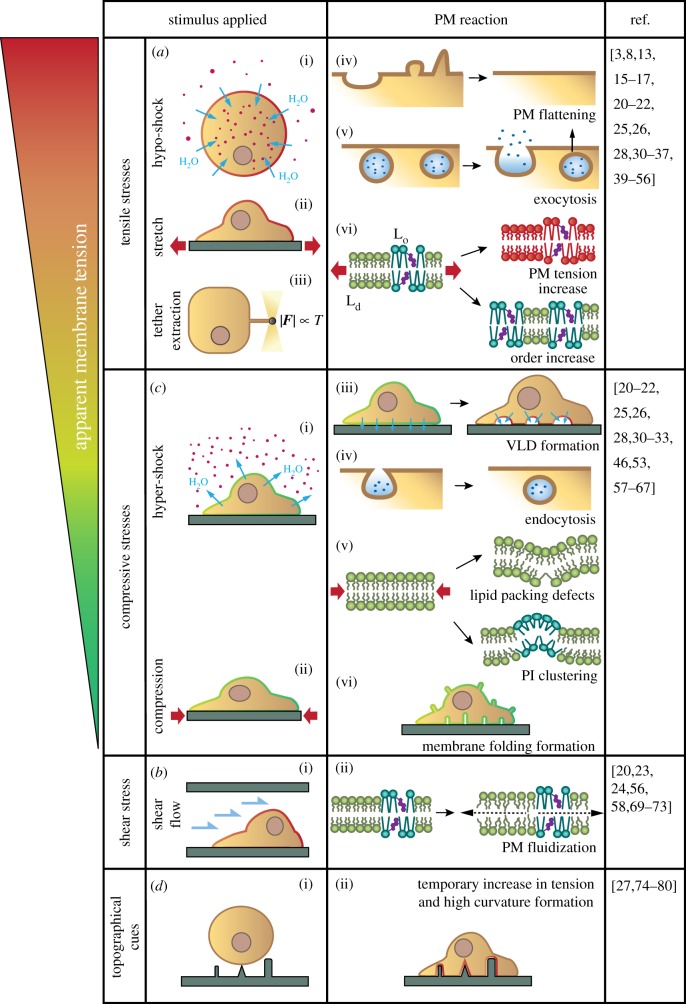
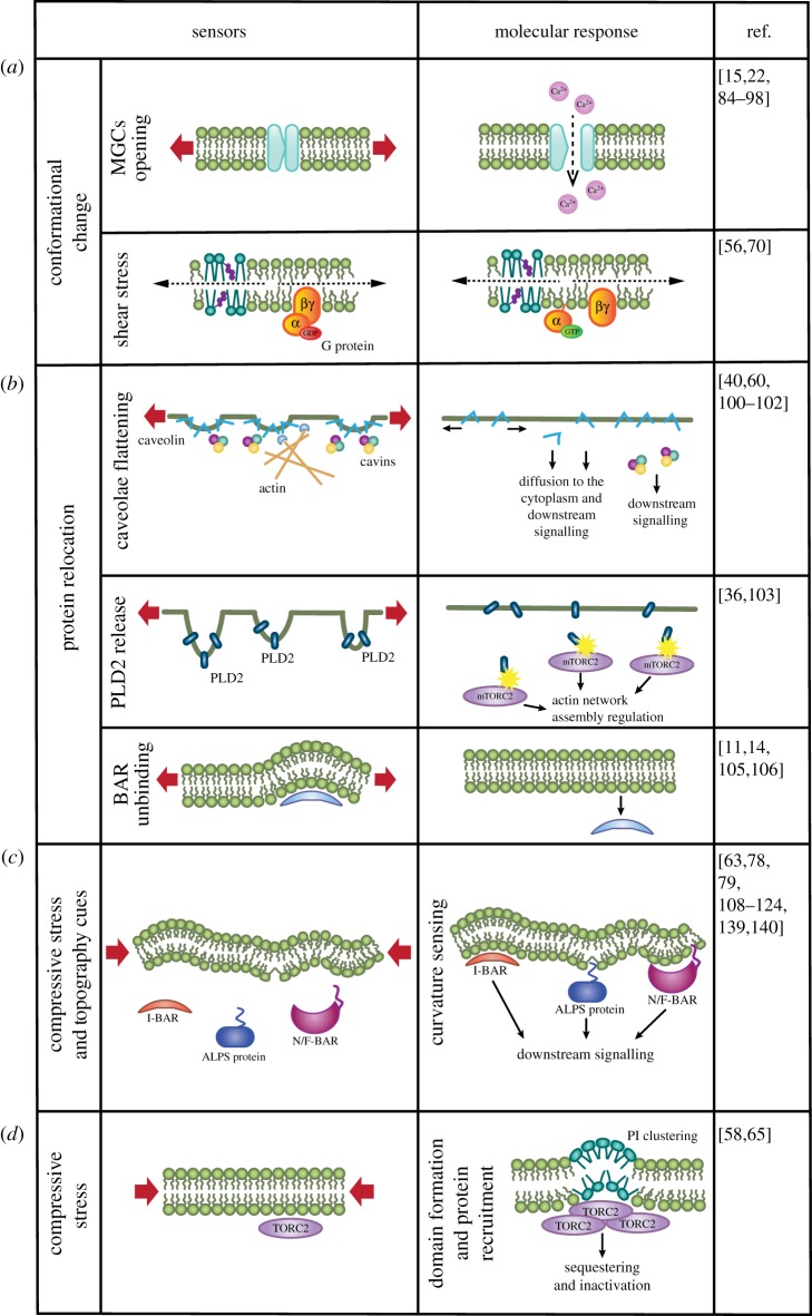
Cells are constantly submitted to external mechanical stresses, which they must withstand and respond to. By forming a physical boundary between cells and their environment that is also a biochemical platform, the plasma membrane (PM) is a key interface mediating both cellular response to mechanical stimuli, and subsequent biochemical responses. Here, we review the role of the PM as a mechanosensing structure. We first analyse how the PM responds to mechanical stresses, and then discuss how this mechanical response triggers downstream biochemical responses. The molecular players involved in PM mechanochemical transduction include sensors of membrane unfolding, membrane tension, membrane curvature or membrane domain rearrangement. These sensors trigger signalling cascades fundamental both in healthy scenarios and in diseases such as cancer, which cells harness to maintain integrity, keep or restore homeostasis and adapt to their external environment. This article is part of a discussion meeting issue 'Forces in cancer: interdisciplinary approaches in tumour mechanobiology'.

Keywords: mechanosensor; mechanotransduction; membrane tension; plasma membrane.


Conflict of interest statement

We declare we have no competing interests.

Figures

Figure 1.
Figure 1.
PM response to applied mechanical stimuli. (a) Tensile stresses are applied experimentally by tether pulling, hypotonic shocks and cell stretching. In response, PM folds flatten and exocytosis increases, buffering the increase in tension. Once lipid reserves have been used, PM tension and order increase. (b) Compressive stresses are applied experimentally through hyper-osmotic shocks and stretch release. In response, PM folds of different shapes and sizes form (vacuole-like dilations (VLDs), tubes), endocytosis increases, lipid packing defects appear in highly curved areas and phosphoinositide (PI) clusters form. (c) Shear stress application results in increased PM fluidity, in both Lo and Ld phases. (d) Upon encountering topographical cues, cells adapt their PM to substrate architecture, likely triggering a temporary increase in PM tension.
Figure 2.
Figure 2.
Molecular sensors of PM mechanical state. (a) Mechanically induced conformational changes include MGC opening upon stretch (thereby enabling ion transport) and G protein dimerization under shear flow. (b) Mechanically induced protein relocation events. (top) With increased PM tension, caveolae flatten and its proteic components relocate by freely diffusing through the PM and/or the cytosol. These molecular players can subsequently activate different signalling pathways. (middle) PLD2 proteins are sequestered at PM invaginations. When they unfold, PLD2 is released and activates its partner mTORC2, which subsequently regulates actin network assembly. (bottom) BAR proteins respond to fold flattening by unbinding from the PM and diffusing into the cytoplasm. (c) Compressive stresses and topographical cues result in the formation of different types of PM curvature that can be recognized by BAR proteins through their positively charged BAR domain. Proteins containing ALPS motifs can also sense curvature by inserting their amphipathic helix in curved PM areas, where lipid packing defects are more abundant. (d) Clustering of PIs due to compressive stresses leads to TORC2 sequestering and inhibition of its activity.

References

    1. Northey JJ, Przybyla L, Weaver VM. 2017. Tissue force programs cell fate and tumor aggression. Cancer Discovery 7, 1224–1237. (10.1158/2159-8290.CD-16-0733) - DOI - PMC - PubMed
    1. Faurobert E, Bouin A-P, Albiges-Rizo C. 2015. Microenvironment, tumor cell plasticity, and cancer. Curr. Opin. Oncol. 27, 64–70. (10.1097/CCO.0000000000000154) - DOI - PubMed
    1. Pontes B, et al. 2017. Membrane tension controls adhesion positioning at the leading edge of cells. J. Cell Biol. 216, 2959 (10.1083/jcb.201611117) - DOI - PMC - PubMed
    1. Chugh P, et al. 2017. Actin cortex architecture regulates cell surface tension. Nat. Cell Biol. 19, 689–697. (10.1038/ncb3525) - DOI - PMC - PubMed
    1. Simon C, Caorsi V, Campillo C, Sykes C. 2018. Interplay between membrane tension and the actin cytoskeleton determines shape changes. Phys. Biol. 15, 65004 (10.1088/1478-3975/aad1ab) - DOI - PubMed

Publication types