

Is cell migration a selectable trait in the natural evolution of cancer development?
- PMID: 31431177
- PMCID: PMC6627024
- DOI: 10.1098/rstb.2018.0224
Is cell migration a selectable trait in the natural evolution of cancer development?
Abstract
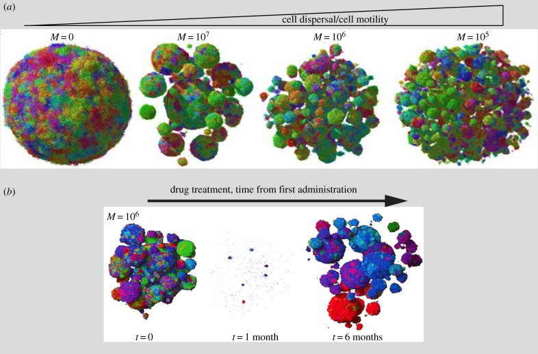
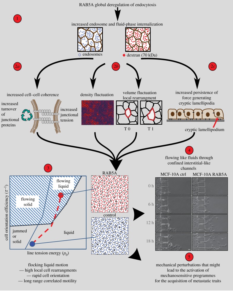
Selective evolutionary pressure shapes the processes and genes that enable cancer survival and expansion in a tumour-suppressive environment. A distinguishing lethal feature of malignant cancer is its dissemination and seeding of metastatic foci. A key requirement for this process is the acquisition of a migratory/invasive ability. However, how the migratory phenotype is selected for during the natural evolution of cancer and what advantage, if any, it might provide to the growing malignant cells remain open issues. In this opinion piece, we discuss three possible answers to these issues. We will examine lines of evidence from mathematical modelling of cancer evolution that indicate that migration is an intrinsic selectable property of malignant cells that directly impacts on growth dynamics and cancer geometry. Second, we will argue that migratory phenotypes can emerge as an adaptive response to unfavourable growth conditions and endow cells not only with the ability to move/invade, but also with specific metastatic traits, including drug resistance, self-renewal and survival. Finally, we will discuss the possibility that migratory phenotypes are coincidental events that emerge by happenstance in the natural evolution of cancer. This article is part of a discussion meeting issue 'Forces in cancer: interdisciplinary approaches in tumour mechanobiology'.
Keywords: cancer evolution; cell migration; collective motility; endocytosis.
Conflict of interest statement
We declare we have no competing interests.
Figures
Similar articles
-
Cancer as an evolutionary process at the cell level: an epidemiological perspective.Carcinogenesis. 2003 Jan;24(1):1-6. doi: 10.1093/carcin/24.1.1. Carcinogenesis. 2003. PMID: 12538342
-
Evolution of cell motility in an individual-based model of tumour growth.J Theor Biol. 2009 Jul 7;259(1):67-83. doi: 10.1016/j.jtbi.2009.03.005. Epub 2009 Mar 12. J Theor Biol. 2009. PMID: 19285513 Free PMC article.
-
The matrix environmental and cell mechanical properties regulate cell migration and contribute to the invasive phenotype of cancer cells.Rep Prog Phys. 2019 Jun;82(6):064602. doi: 10.1088/1361-6633/ab1628. Epub 2019 Apr 4. Rep Prog Phys. 2019. PMID: 30947151 Review.
-
Tracking the evolution of cancer cell populations through the mathematical lens of phenotype-structured equations.Biol Direct. 2016 Aug 23;11(1):43. doi: 10.1186/s13062-016-0143-4. Biol Direct. 2016. PMID: 27550042 Free PMC article.
-
Engineering approaches to studying cancer cell migration in three-dimensional environments.Philos Trans R Soc Lond B Biol Sci. 2019 Aug 19;374(1779):20180219. doi: 10.1098/rstb.2018.0219. Epub 2019 Jul 1. Philos Trans R Soc Lond B Biol Sci. 2019. PMID: 31431175 Free PMC article. Review.
Cited by
-
Evolution of multicellularity by collective integration of spatial information.Elife. 2020 Oct 16;9:e56349. doi: 10.7554/eLife.56349. Elife. 2020. PMID: 33064078 Free PMC article.
-
Direct antitumoral effects of sulfated fucans isolated from echinoderms: a possible role of neuropilin-1/β1 integrin endocytosis and focal adhesion kinase degradation.Glycobiology. 2023 Oct 29;33(9):715-731. doi: 10.1093/glycob/cwad044. Glycobiology. 2023. PMID: 37289485 Free PMC article.
-
The forces of cancer.Philos Trans R Soc Lond B Biol Sci. 2019 Aug 19;374(1779):20190103. doi: 10.1098/rstb.2019.0103. Epub 2019 Jul 1. Philos Trans R Soc Lond B Biol Sci. 2019. PMID: 31431173 Free PMC article. No abstract available.
References
-
- Aguirre-Ghiso JA, Sosa MS. 2018. Emerging topics on disseminated cancer cell dormancy and the paradigm of metastasis. Annu. Rev. Cancer Biol. 2, 377–393. (10.1146/annurev-cancerbio-030617-050446) - DOI
Publication types
MeSH terms
LinkOut - more resources
Full Text Sources
