Review
. 2019 Aug 30;16(157):20190233.
doi: 10.1098/rsif.2019.0233. Epub 2019 Aug 21.

Growth and remodelling of living tissues: perspectives, challenges and opportunities

Affiliations
Review

Growth and remodelling of living tissues: perspectives, challenges and opportunities

Davide Ambrosi et al. J R Soc Interface. .

Abstract

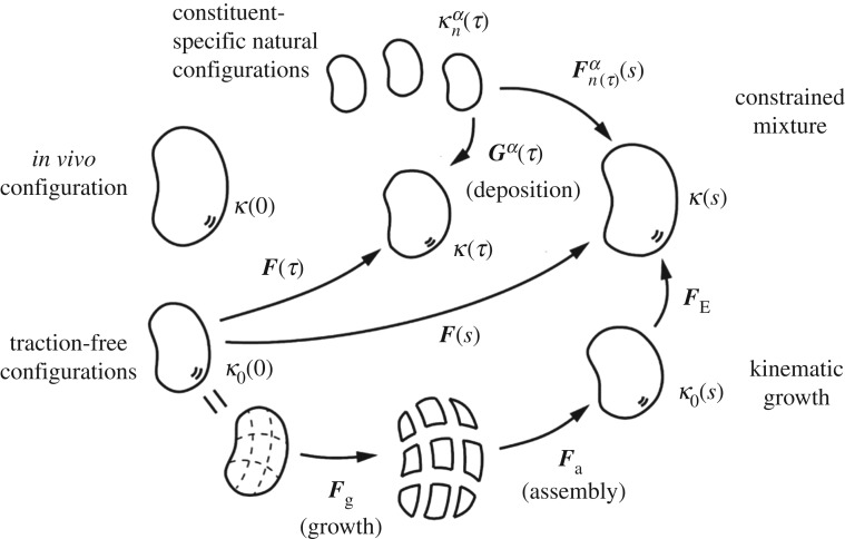
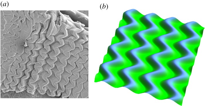
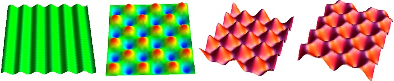
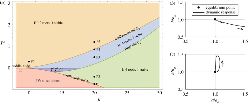
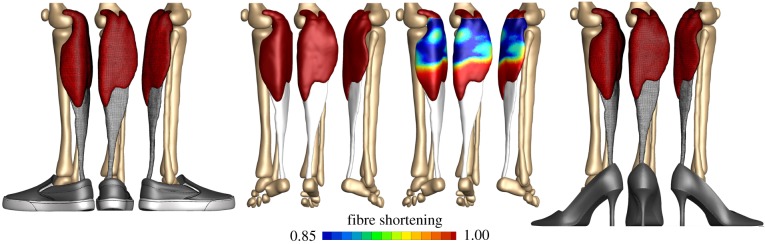
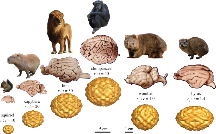
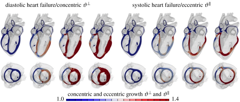
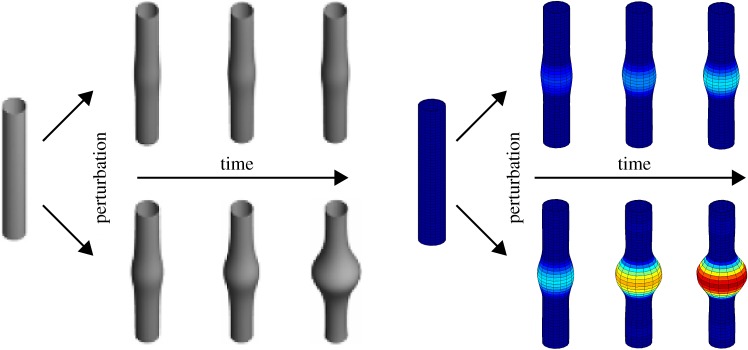
One of the most remarkable differences between classical engineering materials and living matter is the ability of the latter to grow and remodel in response to diverse stimuli. The mechanical behaviour of living matter is governed not only by an elastic or viscoelastic response to loading on short time scales up to several minutes, but also by often crucial growth and remodelling responses on time scales from hours to months. Phenomena of growth and remodelling play important roles, for example during morphogenesis in early life as well as in homeostasis and pathogenesis in adult tissues, which often adapt to changes in their chemo-mechanical environment as a result of ageing, diseases, injury or surgical intervention. Mechano-regulated growth and remodelling are observed in various soft tissues, ranging from tendons and arteries to the eye and brain, but also in bone, lower organisms and plants. Understanding and predicting growth and remodelling of living systems is one of the most important challenges in biomechanics and mechanobiology. This article reviews the current state of growth and remodelling as it applies primarily to soft tissues, and provides a perspective on critical challenges and future directions.

Keywords: growth; instabilities; living systems; morphoelasticity; remodelling.


Conflict of interest statement

We declare we have no competing interests.

Figures

Figure 1.
Figure 1.
A golden age of discovery and invention in growth and remodelling: Wilhelm His’s mechanical analogy between (a) the folding of a rubber tube and (b) the folding of a gut tube during morphogenesis [1]. (c) Wolff’s structural study of a bone [2]. (d) Traction methods by Davis to exploit mechanical homeostasis [3]. (e) Woods’ study of the heart (from Burton [4]). (f) Joseph Nutt’s innovative techniques such as a traction shoe to help elongate the gastrocnemius muscle [5] were based on the idea that stress influences growth and remodelling in soft tissues and bones. Adapted from [6].
Figure 2.
Figure 2.
Schematic drawing of evolving configurations of importance in both a theory of finite kinematic growth (bottom portion) and a constrained mixture theory (top portion). In particular, note the common reference κo(0) and current κ(s) configurations. In the kinematic growth theory, one imagines that infinitesimal stress-free portions of the body grow independently via the transformation Fg, which need not result in compatible growth. An elastic ‘assembly’ transformation Fa ensures a contiguous traction-free body, which typically is residually stressed. Finally, an elastic load-dependent transformation FE yields the current configuration of interest, with Fe=FEFa that part of the deformation that is elastic and determines the stress field. Conversely, in the constrained mixture theory, it is the constituent-specific deformation Fn(τ)α from an individual stress-free configuration that dictates the elastic stress within that constituent. It is easy to show that Fn(τ)α(s)=F(s)F1(τ)Gα(τ), where Gα(τ) is a so-called ‘deposition stretch’ tensor that accounts for cells depositing new extracellular matrix under stress when incorporating it within stressed extant matrix. Both approaches require multiplicative deformations, one in terms of the prescribed growth of stress-free elements and one in terms of a deformation that is built into individual constituents when they are incorporated within extant tissue.
Figure 3.
Figure 3.
Mechanical instability. Various patterns emerge from multi-layered systems during embryogenesis. Zigzag patterning in pre-villus ridges in the jejunum of turkey embryos (a) and simulations using the theory of finite kinematic growth (b) show the importance of mechanical instabilities in governing shape in morphogenesis. Adapted from [31]. (Online version in colour.)
Figure 4.
Figure 4.
Mechanical stability. Only small changes can induce large variations in pattern formation and the generation of shape. Typical undulations obtained after buckling instability include stripes, chessboards, hexagon+ and hexagon− patterns. Adapted from [90]. (Online version in colour.)
Figure 5.
Figure 5.
Mechanobiological stability. Phase diagram (a) showing distinct regions with different equilibrium states and stability behaviours for a growing tubular structure as a function of its homeostatic stress and anisotropy [97]. Phase-plane-type plots (b,c) showing both unstable, for a low value of a parameter governing the rate of matrix synthesis (b), and asymptotically stable, for a higher value of this parameter (c), growth and remodelling responses of an artery (normalized wall thickness h versus luminal radius a) following a transient perturbation in blood pressure [98]. (Online version in colour.)
Figure 6.
Figure 6.
Application to skeletal muscle. Skeletal muscle can lengthen and shorten in response to sustained stretch. A classical everyday example is the chronic shortening of the gastrocnemius muscle in women who frequently wear high heels. The muscle shortens by a chronic loss of sarcomeres, which results in chronic muscle fatigue and an increased injury risk. Adapted from [105]. (Online version in colour.)
Figure 7.
Figure 7.
Application to the brain. Differential growth during development creates the characteristic convoluted surface morphology of our brain. By varying the radius-to-thickness ratio r : t or the degree of ellipticity rz : r, mechanical concepts can help explain the varying degrees of complexity of the mammalian brain, for example the brachycephalic, rounded brain of the wombat and the dolichocephalic, elongated brain of the hyrax. Adapted from [40]. (Online version in colour.)
Figure 8.
Figure 8.
Application to the heart. Our heart responds to a chronic increase in blood pressure by gradual wall thickening and to chronic volume overload by ventricular dilation. Personalized multi-scale models of cardiac growth can predict the time line of cardiac wall thickening in response to local muscle fibre thickening triggered by stresses and of cardiac dilation in response to local muscle fibre lengthening triggered by elevated strains [157]. (Online version in colour.)
Figure 9.
Figure 9.
A circular–cylindrical model blood vessel (left) is perturbed by minor damage to its elastin layer. A healthy blood vessel is mechanobiologically stable, and its growth and remodelling will compensate for the damage over time to ensure just a minor permanent change of geometry (top). A diseased blood vessel may be mechanobiologically unstable, and its growth and remodelling can result in an uncontrolled dilatation over time, possibly resulting in aneurysm formation (bottom), depending on many factors, including matrix turnover rates, values of the deposition stretch and so forth [96,98]. (Online version in colour.)
Figure 10.
Figure 10.
Skin responds to a chronic overstretch by generating new skin, a concept that is frequently used to repair birth defects or burn injuries. Using concepts of multi-view stereo analysis, we can characterize the amount of skin growth from a series of three-dimensional hand-held camera images. Cutting the grown skin into individual pieces in the spirit of figure 2 reveals the effects of differential growth and incompatibility [165]. (Online version in colour.)
Figure 11.
Figure 11.
Finite-element simulations of the fibrous capsule in a two-dimensional neo-Hookean model for both implant and capsule. The aspect ratio reflects an implant of radius R and a capsule of order millimetres. In (ac), the stiffness ratio ρ between capsule and implant is 10; in (d) it is 100. From (a) to (c) relative growth per unit length varies from g = 1.28 for (a), to g = 1.48 for (b), and g = 1.60 for (c). For this choice of ρ and g, the outer boundary does not buckle but flattens. The colour code indicates the maximum in-plane stress and demonstrates that the implant is in tension while the capsule is in compression. In (d), ρ = 100, g = 1.18 and we observe buckling. Magnification on the right shows the resulting deformation more clearly and reveals that stress inhomogeneities occurs only at the interface. (Online version in colour.)
Figure 12.
Figure 12.
Application to tissue equivalents. Schema of a tissue equivalent being remodelled (compacted) by the embedded cells over a short period in a traction-free environment (left) versus a model-based prediction of remodelling-induced stresses in a cell-seeded uniaxial tissue equivalent, with the homeostatic target stress indicated by the horizontal dashed line and the actual response indicated by the solid curve (right). Note the initial build-up of stress as the cells attempt to compact the gel against fixed end constraints and the subsequent ‘relaxation’ of stress back towards homeostatic values following either an abrupt release of (first) or increase in (second) the imposed stress. (Online version in colour.)
Figure 13.
Figure 13.
Application to tissue engineering. Characterization of growth during the early and late stages of human heart valve development reveals the amount of leaflet growth that is a mandatory mechanism to prevent regurgitation. Adapted from [190]. (Online version in colour.)

References

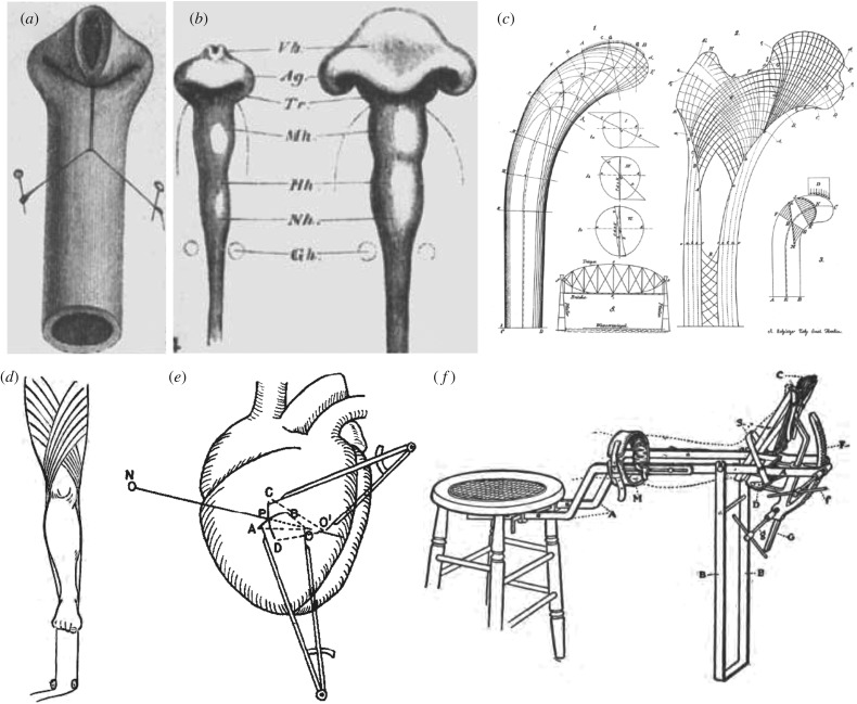
    1. His W. 1888. On the principles of animal morphology. Proc. R. Soc. Edin. 15, 287–298.
    1. Wolff J. 1870. Über die innere architektur der knochen und ihre bedeutung für die frage von knochenwachstum. Arch. Pathol. Anat. Physiol. Klin. Med. 50, 389–453. (10.1007/BF01944490) - DOI
    1. Davis HG. 1867. Conservative surgery. New York, NY: D. Appleton & Company.
    1. Burton AC. 1957. The importance of the shape and size of the heart. American Heart J. 54, 801–810. (10.1016/0002-8703(57)90186-2) - DOI - PubMed
    1. Nutt JJ. 1913. Diseases and deformities of the foot. London, UK: Forgotten Books.

LinkOut - more resources