. 2019 Oct;213(2):395-409.
doi: 10.1534/genetics.119.302625. Epub 2019 Aug 20.

Synaptonemal Complex Central Region Proteins Promote Localization of Pro-crossover Factors to Recombination Events During Caenorhabditis elegans Meiosis

Affiliations

Synaptonemal Complex Central Region Proteins Promote Localization of Pro-crossover Factors to Recombination Events During Caenorhabditis elegans Meiosis

Cori K Cahoon et al. Genetics. 2019 Oct.

Abstract

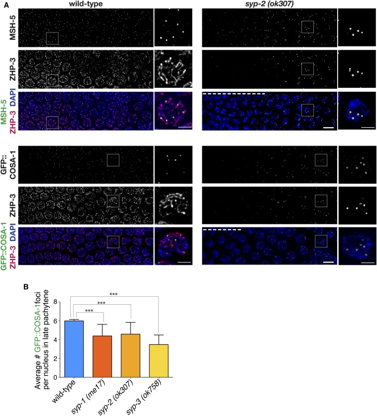
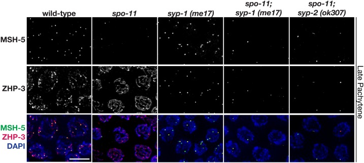
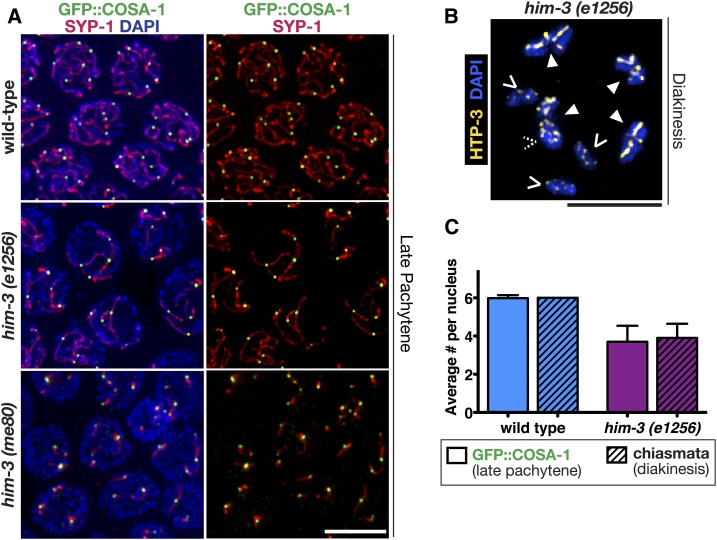
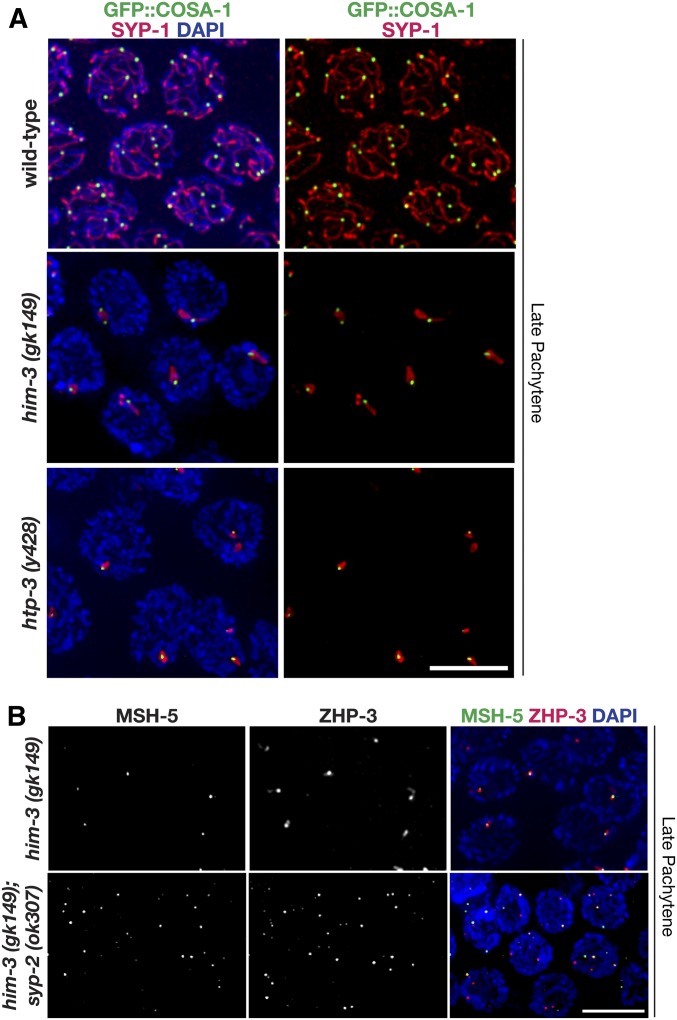
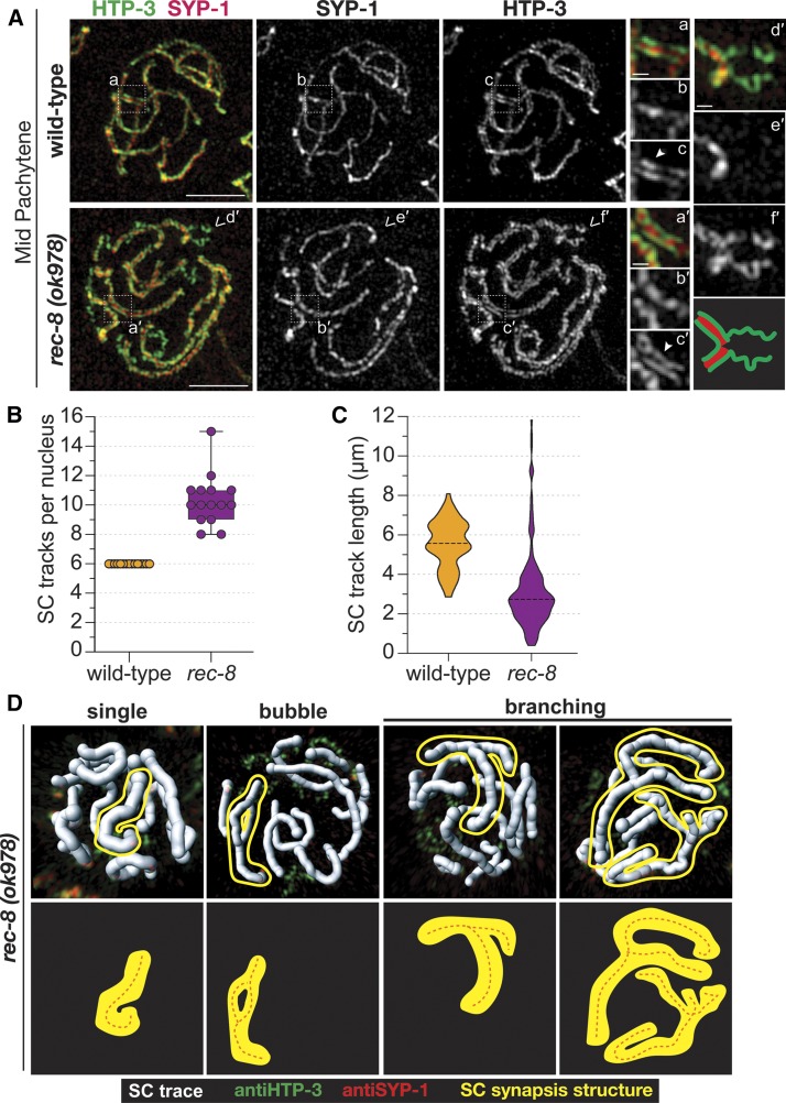
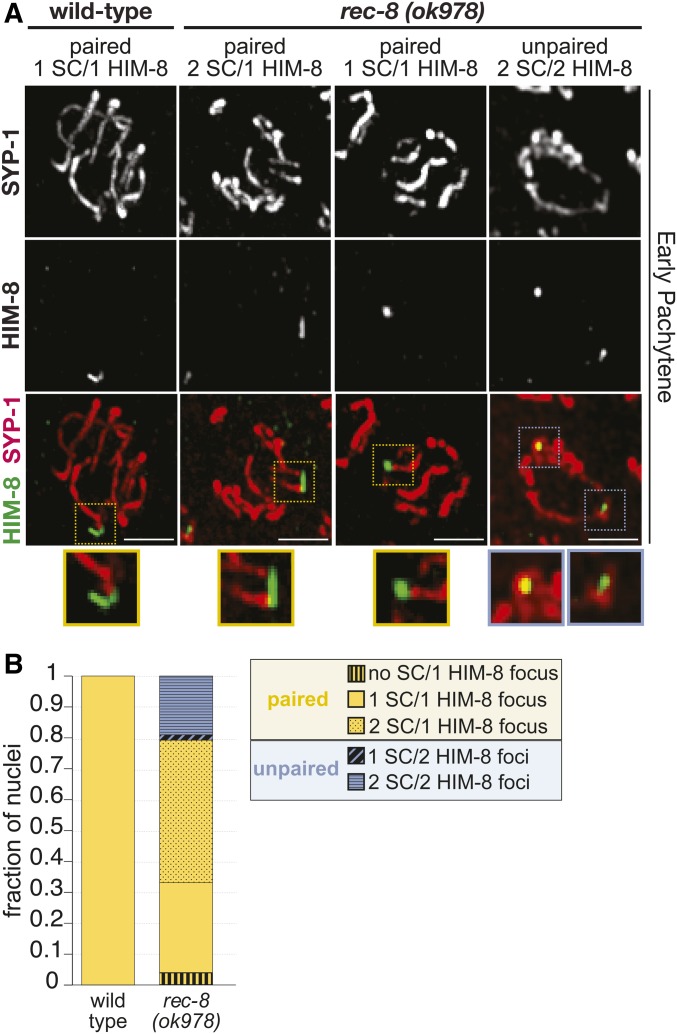
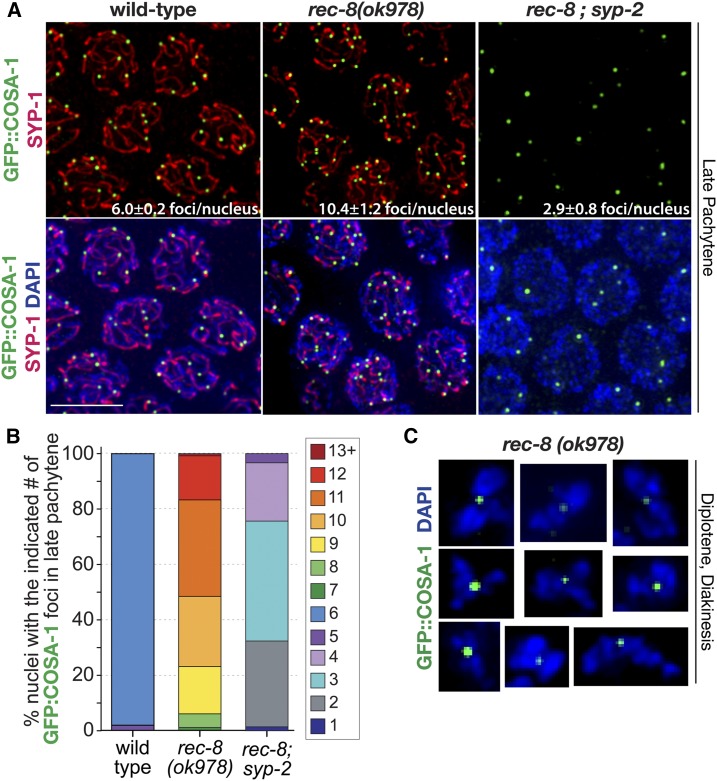
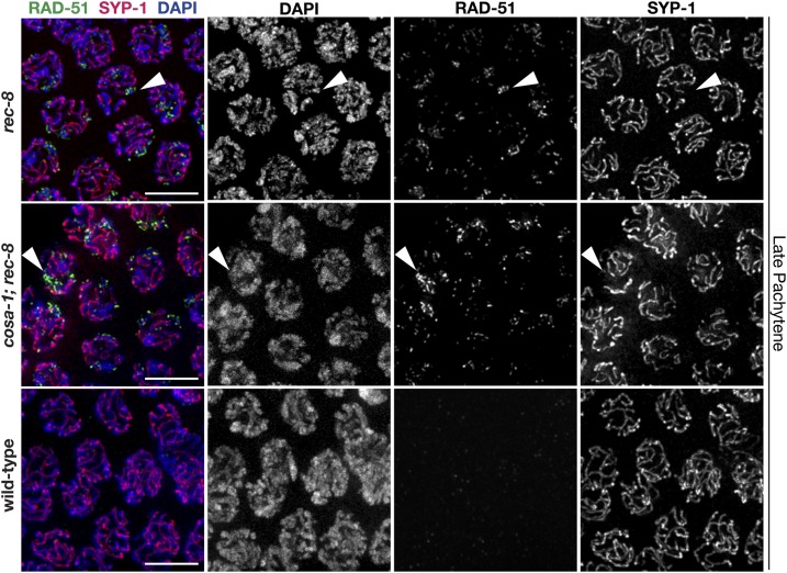
Crossovers (COs) between homologous chromosomes are critical for meiotic chromosome segregation and form in the context of the synaptonemal complex (SC), a meiosis-specific structure that assembles between aligned homologs. During Caenorhabditis elegans meiosis, central region components of the SC (SYP proteins) are essential to repair double-strand DNA breaks (DSBs) as COs. Here, we investigate the relationships between the SYP proteins and conserved pro-CO factors by examining the immunolocalization of these proteins in meiotic mutants where SYP proteins are absent, reduced, or mislocalized. Although COs do not form in syp null mutants, pro-CO factors COSA-1, MSH-5, and ZHP-3 nevertheless colocalize at DSB-dependent sites during late prophase, reflecting an inherent affinity of these factors for DSB repair sites. In contrast, in mutants where SYP proteins are present but form aggregates or display abnormal synapsis, pro-CO factors consistently track with SYP-1 localization. Further, pro-CO factors usually localize to a single site per SYP-1 structure, even in SYP aggregates or in mutants where the SC forms between sister chromatids, suggesting that CO regulation occurs within these aberrant SC structures. Moreover, we find that the meiotic cohesin REC-8 is required to ensure that SC formation occurs between homologs and not sister chromatids. Taken together, our findings support a model in which SYP proteins promote CO formation by promoting the localization of pro-CO factors to recombination events within an SC compartment, thereby ensuring that pro-CO factors identify a recombination event within an SC structure and that CO maturation occurs only between properly aligned homologous chromosomes.

Keywords: C. elegans; chromosome axis; cohesin; crossovers; meiosis; recombination; synaptonemal complex.


Figures

Figure 1
Figure 1
DSB-dependent colocalization of pro-CO factors to late pachytene foci in syp mutants. (A) Immunofluorescence images of mid-to-late pachytene region of germ lines from wild-type and syp-2(ok307) mutant worms, with meiotic prophase progressing from left to right. In wild-type nuclei at the midpachytene stage (left sides of wild-type panels), ZHP-3 is localized along the lengths of the chromosomes, MSH-5 is detected as foci in excess of the eventual number of COs, and COSA-1 foci are not detected. Upon transition to late pachytene, COSA-1 foci are detected at nascent CO sites, colocalized with MSH-5, and ZHP-3 tracks gradually reduce and retract toward the COSA-1 foci. In the syp-2 mutant panels, nuclei at the left sides of the images exhibit characteristic DAPI signals reflecting prolonged persistence of chromosome clustering and chromosome movement (dashed white line). However, upon eventual release from chromosome clustering and transition to a late pachytene-like dispersed chromosome organization, ZHP-3 is detected as foci, most of which colocalize with MSH-5 (top) or GFP::COSA-1 (bottom). Both Jantsch et al. (2004) and Zhang et al. (2018) have also published ZHP-3 localization in syp null mutants. As the ZHP-3, MSH-5, and COSA-1 foci in the syp mutants are of weaker intensity than in wild-type, signal intensities in the syp-2 images were boosted relative to controls to enable visualization of the foci (see Figure S1 for unadjusted images). Dashed box indicates the nucleus that is enlarged in the adjacent image and scale bars on the enlarged images represent 2 µm. All other bars, 5 μm. (B) Quantitation of GFP::COSA-1 foci in late pachytene nuclei for syp null mutants (Table S1). Number of asterisks represents degree of statistical significance from a Mann–Whitney U-test (*** = P < 0.0001). Error bars represent SD. Number of nuclei scored for GFP::COSA-1: wild-type, n = 505; syp-1(me17), n = 223; syp-2(ok307), n = 101; syp-3(ok758), and n = 99. CO, crossover; DSB, DNA double-strand break.
Figure 2
Figure 2
Colocalization of pro-CO factors in syp mutants is DSB-dependent. Immunolocalization of MSH-5 and ZHP-3 in representative late pachytene nuclei from wild-type, spo-11, syp-1(me17), spo-11; syp-1(me17), and spo-11; syp-2(ok307). Similar to the spo-11 single mutant, late pachytene nuclei in spo-11; syp-1 and spo-11; syp-2 double mutants typically have only an occasional MSH-5 focus (zero-to-one), indicating that the presence of multiple foci in the syp single mutants is DSB-dependent. Representative images of the syp-2 single mutant are in Figure 1. Bar, 5 μm. CO, crossover; DSB, DNA double-strand break.
Figure 3
Figure 3
GFP::COSA-1 specifically associates with synapsed chromosome segments in mutants with limited synapsis. (A) Immunofluorescence images of representative nuclei in the late pachytene regions of germ lines from worms of the indicated genotypes, in which the synaptonemal complex central region protein SYP-1 (red) localizes: along the lengths of paired homologs (wild-type), along the lengths of a subset of chromosomes [him-3(e1256) mutant], or in several short stretches [him-3(me80) mutant]. All GFP::COSA-1 (green) are associated with the chromosomes or chromosome segments where SYP-1 localizes. (B) Immunofluorescence image of a representative diakinesis nucleus from the him-3(e1256) mutant, shown with DAPI (blue) and chromosome axis component HTP-3 (yellow) to visualize the chiasmata. Chiasmata were visualized and counted using three-dimensional rotations; solid arrowheads indicate bivalents connected by chiasmata, while carets indicate achiasmate chromosomes (univalent) (dashed caret indicates a univalent hidden in this projection). (C) Bar graph depicting quantitation of GFP::COSA-1 foci in late pachytene nuclei (bars without a pattern), and chiasmata (bars with diagonal lines), in diakinesis nuclei for wild-type (blue bars) and the him-3(e1256) (purple bars) partial loss-of-function chromosome axis mutant (Table S1); error bars indicate SD (P < 0.0001, Mann–Whitney U-test). Number of late pachytene nuclei scored for COSA-1 foci: wild-type, n = 505 and him-3(e1256), n = 161. Number of nuclei scored for chiasmata: wild-type, n = 28 and him-3(e1256), n = 40. Bars, 5 μm.
Figure 4
Figure 4
Pro-CO factors associate with SYP-1 aggregates in mutants lacking meiotic chromosome axis components. (A) Immunolocalization of SYP-1 (red) and GFP::COSA-1 (green) in nuclei from the late pachytene regions of null mutants lacking chromosome axis components HIM-3 or HTP-3. SC assembly is severely impaired in both the him-3(gk149) and htp-3(y428) null mutants, and SYP proteins instead assemble into abnormal aggregates known as polycomplexes. GFP::COSA-1 localization is consistently associated with these abnormal SYP-1 structures in both mutants. The representative image of late pachytene in wild-type is repeated from Figure 3. Rog et al. (2017) also showed that ZHP-3 and COSA-1 also localize to SC aggregates in htp-3(tm3655) null mutants. (B) Immunolocalization of MSH-5 (green) and ZHP-3 (red) in nuclei from late pachytene from him-3(gk149) and the him-3(gk149); syp-2(ok307) double mutant. Whereas MSH-5 and ZHP-3 are usually detected together at a single site per nucleus in the him-3(gk149) mutant, multiple foci are detected in nuclei in the him-3(gk149); syp-2(ok307) double mutant (as in the syp-2 single mutant, see Figure 1). Bars, 5 μm. CO, crossover; SC, synaptonemal complex.
Figure 5
Figure 5
Synapsis occurs between sister chromatid pairs in rec-8 mutants. (A) Structured illumination microscopy images of SYP-1 (red) and chromosome axis component HTP-3 (green) in representative midpachytene nuclei, showing that SYP-1 localizes between pairs of HTP-3 tracks in both wild-type and rec-8(ok978); this indicates that synapsis occurs between sister chromatid pairs in the rec-8 mutant. White dashed box indicates the enlarged region of the SC depicted in the smaller images on the right. The solid arrowhead identifies a region where both lateral elements of the SC are visible, indicated by the two tracks of HTP-3. The carets indicate a region of unsynapsed HTP-3, which is enlarged in (d′–f′) with a cartoon diagram of the unsynapsed region below (f′) (red, SYP-1 and green, HTP-3). Bars for whole-nucleus images represent 2 µm and scale bars for smaller enlarged SC segments represent 250 nm. (B) Box plot depicting the number of SC tracks per nucleus showing that rec-8(ok978) (purple) mutants display on average 10 SC tracks per nucleus, while wild-type (yellow) only has 6 SC tracks per nucleus. (C) Violin plots showing the distribution of the SC track length in micrometers from wild-type (yellow) and rec-8(ok978) (purple). Number of midpachytene nuclei traced for the SC: wild-type, n = 15 (three total gonads) and rec-8(ok978), n = 15 (three total gonads). (D) 3D surfaces, generated in IMARIS, showing the SC traces (white) in each nucleus from rec-8(ok978). Each SC trace contains both HTP-3 (green) and SYP-1 (red). Multiple SC synapsis structures were observed in rec-8(ok978) mutants: single SC track, bubble SC track, Y-shaped branching SC track, and multi-branching SC track. A representative example of each SC synapsis structure is outlined in yellow and depicted as a diagram below each 3D surface image with the orange dashed line representing the SC trace. 3D, three-dimensional; SC, synaptonemal complex.
Figure 6
Figure 6
Paired X chromosomes in rec-8 mutants exhibit two stretches of SC. (A) Immunolocalization of HIM-8 (green) and SYP-1 (red) in early pachytene nuclei from rec-8(ok978) and wild-type. As some SCs from the top and bottom halves of the nuclei are superimposed in the full projections encompassing whole nuclei, partial projections showing half nuclei are shown. Colored dashed boxes indicate the enlarged regions of the HIM-8 focus and SC depicted in the smaller images below, with the color indicating paired (yellow) or unpaired (blue) HIM-8 foci. Bars, 5 µm. (B) Stacked bar plot showing the fraction of nuclei displaying paired (yellow) or unpaired (blue) HIM-8 foci. HIM-8 foci were considered paired if the distance between the foci was ≤ 0.7 µm (see Materials and Methods). All HIM-8 foci in wild-type are paired, and in rec-8(ok978) mutants 80% of the HIM-8 foci are paired. In wild-type, all of the paired HIM-8 foci are associated with one SC track (solid bar) indicating SCs between homologous chromosomes. However, within rec-8 mutant nuclei, we observed differences in the number of SC tracks associating with either the paired or unpaired HIM-8 focus/foci. Among the paired HIM-8 foci (yellow), the majority of the rec-8 mutant nuclei displayed HIM-8 foci associated with two SC tracks (dotted bar) suggesting SC assembly between sister chromatids. Paired HIM-8 foci were also observed to not be associated with SC tracks (vertical striped bar) or associated with one SC track (solid bar). Among the unpaired (blue) HIM-8 foci in rec-8 mutants, the majority of the nuclei displayed HIM-8 foci where each was associated with an SC track (horizontal striped bar), also suggesting that the SC is assembling between sister chromatids in this context. Unpaired HIM-8 foci were also observed where only one focus was associated with an SC track (diagonal striped bar). Number of early pachytene nuclei scored for HIM-8: wild-type, n = 90 and rec-8(ok798), n = 116. SC, synaptonemal complex.
Figure 7
Figure 7
Pro-CO factors associate with SYP-1 stretches and between sister chromatid pairs in rec-8 mutants. (A) Immunolocalization of SYP-1 and GFP::COSA-1 in fields of nuclei from the late pachytene regions of wild-type, rec-8(ok978), and rec-8; syp-2 germ lines. Average number of COSA-1 foci per nucleus and SD is labeled on the image for each genotype. Bar, 5 μm. (B) Stacked bar graph showing percentages of nuclei with indicated numbers of GFP::COSA-1 foci in late pachytene for wild-type, rec-8(ok978), and rec-8; syp-2 (Table S1). Number of late pachytene nuclei scored for COSA-1 foci: wild-type, n = 505; rec-8(ok978), n = 245; and rec-8; syp-2, n = 204. (C) Immunolocalization of GFP::COSA-1 in DAPI-stained diplotene and diakinesis bivalents from rec-8(ok978) germ lines. CO, crossover.
Figure 8
Figure 8
Regions of desynapsis in rec-8 null mutants fail to repair DSBs. Immunolocalization of RAD-51 and SYP-1 in the late pachytene regions of wild-type, rec-8(ok978), and cosa-1; rec-8(ok978) germ lines. Arrowheads indicate chromatids that failed to load SYP-1 and have accumulated RAD-51 foci. Bars, 5 µm. DSB, DNA double-strand break.

Similar articles

Cited by

References

    1. Alpi A., Pasierbek P., Gartner A., and Loidl J., 2003. Genetic and cytological characterization of the recombination protein RAD-51 in Caenorhabditis elegans. Chromosoma 112: 6–16. 10.1007/s00412-003-0237-5 - DOI - PubMed
    1. Bhalla N., Wynne D. J., Jantsch V., and Dernburg A. F., 2008. ZHP-3 acts at crossovers to couple meiotic recombination with synaptonemal complex disassembly and bivalent formation in C. elegans. PLoS Genet. 4: e1000235 10.1371/journal.pgen.1000235 - DOI - PMC - PubMed
    1. Cahoon C. K., and Hawley R. S., 2016. Regulating the construction and demolition of the synaptonemal complex. Nat. Struct. Mol. Biol. 23: 369–377. 10.1038/nsmb.3208 - DOI - PubMed
    1. Cahoon C. K., and Libuda D. E., 2019. Leagues of their own: sexually dimorphic features of meiotic prophase I. Chromosoma. DOI: 10.1007/s00412-019-00692-x. 10.1007/s00412-019-00692-x - DOI - PMC - PubMed
    1. Cahoon C. K., Yu Z., Wang Y., Guo F., Unruh J. R. et al. , 2017. Superresolution expansion microscopy reveals the three-dimensional organization of the Drosophila synaptonemal complex. Proc. Natl. Acad. Sci. USA 114: E6857–E6866. 10.1073/pnas.1705623114 - DOI - PMC - PubMed

Publication types

MeSH terms