. 2019 Aug 20;2(4):e201800213.
doi: 10.26508/lsa.201800213. Print 2019 Aug.

A molecule inducing androgen receptor degradation and selectively targeting prostate cancer cells

Affiliations

A molecule inducing androgen receptor degradation and selectively targeting prostate cancer cells

Serge Auvin et al. Life Sci Alliance. .

Abstract

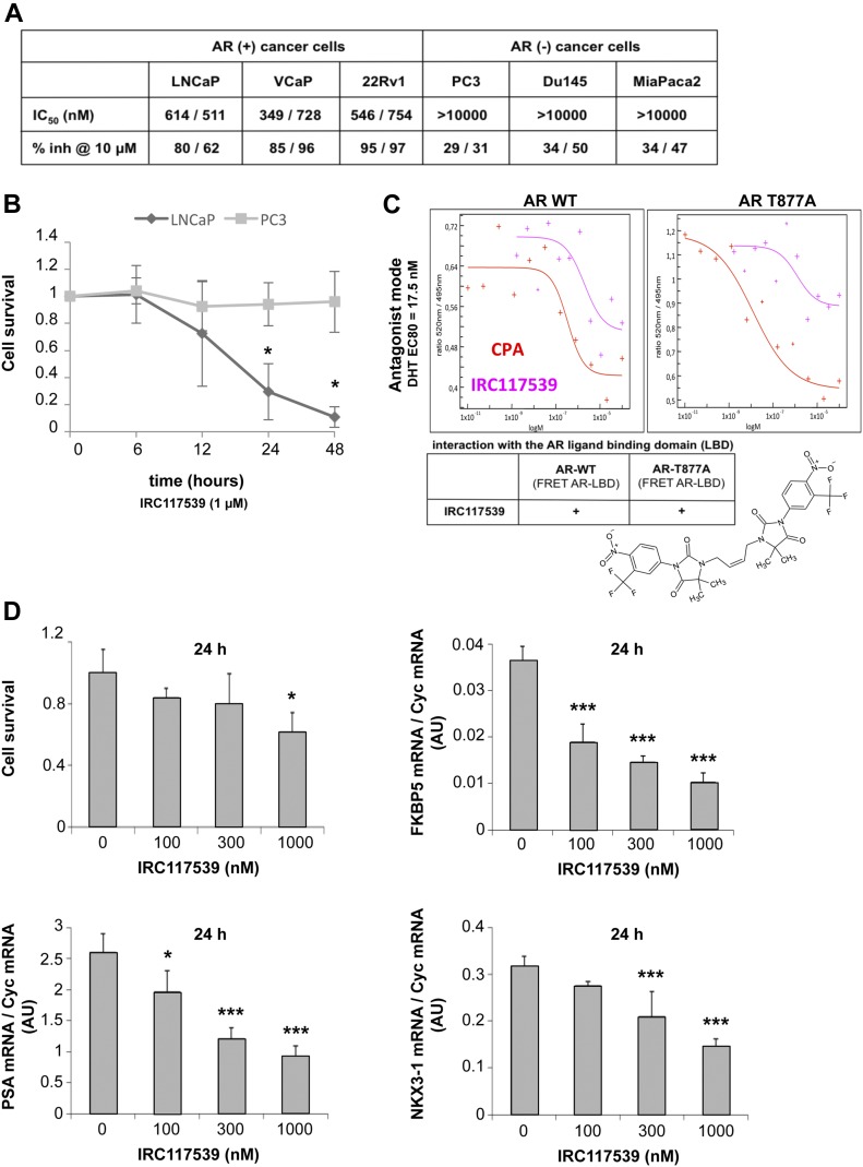
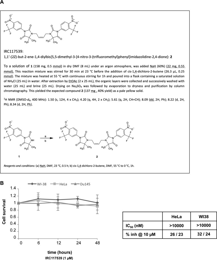
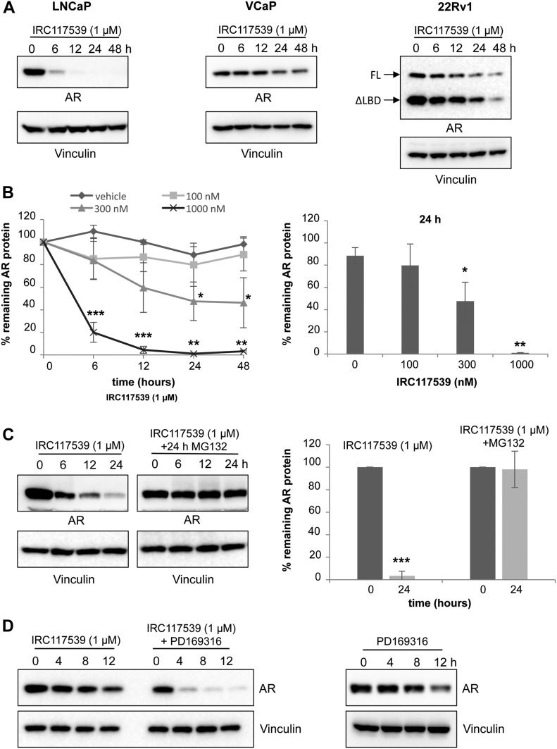
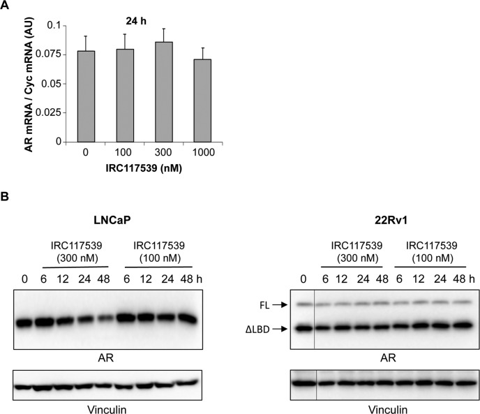
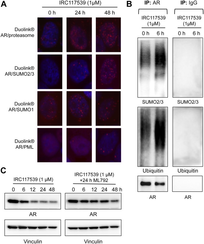
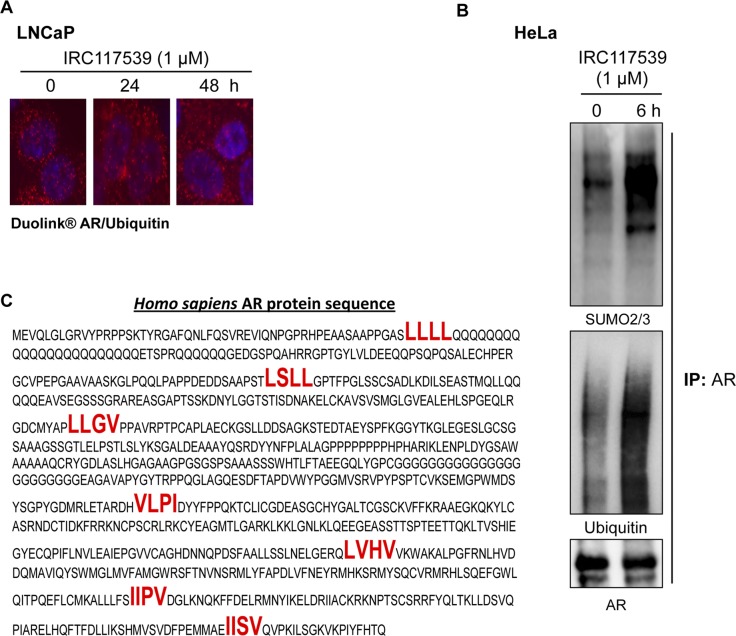
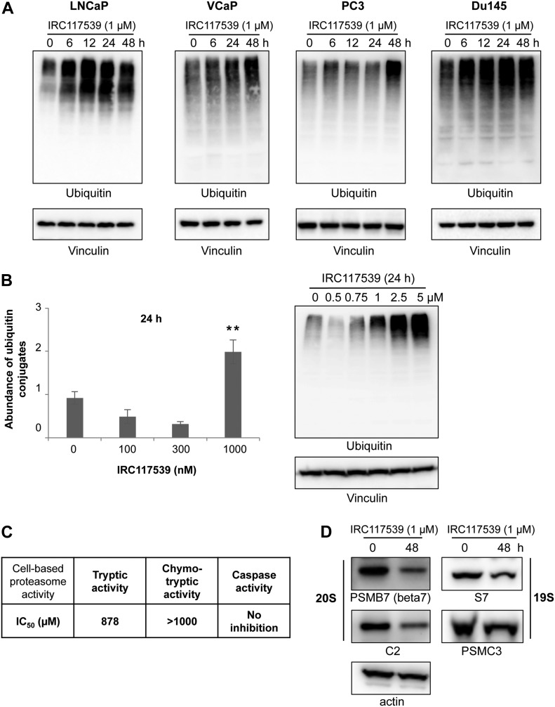
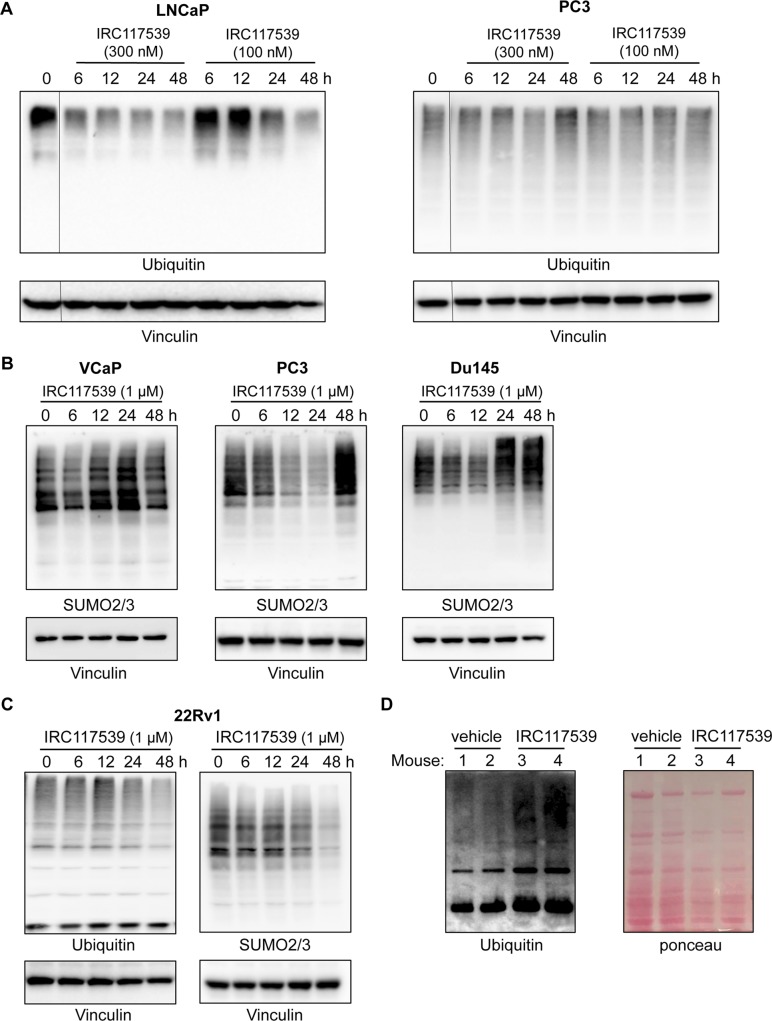
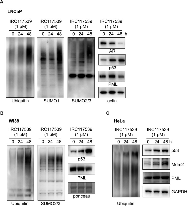
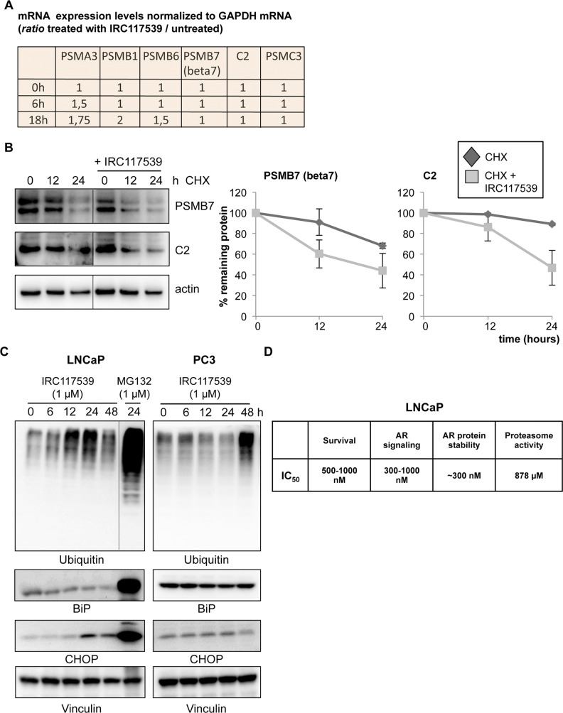
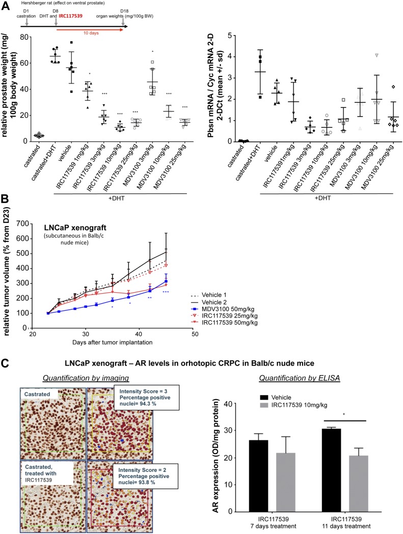
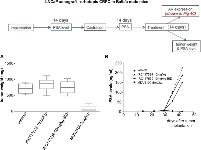
Aberrant androgen signaling drives prostate cancer and is targeted by drugs that diminish androgen production or impede androgen-androgen receptor (AR) interaction. Clinical resistance arises from AR overexpression or ligand-independent constitutive activation, suggesting that complete AR elimination could be a novel therapeutic strategy in prostate cancers. IRC117539 is a new molecule that targets AR for proteasomal degradation. Exposure to IRC117539 promotes AR sumoylation and ubiquitination, reminiscent of therapy-induced PML/RARA degradation in acute promyelocytic leukemia. Critically, ex vivo, IRC117539-mediated AR degradation induces prostate cancer cell viability loss by inhibiting AR signaling, even in androgen-insensitive cells. This approach may be beneficial for castration-resistant prostate cancer, which remains a clinical issue. In xenograft models, IRC117539 is as potent as enzalutamide in impeding growth, albeit less efficient than expected from ex vivo studies. Unexpectedly, IRC117539 also behaves as a weak proteasome inhibitor, likely explaining its suboptimal efficacy in vivo. Our studies highlight the feasibility of AR targeting for degradation and off-target effects' importance in modulating drug activity in vivo.


Conflict of interest statement

S Auvin, G Mautino, F Meyer-Losic, and T Bashir are employees of IPSEN that also paid US salary. This study was funded by IPSEN.

Figures

Figure 1.
Figure 1.. The compound IRC117539 inhibits AR signaling axis and selectively induces loss of viability in AR-positive prostate cancer cells.
(A) The effect and specificity of IRC117539 on prostate cancer cell proliferation. Three AR-positive prostate cancer and three different AR-negative cancer cell lines are shown for comparison. (B) Kinetics of IRC117539-induced loss of viability in AR-dependent LNCaP prostate cancer cells. AR-negative PC3 cells are also shown for comparison (IRC117539: 1 μM). (C) Testing antagonist mode of IRC117539 (chemical structure is shown) by FRET assay. The assay was performed as described in the Materials and Methods section using cyproterone acetate (CPA) as a positive control. IRC117539 binds AR in the LBD, both wt and T877A. (D) Dose-dependent reversal of AR target gene expression in LNCaP prostate cancer cells by IRC117539. FKBP5, PSA, and NKX3-1 mRNA levels were analyzed by Q-PCR and normalized to Cyc mRNA expression. Upper left graph shows cell viability at indicated doses of IRC117539. Data information: in (B and D), data are presented as mean ± SEM (n = 3). Asterisks denote statistical significance (*P < 0.05, ***P < 0.001, using t test assuming unequal variances).
Figure S1.
Figure S1.. The compound IRC117539 does not induce loss of viability in AR-negative cells.
(A) Synthesis of IRC117539 is described in detail. (B) IRC117539 does not affect survival of AR-negative Du145 prostate cancer, WI38 primary human fibroblast or HeLa cervical cancer cells (compare this with LNCaP data in Fig 1B). Data are presented as mean ± SEM (n = 3), IRC117539: 1 μM. For HeLa and WI38 cells, IC50 values are also indicated (compare with Fig 1A).
Figure 2.
Figure 2.. IRC117539 induces AR protein degradation by the proteasome.
(A) Western blot analysis of AR protein levels in LNCaP, VCaP, and 22Rv1 prostate cancer cells treated with 1 μM IRC117539 compound for indicated times. Note two different AR isoforms (FL, full-length retaining LBD; ΔLBD, the truncated variant) in 22Rv1 cells, which are both degraded. (B) Dose-dependent kinetics of IRC117539-induced AR degradation in LNCaP cells (left panel). AR protein levels were quantified after Western blot and normalized to vinculin (n = 3, representative blots are shown in Figs 2A and S2B). A dose–response graphic for the 24-h time point is shown in the right panel. (C) Proteasome-dependent degradation of AR. Western blot (left) shows AR protein levels in LNCaP cells exposed to 1 μM IRC117539, with or without proteasome inhibition. Right panel shows quantification of AR protein levels at 24 h (n = 3). (D) Western blot analysis of AR protein levels in LNCaP cells co-treated with 10 μM PD169316, a small molecule proteasome activator. Note accelerated kinetics of IRC117539-induced AR degradation. Data information: in (B and C), data are presented as mean ± SEM (n = 3). Asterisks denote statistical significance (*P < 0.05, **P < 0.01, ***P < 0.001, using t test assuming unequal variances).
Figure S2.
Figure S2.. IRC117539 induces AR protein degradation in prostate cancer cells.
(A) Q-PCR analyses show AR transcript levels in LNCaP prostate cancer cells. AR mRNA levels were normalized to Cyc mRNA expression. Data are presented as mean ± SEM (n = 3). (B) Western blot analysis of AR protein levels in LNCaP and 22Rv1 prostate cancer cells treated with lower doses (100 nM and 300 nM) of the IRC117539 compound (for comparison with compound’s effect at 1 μM, as shown in Fig 2A).
Figure 3.
Figure 3.. IRC117539 induces AR posttranslational modifications before degradation.
(A) Proximity ligation (Duolink) analyses probe AR physical interaction with the proteasome (20S particle) or with PML, as well as AR modification by SUMO1 or SUMO2/3 peptides at indicated times of IRC117539 treatment (1 μM) in LNCaP cells. (B) Immunoprecipitation of endogenous AR from LNCaP cells confirms IRC117539-induced AR conjugation by SUMO2/3 and ubiquitin. IRC117539 was used at 1 μM. A nonspecific IgG was used as a negative control (right panel). (C) Sumoylation is required for IRC117539-induced AR degradation. AR protein levels were assessed by Western blot in LNCaP cells co-treated with ML792 (1 μM), a small molecule sumoylation inhibitor.
Figure S3.
Figure S3.. IRC117539 induces AR sumoylation and ubiquitination.
(A) IRC117539-induced increase in AR and ubiquitin interactions in LNCaP cells as detected by PLA. Note the high levels of AR/ubiquitin PLA signals in resting cells, consistent with previous reports that AR is heavily ubiquitinated. IRC117539 was used at 1 μM. (B) Immunoprecipitation of transfected AR from IRC117539-treated HeLa cells confirms massive AR conjugation by SUMO2/3 and ubiquitin. IRC117539 was used at 1 μM. (C) SIMs (indicated in bold red) on AR protein may promote its association with PML (Sahin et al, 2014c).
Figure 4.
Figure 4.. IRC117539 impairs proteasome function.
(A) Accumulation of global ubiquitin conjugates in AR-positive (LNCaP and VCaP) and AR-negative (PC3 and Du145) cells treated with 1 μM IRC117539. (B) Dose-dependent accumulation of ubiquitinated proteins in IRC117539-treated LNCaP cells at 24 h. Global protein ubiquitination was quantified after Western blot and normalized to vinculin (n = 3, representative blots are shown in right panel, in Fig 4A and in Fig S4A). The slight decrease in cellular ubiquitination at 100 nM, 300 nM, and 500 nM is likely due to enhanced proteasome activity and consistent with biphasic/hormetic cellular response (hormesis) to lower doses of a drug (Calabrese, 2008; Kendig et al, 2010). Data are presented as mean ± SEM. Asterisks denote statistical significance (**P < 0.01, compared with the 0 nM bar, using t test assuming unequal variances). (C) IRC117539’s cell-based IC50 values are indicated for proteasome’s tryptic and chymotryptic activities. Analysis was performed in LNCaP cells. (D) Western blot analysis of various proteasome subunits in LNCaP cells treated with 1 μM IRC117539.
Figure S4.
Figure S4.. IRC117539 interferes with global proteolysis.
(A) Western blot analysis of global ubiquitin conjugates in AR-positive LNCaP and AR-negative PC3 prostate cancer cells treated with lower doses (100 nM and 300 nM) of the IRC117539 compound (for comparison with compound’s effect at 1 μM, as shown in Fig 4A). (B) Accumulation of global SUMO conjugates in AR-positive (VCaP) and AR-negative (PC3 and Du145) cells treated with 1 μM IRC117539. (C) Western blot analysis of global ubiquitin and SUMO conjugates in AR-positive 22Rv1 cells treated with 1 μM IRC117539. (D) Accumulation of global ubiquitin conjugates in vivo in IRC117539-treated mice. Mice were euthanized after 7 d of treatment (vehicle or IRC117539, 50 mg/kg). Western blot was performed after protein extraction from liver tissue. Two mice are shown for each group (#1 and #2: treated with vehicle, #3 and #4: treated with IRC117539).
Figure S5.
Figure S5.. IRC117539’s effect on protein turnover in LNCaP, HeLa, and WI38 cells.
(A–C) Accumulation of global SUMO and ubiquitin conjugates and p53 protein in LNCaP (A) and primary human WI38 cells (B) exposed to 1 μM IRC117539 and stabilization of global ubiquitin conjugates, p53 and Mdm2, in HeLa cells treated with 1 μM IRC117539 (C).
Figure S6.
Figure S6.. IRC117539’s effect on proteasomes, its subunits and ER stress.
(A) mRNA levels of various proteasome subunits were determined by q-PCR in LNCaP cells treated with the IRC117539 (1 μM) compound for indicated times. Expression levels were normalized to GAPDH mRNA. (B) Cycloheximide (CHX) chase experiment indicates enhanced turnover and diminished half-life of proteasome subunits in cells treated with IRC117539 (1 μM). Western blot analyses of protein levels and quantifications are shown (n = 2). (C) Western blot analyses of CHOP and BiP expression in AR-positive LNCaP and AR-negative PC3 cells treated with 1 μM IRC117539. Global ubiquitin conjugates are also shown. MG132, a potent ER stress inducer, was used at 1 μM as a positive control. (D) IC50 values are shown for comparison of IRC117539-induced changes in LNCaP cells (treatment: 24 h). IRC117539 achieves AR degradation and impairs AR signaling and prostate cancer cell survival at comparable doses.
Figure 5.
Figure 5.. In vivo efficacy of the IRC117539 compound.
(A) Effect of IRC117539 on ventral prostate weight in a Hershberger rat after 10 d of treatment in vivo at increasing doses (left panel). Transcript levels of probasin (Pbsn), a protein abundantly expressed in prostate epithelia, were determined in a Hershberger rat treated as indicated (right panel). (B) Decrease in tumor volume in LNCaP prostate cancer xenograft models after 46 d of treatment with IRC117539. The effect of enzalutamide (MDV3100) is also shown for comparison. (C). AR expression levels in orthotopically implanted LNCaP tumors. Animals were castrated 14 d after tumor implantation, and another 14 d after castration treatments were administered for 14 d. Tumors were harvested and analyzed for AR protein expression by immunostaining and ELISA. Data information: data are presented as mean ± SEM (n > 3). Asterisks denote statistical significance (*P < 0.05, **P < 0.01, ***P < 0.001, using t test assuming unequal variances).
Figure S7.
Figure S7.. Suboptimal efficacy of IRC117539 in vivo in CRPC models.
(A, B) Tumor weight (A) and PSA levels in peripheral blood (B) of castration-resistant LNCaP xenograft mouse models treated as indicated. MDV3100: enzalutamide. Data are presented as mean ± SEM (n > 3).

Similar articles

Cited by

References

    1. Ablain J, Nasr R, Bazarbachi A, de The H (2011) The drug-induced degradation of oncoproteins: An unexpected Achilles’ heel of cancer cells? Cancer Discov 1: 117–127. 10.1158/2159-8290.CD-11-0087 - DOI - PubMed
    1. An J, Wang C, Deng Y, Yu L, Huang H (2014) Destruction of full-length androgen receptor by wild-type SPOP, but not prostate-cancer-associated mutants. Cell Rep 6: 657–669. 10.1016/j.celrep.2014.01.013 - DOI - PMC - PubMed
    1. Anbalagan M, Huderson B, Murphy L, Rowan BG (2012) Post-translational modifications of nuclear receptors and human disease. Nucl Recept Signal 10: e001 10.1621/nrs.10001 - DOI - PMC - PubMed
    1. Armstrong CM, Gao AC (2015) Drug resistance in castration resistant prostate cancer: Resistance mechanisms and emerging treatment strategies. Am J Clin Exp Urol 3: 64–76. - PMC - PubMed
    1. Balbas MD, Evans MJ, Hosfield DJ, Wongvipat J, Arora VK, Watson PA, Chen Y, Greene GL, Shen Y, Sawyers CL (2013) Overcoming mutation-based resistance to antiandrogens with rational drug design. Elife 2: e00499 10.7554/eLife.00499 - DOI - PMC - PubMed

Publication types

MeSH terms