. 2019 Aug 20;10(4):e01210-19.
doi: 10.1128/mBio.01210-19.

How Thermophilic Gram-Positive Organisms Perform Extracellular Electron Transfer: Characterization of the Cell Surface Terminal Reductase OcwA

Affiliations

How Thermophilic Gram-Positive Organisms Perform Extracellular Electron Transfer: Characterization of the Cell Surface Terminal Reductase OcwA

N L Costa et al. mBio. .

Abstract

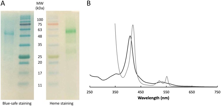
Extracellular electron transfer is the key process underpinning the development of bioelectrochemical systems for the production of energy or added-value compounds. Thermincola potens JR is a promising Gram-positive bacterium to be used in these systems because it is thermophilic. In this paper, we describe the structural and functional properties of the nonaheme cytochrome OcwA, which is the terminal reductase of this organism. The structure of OcwA, determined at 2.2-Å resolution, shows that the overall fold and organization of the hemes are not related to other metal reductases and instead are similar to those of multiheme cytochromes involved in the biogeochemical cycles of nitrogen and sulfur. We show that, in addition to solid electron acceptors, OcwA can also reduce soluble electron shuttles and oxyanions. These data reveal that OcwA can work as a multipurpose respiratory enzyme allowing this organism to grow in environments with rapidly changing availability of terminal electron acceptors without the need for transcriptional regulation and protein synthesis.IMPORTANCE Thermophilic Gram-positive organisms were recently shown to be a promising class of organisms to be used in bioelectrochemical systems for the production of electrical energy. These organisms present a thick peptidoglycan layer that was thought to preclude them to perform extracellular electron transfer (i.e., exchange catabolic electrons with solid electron acceptors outside the cell). In this paper, we describe the structure and functional mechanisms of the multiheme cytochrome OcwA, the terminal reductase of the Gram-positive bacterium Thermincola potens JR found at the cell surface of this organism. The results presented here show that this protein can take the role of a respiratory "Swiss Army knife," allowing this organism to grow in environments with soluble and insoluble substrates. Moreover, it is shown that it is unrelated to terminal reductases found at the cell surface of other electroactive organisms. Instead, OcwA is similar to terminal reductases of soluble electron acceptors. Our data reveal that terminal oxidoreductases of soluble and insoluble substrates are evolutionarily related, providing novel insights into the evolutionary pathway of multiheme cytochromes.

Keywords: Gram-positive bacteria; Thermincola; bioelectrochemical systems; extracellular electron transfer; multiheme cytochromes; terminal oxidoreductases.


Figures

FIG 1
FIG 1
Isolation of OcwA from T. potens JR. (A) Blue-safe and heme-stained SDS-PAGE of purified OcwA. (B) UV-visible spectra of OcwA obtained in the reduced (gray) and oxidized (black) states.
FIG 2
FIG 2
Magnetic spectroscopic properties of OcwA from T. potens JR. (A) 1H NMR spectrum of OcwA obtained at 25°C in the oxidized state (B) One-dimensional (1D) 1H NMR spectrum of OcwA in the reduced state obtained at 25°C by the addition of sodium dithionite. (C) EPR spectra of OcwA at 9.39 GHz in the oxidized (black line) and reduced (gray line) states at 7 K. The unlabeled signal at g of 4.3 likely corresponds to a small amount of high-spin ferric iron adventitiously present in the sample.
FIG 3
FIG 3
Structure of OcwA from T. potens JR. (A) Dimer structure of OcwA. The right monomer is colored from blue at the N terminus to red at the C terminus. Heme groups are depicted in a stick representation. (B) Close-up of the active-site heme groups 2 (left) and 5 (right) highlighting the similar environment of the two centers and depicting the water molecules found on the distal side. (C and D) Comparison of the heme cores of OcwA (C) and MtrC (D) (PDB 4LM8). (E) Stereo representation of the 2Fo–Fc electron density map surrounding heme group 2, contoured at the 1 σ level.
FIG 4
FIG 4
Structural and sequence similarities within the NrfA family of MHC. (A) Structure of the OcwA monomer and heme arrangement (below), with numbering of cofactors according to their occurrence in the protein sequence. (B) Structural alignment with Nitrosomonas europaea cytochrome c554 (PDB 1FT5) showing the heme numbering of this cytochrome. (C) Structural alignment with Wollinella succinogenes NrfA (PDB 1FS7) showing the heme numbering of this pentaheme cytochrome. (D) Structural alignment with W. succinogenes MccA (PDB 4RKM) showing the numbering of this octaheme cytochrome. (E) Schematic presentation of a structure-based amino acid sequence alignment of OcwA, NrfA, and c554, highlighting axial heme ligands (orange circles) and distal residues of the active site(s) (black boxes). Hatched boxes represent areas with high structural homology. Heme motifs are shown with red boxes.
FIG 5
FIG 5
Cyclic voltammetry of OcwA. (A) Raw voltammogram obtained at a scan rate of 200 mV/s at pH 7.5. (B) Baseline-subtracted data of the voltammograms obtained at a scan rate of 200 mV/s at different pH values.
FIG 6
FIG 6
Reactivity of OcwA with electron shuttles. (A) Kinetic data obtained by mixing OcwA (0.43 μM) with excess of AQDS (36 μM), PMS (9 μM), FMN (19 μM), and RF (14 μM). (B) Midpoint reduction potentials of the different electron shuttles versus the redox-active range of OcwA at pH 7.6 (C) 1D 31P-NMR spectra of FMN in the presence of increasing amounts of OcwA obtained at 25°C.

References

    1. Melton ED, Swanner ED, Behrens S, Schmidt C, Kappler A. 2014. The interplay of microbially mediated and abiotic reactions in the biogeochemical Fe cycle. Nat Rev Microbiol 12:797–808. doi:10.1038/nrmicro3347. - DOI - PubMed
    1. Iverson TM, Arciero DM, Hooper AB, Rees DC. 2001. High-resolution structures of the oxidized and reduced states of cytochrome c554 from Nitrosomonas europaea. J Biol Inorg Chem 6:390–397. doi:10.1007/s007750100213. - DOI - PubMed
    1. Iverson TM, Arciero DM, Hsu BT, Logan MSP, Hooper AB, Rees DC. 1998. Heme packing motifs revealed by the crystal structure of the tetra-heme cytochrome c554 from Nitrosomonas europaea. Nat Struct Mol Biol 5:1005–1012. doi:10.1038/2975. - DOI - PubMed
    1. Costa NL, Clarke TA, Philipp LA, Gescher J, Louro RO, Paquete CM. 2018. Electron transfer process in microbial electrochemical technologies: the role of cell-surface exposed conductive proteins. Bioresour Technol 255:308–317. doi:10.1016/j.biortech.2018.01.133. - DOI - PubMed
    1. Karthikeyan R, Singh R, Bose A. 2019. Microbial electron uptake in microbial electrosynthesis : a mini‑review. J Ind Microbiol Biotechnol, in press. - PubMed

Publication types

Supplementary concepts

LinkOut - more resources