. 2019 Aug 20;10(4):e01659-19.
doi: 10.1128/mBio.01659-19.

Myeloid and CD4 T Cells Comprise the Latent Reservoir in Antiretroviral Therapy-Suppressed SIVmac251-Infected Macaques

Affiliations

Myeloid and CD4 T Cells Comprise the Latent Reservoir in Antiretroviral Therapy-Suppressed SIVmac251-Infected Macaques

Celina M Abreu et al. mBio. .

Abstract

Human immunodeficiency virus (HIV) eradication or long-term suppression in the absence of antiretroviral therapy (ART) requires an understanding of all viral reservoirs that could contribute to viral rebound after ART interruption. CD4 T cells (CD4s) are recognized as the predominant reservoir in HIV type 1 (HIV-1)-infected individuals. However, macrophages are also infected by HIV-1 and simian immunodeficiency virus (SIV) during acute infection and may persist throughout ART, contributing to the size of the latent reservoir. We sought to determine whether tissue macrophages contribute to the SIVmac251 reservoir in suppressed macaques. Using cell-specific quantitative viral outgrowth assays (CD4-QVOA and MΦ-QVOA), we measured functional latent reservoirs in CD4s and macrophages in ART-suppressed SIVmac251-infected macaques. Spleen, lung, and brain in all suppressed animals contained latently infected macrophages, undetectable or low-level SIV RNA, and detectable SIV DNA. Silent viral genomes with potential for reactivation and viral spread were also identified in blood monocytes, although these cells might not be considered reservoirs due to their short life span. Additionally, virus produced in the MΦ-QVOA was capable of infecting healthy activated CD4s. Our results strongly suggest that functional latent reservoirs in CD4s and macrophages can contribute to viral rebound and reestablishment of productive infection after ART interruption. These findings should be considered in the design and implementation of future HIV cure strategies.IMPORTANCE This study provides further evidence that the latent reservoir is comprised of both CD4+ T cells and myeloid cells. The data presented here suggest that CD4+ T cells and macrophages found throughout tissues in the body can contain replication-competent SIV and contribute to rebound of the virus after treatment interruption. Additionally, we have shown that monocytes in blood contain latent virus and, though not considered a reservoir themselves due to their short life span, could contribute to the size of the latent reservoir upon entering the tissue and differentiating into long-lived macrophages. These new insights into the size and location of the SIV reservoir using a model that is heavily studied in the HIV field could have great implications for HIV-infected individuals and should be taken into consideration with the development of future HIV cure strategies.

Keywords: HIV; SIV; latency; macrophages; monocytes.


Figures

FIG 1
FIG 1
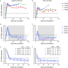
Viral load in plasma and CSF in untreated and ART-suppressed SIVmac251-infected macaques. Eight rhesus macaques were infected with SIVmac251; four were left untreated, and four were treated with ART starting at 14 dpi. Viral load was measured longitudinally in the plasma and CSF samples from the untreated (A and D) and ART-treated (B and E) groups. Decay from peak viremia in plasma (C) and CSF (F) for the four ART-treated animals was determined using a biphasic two-exponential decay model. Solid lines indicate the best-fit biphasic model for each animal. Graphs display two limits of detection (dashed lines), depending on the assay, as described in the text; filled symbols indicate measurements above the limit of quantitation for that measurement; open symbols indicate measurements below the limit of quantitation. Insets display half-lives for both phases of decay.
FIG 2
FIG 2
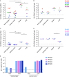
Detection of SIV RNA and DNA in tissues of untreated and ART-suppressed SIVmac251-infected macaques. SIV RNA and DNA were measured in independent tissue samples from the brain s(basal ganglia and parietal cortex), spleens, and lungs from untreated animals (A and B) and ART-treated animals (C and D). Select samples from the brain were used to measure SIV RNA and DNA simultaneously in ART-treated animals (E) to determine if provirus was present despite the absence of SIV RNA. Multiple measurements from the same animal and same tissue were averaged. Statistics were then calculated using a one-way ANOVA with Tukey’s multiple-comparison test. Values that are significantly different are indicated by a bar and asterisks as follows: *, P < 0.05; **, P < 0.01; ***, P < 0.001.
FIG 3
FIG 3
Schematic of cell isolation methods and purity assessments for the macrophage and CD4 QVOAs. (A) Single cells were isolated from brain, lung, spleen, and blood from four SIV-infected ART-suppressed macaques. (B) Monocytes from blood and tissue macrophages from brain, lung, and spleen were purified from single-cell suspensions using CD11b-positive selection. (C) Flowthrough samples from the CD11b selection were then used to isolate untouched CD4 T cells. (D) CD11b+ cells were saved for purity checks by qPCR and FACS assessment as well as SIV RNA and DNA measurements. For further purification by adherence, the remaining CD11b+ cells were plated at limiting dilutions in the presence of ART and allowed to differentiate for 2 to 7 days, depending on tissue of origin. Nonadherent cells and ART were removed prior to activation with TNF and coculture with CEMx174 cells. Supernatants were sampled every 2 days for 12 days. On day 12, all supernatants and cells were harvested and assessed for the presence of SIV RNA and DNA, as well as T cell contamination by TCRβ RNA. (E) CD4 T cells were saved for SIV RNA and DNA measurements by qPCR. The remaining CD4 cells were plated at limiting dilutions and cocultured with CEMx174 cells for 10 days. On day 10, all supernatants and cells were harvested and assessed for the presence of SIV RNA and DNA.
FIG 4
FIG 4
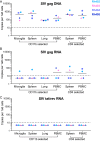
Comparison of cellular SIV DNA and RNA levels in CD11b+ macrophages and CD4+ T cells isolated from tissues of ART-suppressed SIVmac251-infected macaques. CD11b+ cells were isolated from brains, spleens, lungs, and PBMCs, and CD4+ T cells were isolated from spleens and PBMCs from four SIV-infected ART-suppressed macaques. Cellular DNA and RNA were then extracted and analyzed for SIV gag DNA (A), SIV gag RNA (B), and SIV tat/rev RNA (C) by qPCR. The dashed line represents the limit of quantification (LOQ) for each qPCR assay.
FIG 5
FIG 5
Functional latent reservoirs detected in CD4 T cells, monocytes, and macrophages isolated from SIVmac251-infected ART-suppressed macaques. Infectious units per million cells (IUPMs) were calculated for CD4 T cell (A) and monocyte/macrophage (B) QVOAs. Cells were isolated from blood, spleen, lung, and brain from ART-treated SIV-infected macaques and plated at limiting dilutions. Supernatants were sampled every 2 days and measured for SIV RNA. Comparison of blood CD4 T cell and monocyte-derived macrophage (MDM) (C) and splenic CD4 T cell and macrophage (D) IUPM values. Samples with IUPM values below the limit of detection are not shown. (A and C) Rh402 CD4 from PBMCs, (B) Rh402 macrophages from lung and B cell QVOAs from all animals.
FIG 6
FIG 6
Macrophage-produced virus is capable of establishing de novo infection. Activated CD4 T cells from a healthy rhesus macaque were spinoculated with culture supernatant from brain macrophages (A), splenic macrophages (B), lung macrophages (C), and monocyte-derived macrophages (D) QVOAs. All infections were normalized, One hundred copies of gag RNA as measured by RT-qPCR was used for the initial input. After spinoculation, the cells were washed and cultured for 21 days. Supernatant samples were taken every 3 days and measured for SIV RNA.

Similar articles

Cited by

References

    1. Finzi D, Blankson J, Siliciano JD, Margolick JB, Chadwick K, Pierson T, Smith K, Lisziewicz J, Lori F, Flexner C, Quinn TC, Chaisson RE, Rosenberg E, Walker B, Gange S, Gallant J, Siliciano RF. 1999. Latent infection of CD4+ T cells provides a mechanism for lifelong persistence of HIV-1, even in patients on effective combination therapy. Nat Med 5:512–517. doi:10.1038/8394. - DOI - PubMed
    1. Siliciano JD, Kajdas J, Finzi D, Quinn TC, Chadwick K, Margolick JB, Kovacs C, Gange SJ, Siliciano RF. 2003. Long-term follow-up studies confirm the stability of the latent reservoir for HIV-1 in resting CD4+ T cells. Nat Med 9:727–728. doi:10.1038/nm880. - DOI - PubMed
    1. Avalos CR, Abreu CM, Queen SE, Li M, Price S, Shirk EN, Engle EL, Forsyth E, Bullock BT, Mac Gabhann F, Wietgrefe SW, Haase AT, Zink MC, Mankowski JL, Clements JE, Gama L. 2017. Brain macrophages in simian immunodeficiency virus-infected, antiretroviral-suppressed macaques: a functional latent reservoir. mBio 8:e01186-17. doi:10.1128/mBio.01186-17. - DOI - PMC - PubMed
    1. Avalos CR, Price SL, Forsyth ER, Pin JN, Shirk EN, Bullock BT, Queen SE, Li M, Gellerup D, O’Connor SL, Zink MC, Mankowski JL, Gama L, Clements JE. 2016. Quantitation of productively infected monocytes and macrophages of simian immunodeficiency virus-infected macaques. J Virol 90:5643–5656. doi:10.1128/JVI.00290-16. - DOI - PMC - PubMed
    1. Abreu CM, Veenhuis RT, Avalos CR, Graham S, Queen SE, Shirk EN, Bullock BT, Li M, Metcalf Pate KA, Beck SE, Mangus LM, Mankowski JL, Clements JE, Gama L. 2019. Infectious virus persists in CD4+ T cells and macrophages in antiretroviral therapy-suppressed simian immunodeficiency virus-infected macaques. J Virol 93:e00065-19. doi:10.1128/JVI.00065-19. - DOI - PMC - PubMed

Publication types

Substances