. 2019 Aug 20;10(1):3745.
doi: 10.1038/s41467-019-11730-8.

Syringeable immunotherapeutic nanogel reshapes tumor microenvironment and prevents tumor metastasis and recurrence

Affiliations

Syringeable immunotherapeutic nanogel reshapes tumor microenvironment and prevents tumor metastasis and recurrence

Chanyoung Song et al. Nat Commun. .

Abstract

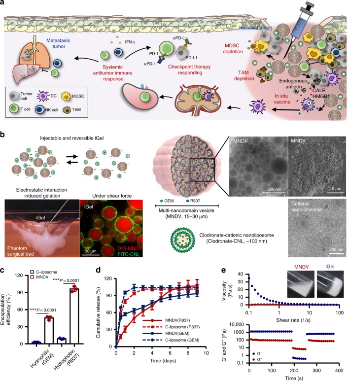
The low response rate of current cancer immunotherapy suggests the presence of few antigen-specific T cells and a high number of immunosuppressive factors in tumor microenvironment (TME). Here, we develop a syringeable immunomodulatory multidomain nanogel (iGel) that overcomes the limitation by reprogramming of the pro-tumoral TME to antitumoral immune niches. Local and extended release of immunomodulatory drugs from iGel deplete immunosuppressive cells, while inducing immunogenic cell death and increased immunogenicity. When iGel is applied as a local postsurgical treatment, both systemic antitumor immunity and a memory T cell response are generated, and the recurrence and metastasis of tumors to lungs and other organs are significantly inhibited. Reshaping of the TME using iGel also reverts non-responding groups to checkpoint blockade therapies into responding groups. The iGel is expected as an immunotherapeutic platform that can reshape immunosuppressive TMEs and synergize cancer immunotherapy with checkpoint therapies, with minimized systemic toxicity.


Conflict of interest statement

The authors declare no competing interests.

Figures

Fig. 1
Fig. 1
MNDV and syringeable iGel for post surgical local treatment. a Schematic depiction of iGel for the prevention of post operative tumor recurrence and metastasis. Gemcitabine (GEM) and clodronate-CNLs act as MDSC-, and TAM-depleting drugs to revert the immunosuppressive microenvironment. Release of the vaccine, R837, and an endogenous antigen provide immunostimulation and elicit an antitumor immune response. Moreover, combination treatment with checkpoint therapy can help to boost the immune response against cancer cells. b Left: schematic illustration of injectable and reversible iGel. A photograph image represents the characteristic of iGel in easy injectability onto irregular cavity in phantom surgical bed and fluorescent images of the gel showing that DID-loaded MNDVs were interconnected by FITC-labeled CNLs. Right: schematic of MNDV and CNL. The inset shows a transmission and scanning electron cryomicrographs of the structure of MNDVs and CNLs. c Comparison of hydrophilic (GEM) and hydrophobic (R837) drug encapsulation efficiency in MNDVs versus conventional liposomes (C-liposomes). d Kinetics of drug release from MNDVs and C-liposomes. P-values were analyzed by Student’s t-test (n = 3). e Rheological properties of iGel. Viscosity over shear rates of MNDVs versus the gel (upper). Recovery of the gel undergoing cyclic deformation of 0.2 and 500% strain with G′ and G″ (lower). The results are representative of one of three independent experiments. Source data are provided as a Source Data file
Fig. 2
Fig. 2
Extended release of drugs from localized iGel can reduce systemic toxicity. a Fluorescence imaging depicting the retention of IR dye-labeled MNDVs after treatment. b Serum levels of IL-6 and c percentage of body weight change following subcutaneous administration of different treatment groups. Data are presented as the means ± S.D. (n = 4). P-values were analyzed by one-way ANOVA and Tukey’s test (serum IL-6 data at 3 h and body weight data at day 1). The results are representative of one of two independent experiments. Source data are provided as a Source Data file
Fig. 3
Fig. 3
In vitro test of induction of an in situ cancer vaccine and depletion of immunosuppressive cells. a In vitro test of MNDV(GEM)-induced apoptosis. Percentage of apoptotic 4T1 cells (upper) and MDSCs (lower) (Annexin-V and PI double-positive cells). b MNDV(GEM) induced immunogenic changes in 4T1 cells. Representative fluorescence images showing the induction of CALR expression in 4T1 cells in the presence of MNDV(GEM) for 4 h. The cell nuclei and CALR were detected by Hoechst and anti-CALR/FITC-conjugated anti-IgG antibodies staining, respectively. Flow cytometry analysis of CALR+ 4T1 cells. HMGB1 release into the supernatants of MNDV(GEM)-treated 4T1 cell cultures was examined by ELISA 24 h after treatment. c Histograms representing the cell surface expression of activation and maturation markers in BMDCs following treatment with MNDV(R837), MNDV(GEM) (MNDV(GEM)-treated 4T1 cell-conditioned media), or Combo. d Quantification of IL-6 and TNF-α production via ELISA following the treatment. P-values were determined by one-way ANOVA and Tukey’s test. e Concentration-dependent cytotoxicity of clodronate-CNLs to BMDMs. f Cytotoxicity of clodronate-CNLs to BMDMs and 4T1 cells. P-values were determined by Student’s t-test. g Concentration-dependent cytotoxicity of clodronate-CNLs to human M2 macrophage-like THP-1 cells. Viability of the treated cells was assessed by MTS assay. Data are each pooled from three independent experiments and presented as the mean ± SD (n = 9). Source data are provided as a Source Data file
Fig. 4
Fig. 4
Antitumor effects of syringeable iGel in post surgical 4T1 tumor models. Tumors were resected when the tumor reached 300 mm3 in size and were subsequently treated as follows: G1, surgery only; G2, blank gel; G3, MNDV(GEM/R837)/CNL; G4, blank MNDV/clodronate-CNL; G5, MNDV(GEM/R837)/clodronate-CNL; and G6, MNDV(GEM/R837)/clodronate-ANL. a Survival rate of mice after treatment. Differences in survival were determined for each group (n = 11) by the Kaplan–Meier method, and the overall P-value was calculated by the log-rank test. b Recurrent tumor weight (n = 10). c FACSs analysis demonstrating infiltrating CD4+, CD8+ T cells, NK cells, M2 macrophages, and MDSCs in recurring tumors at day 7 post surgery (n = 10). d Production of cytokines related to the antigen-specific response in spleen and tumor-draining lymph nodes (n = 6). Lymphocytes isolated from the spleen and tumor-draining lymph nodes were stimulated with 4T1 tumor antigens for 72 h. After incubation, the supernatants were collected. IL-2, TNF-α, and IFN-γ production was measured by ELISA. Data are presented as the mean ± SD. Statistical significance was calculated by one-way ANOVA and Tukey’s test. Data are each pooled from two independent experiments for bd. Source data are provided as a Source Data file
Fig. 5
Fig. 5
Antitumor effects of syringeable iGel in post surgical TC1 tumor models. Tumors were resected when the tumor reached 300 mm3 in size and subsequently treated as indicated in 4T1 tumor model. a Survival rate of mice after treatment. Differences in survival were determined for each group (n = 10) by the Kaplan–Meier method, and the overall P-value was calculated by the log-rank test. b Recurrent tumor weight (n = 10). c FACSs analysis demonstrating infiltrating CD4+, CD8+ T cells, NK cells, M2 macrophages, and MDSCs in recurring tumors at day 7 post surgery (n = 8). d Production of cytokines related to the antigen-specific response in spleen and tumor-draining lymph nodes (n = 6). Lymphocytes isolated from the spleen and tumor-draining lymph nodes were stimulated with TC1 tumor antigens for 72 h. After incubation, the supernatants were collected. IL-2, TNF-α, and IFN-γ production was measured by ELISA. Data are presented as the mean ± SD. Statistical significance was calculated by one-way ANOVA and Tukey’s test. Data are each pooled from two independent experiments for bd. Source data are provided as a Source Data file
Fig. 6
Fig. 6
Generation of a systemic antitumor immune response by iGel treatment. a Schedule for the systemic antitumor immune response test (n = 9, 6 mice were used for b and 3 mice were used for ce). b Secondary tumor growth curves. c Tumor weight and d representative photographs of mice on day 14 after treatment. Statistical analysis was performed by Student’s t-test. e Percentages and representative dot plots of CD4+ and CD8+ T cells in secondary tumors of untreated and treated mice. f Representative images of the intestine, liver, and spleen collected from mice 21 days after surgery. g Images of lungs collected from mice 21 days after surgery. Number of metastatic lung nodules (n = 4). Statistical analysis was performed by Mann–Whitney test. h MRI scans images showing tumor metastasis. Yellow arrows in coronal and axial images indicate the tumors in lung. Blue arrows in coronal image of untreated group indicate tumors in other body parts. Another hyper-intense white signal that is not indicated by arrows is related to massive ascites, which reflected the aggressive features of peritoneal spread tumor (n = 3). The results are representative of one of two independent experiments. Source data are provided as a Source Data file
Fig. 7
Fig. 7
Syringeable iGel for memory T-cell response. a Splenocytes isolated from tumor-bearing mice were analyzed for the presence of memory T cells gated on CD4+ and CD8+ cells (n = 3). b Treatment schedule for the tumor rechallenge experiment. c Tumor image and d tumor volume of naive and treated (tumor-free mice after iGel treatment, G5 group) mice. e Representative images of lungs collected from mice 30 days after tumor rechallenge. White nodules indicate metastatic tumors in the lungs. Number of metastatic lung nodules (n = 5). f Weight of recurring tumors after treatment with iGel. Specific immune cell subsets were depleted using anti-CD4 (αCD4), anti-CD8 (αCD8) and anti-CD49b (αNK) antibodies to reveal their relative contributions (n = 5). g Number of metastatic lung nodules. Data are presented as the mean ± SD (n = 5). For a and f, P-values were determined by one-way ANOVA and Tukey’s test. For e and g, P-values were determined by Mann–Whitney test. The results are representative of one of two independent experiments. Source data are provided as a Source Data file
Fig. 8
Fig. 8
Therapeutic effects of iGel in enhancing antitumor immune response to checkpoint inhibitors in vivo. a Schematic depiction of utilizing a syringeable synthetic immune niche based on iGel that can modulate tumor-induced immunosuppressive TMEs in a spatiotemporal manner, enhance antitumor immune priming and turn checkpoint therapy-non-responding tumors into checkpoint therapy-responding tumors. The therapeutic response was tested in the 4T1 (bf) and TC1 (gk) models. b and g Upregulation of PD-1 and PD-L1 expression in recurring tumors after treatment (n = 5 for 4T1 model n = 3 for TC1 model). c and h Treatment schedule for combination with a checkpoint inhibitor. d and i Survival curves for treated and control mice (n = 10). e and j Infiltrating CD8+ T cells (n = 5 for 4T1 model n = 4 for TC1 model), and f and k IFN-γ secretion after combination with checkpoint inhibitors (n = 3). Data are presented as the mean ± SD. P-values were determined by one-way ANOVA and Tukey’s test. The results are representative of one of two independent experiments. Source data are provided as a Source Data file

Similar articles

Cited by

References

    1. Takayama T, et al. Adoptive immunotherapy to lower postsurgical recurrence rates of hepatocellular carcinoma: a randomised trial. Lancet. 2000;356:802–807. doi: 10.1016/S0140-6736(00)02654-4. - DOI - PubMed
    1. Bakos O, Lawson C, Rouleau S, Tai LH. Combining surgery and immunotherapy: turning an immunosuppressive effect into a therapeutic opportunity. J. Immunother. Cancer. 2018;6:86. doi: 10.1186/s40425-018-0398-7. - DOI - PMC - PubMed
    1. Sharma P, Allison JP. The future of immune checkpoint therapy. Science. 2015;348:56–61. doi: 10.1126/science.aaa8172. - DOI - PubMed
    1. Emens LA, et al. Cancer immunotherapy: opportunities and challenges in the rapidly evolving clinical landscape. Eur. J. Cancer. 2017;81:116–129. doi: 10.1016/j.ejca.2017.01.035. - DOI - PubMed
    1. Ribas A, Wolchok JD. Cancer immunotherapy using checkpoint blockade. Science. 2018;359:1350–1355. doi: 10.1126/science.aar4060. - DOI - PMC - PubMed

Publication types

MeSH terms