. 2020 Feb;41(2):229-236.
doi: 10.1038/s41401-019-0289-6. Epub 2019 Aug 20.

Artesunate attenuates LPS-induced osteoclastogenesis by suppressing TLR4/TRAF6 and PLCγ1-Ca2+-NFATc1 signaling pathway

Affiliations

Artesunate attenuates LPS-induced osteoclastogenesis by suppressing TLR4/TRAF6 and PLCγ1-Ca2+-NFATc1 signaling pathway

Xiang-Zhou Zeng et al. Acta Pharmacol Sin. 2020 Feb.

Abstract

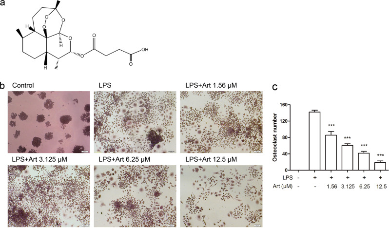
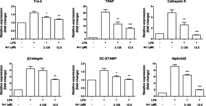
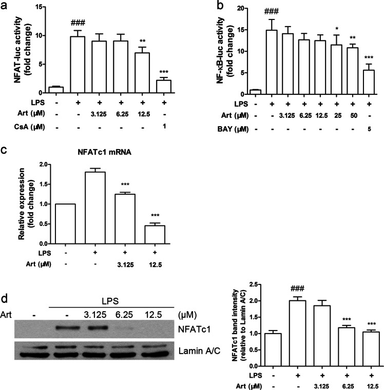
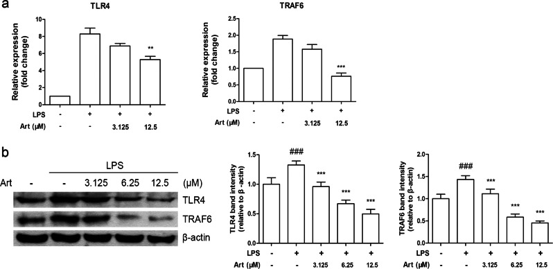
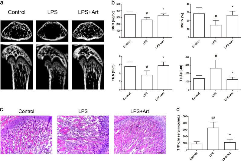
In chronic infectious diseases caused by gram-negative bacteria, such as osteomyelitis, septic arthritis, and periodontitis, osteoclastic activity is enhanced with elevated inflammation, which disturbs the bone homeostasis and results in osteolysis. Lipopolysaccharide (LPS), as a bacteria product, plays an important role in this process. Recent evidence shows that an antimalarial drug artesunate attenuates LPS-induced osteolysis independent of RANKL. In this study we evaluated the effects of artesunate on LPS-induced osteoclastogenesis in vitro and femur osteolysis in vivo, and explored the mechanisms underlying the effects of artesunate on LPS-induced osteoclast differentiation independent of RANKL. In preosteoclastic RAW264.7 cells, we found that artesunate (1.56-12.5 μM) dose dependently inhibited LPS-induced osteoclast formation accompanied by suppressing LPS-stimulated osteoclast-related gene expression (Fra-2, TRAP, Cathepsin K, β3-integrin, DC-STAMP, and Atp6v0d2). We showed that artesunate (3.125-12.5 µM) inhibited LPS-stimulated nuclear factor of activated T cells c1 (NFATc1) but not NF-κB transcriptional activity; artesunate (6.25, 12.5 μM) significantly inhibited LPS-stimulated NFATc1 protein expression. Furthermore, artesunate treatment markedly suppressed LPS-induced Ca2+ influx, and decreased the expression of PP2B-Aα (calcineurin) and pPLCγ1 in the cells. In addition, artesunate treatment significantly decreased the expression of upstream signals TLR4 and TRAF6 during LPS-induced osteoclastogenesis. Administration of artesunate (10 mg/kg, ip) for 8 days effectively inhibited serum TNF-α levels and ameliorated LPS (5 mg/kg, ip)-induced inflammatory bone loss in vivo. Taken together, artesunate attenuates LPS-induced inflammatory osteoclastogenesis by inhibiting the expression of TLR4/TRAF6 and the downstream PLCγ1-Ca2+-NFATc1 signaling pathway. Artesunate is a valuable choice to treat bone loss induced by gram-negative bacteria infection or inflammation in RANKL-independent pathway.

Keywords: Ca2+; LPS; NFATc1; PP2B-Aα; RAW264.7 cells; TLR4; artesunate; chronic infectious diseases; osteoclast.


Conflict of interest statement

The authors declare no competing interests.

Figures

Fig. 1
Fig. 1
Artesunate inhibits LPS-induced osteoclastogenesis. a The chemical structure of artesunate. b Artesunate inhibits LPS-induced osteoclast formation as assessed by a TRAP assay in preosteoclastic RAW264.7 cells. c The osteoclast number was counted per well of each group. Bars show the mean ± SEM from three independent experiments. ***P< 0.001, compared with the LPS-treated group
Fig. 2
Fig. 2
Artesunate suppresses LPS-induced osteoclast-related gene expression. The expression of osteoclast-specific genes was measured by real-time PCR in preosteoclastic RAW264.7 cells after artesunate and LPS administration. Bars represent the mean ± SEM. *P < 0.05, **P < 0.01, ***P < 0.001, compared with the LPS-treated group
Fig. 3
Fig. 3
Artesunate inhibits LPS-induced activation of NFATc1. a The NFAT luciferase activity of NFAT–luc-RAW264.7 cells and b the NF-κB luciferase activity of NF-κB-luc-RAW264.7 cells were measured. c mRNA levels of NFATc1 were assessed by real-time PCR. d Protein levels of NFATc1 were detected by Western blot (left panel). The relative density of nuclear NFATc1 was calculated (right panel). Bars represent the mean ± SEM from three independent experiments. ###P< 0.001, compared with the untreated group; *P < 0.05, **P < 0.01, ***P< 0.001, compared with the LPS-treated group
Fig. 4
Fig. 4
Artesunate suppresses LPS-increased Ca2+ levels and calcineurin expression by decreasing the phosphorylation of PLCγ1. a LPS-induced Ca2+ influx was determined by confocal microscopy. b The fluorescence intensity in 200 s was analyzed to indicate intracellular [Ca2+]. Protein levels of c PP2B-Aα and d phosphorylated PLCγ1 were analyzed by Western blot. Bars show the mean ± SEM from three independent experiments. ##P< 0.01, ###P< 0.001, compared with the untreated group; *P < 0.05, **P < 0.01, ***P< 0.001, compared with the LPS-treated group
Fig. 5
Fig. 5
Artesunate suppresses LPS-induced expression of TLR4 and TRAF6. a mRNA levels of TLR4 and TRAF6 were determined by real-time PCR. b Protein levels of TLR4 and TRAF6 were detected by Western blot (left panel). The relative TLR4 and TRAF6 densities were calculated (right panel). Bars show the mean ± SEM from three independent experiments. ###P< 0.001, compared with the untreated group; **P < 0.01, ***P < 0.001, compared with the LPS-treated group
Fig. 6
Fig. 6
Artesunate prevents LPS-induced osteolysis in vivo. a Distal femur images of micro-CT analysis. b Graphical depiction of trabecular bone parameters, including BMD, BV/TV, and Tb.N. and Tb.Sp. by micro-CT. c Histological changes of tibial sections were assessed by H&E staining (×100). d Serum levels of TNF-α were measured using an ELISA kit. Data are representative of two independent experiments, and bars show the mean ± SEM (n = 5). #P< 0.05, ##P< 0.01, compared with the control group; *P < 0.05, **P < 0.01, compared with the LPS-injected group

References

    1. Zhao C, Irie N, Takada Y, Shimoda K, Miyamoto T, Nishiwaki T, et al. Bidirectional ephrinB2-EphB4 signaling controls bone homeostasis. Cell Metab. 2006;4:111–21. doi: 10.1016/j.cmet.2006.05.012. - DOI - PubMed
    1. Nair SP, Meghji S, Wilson M, Reddi K, White P, Henderson B. Bacterially induced bone destruction: mechanisms and misconceptions. Infect Immun. 1996;64:2371–80. doi: 10.1128/IAI.64.7.2371-2380.1996. - DOI - PMC - PubMed
    1. Keestra JAJ, Grosjean I, Coucke W, Quirynen M, Teughels W. Non‐surgical periodontal therapy with systemic antibiotics in patients with untreated aggressive periodontitis: a systematic review and meta‐analysis. J Periodontal Res. 2014;50:689–706. doi: 10.1111/jre.12252. - DOI - PubMed
    1. Bohannon JK, Hernandez A, Enkhbaatar P, Adams WL, Sherwood ER. The immunobiology of toll-like receptor 4 agonists: from endotoxin tolerance to immunoadjuvants. Shock. 2013;40:451–62. doi: 10.1097/SHK.0000000000000042. - DOI - PMC - PubMed
    1. Suda K, Udagawa N, Sato N, Takami M, Itoh K, Woo JT, et al. Suppression of osteoprotegerin expression by prostaglandin E2 is crucially involved in lipopolysaccharide-induced osteoclast formation. J Immunol. 2004;172:2504–10. doi: 10.4049/jimmunol.172.4.2504. - DOI - PubMed

MeSH terms