. 2020 Jan;41(1):22-33.
doi: 10.1038/s41401-019-0284-y. Epub 2019 Aug 20.

The lncRNA Malat1 functions as a ceRNA to contribute to berberine-mediated inhibition of HMGB1 by sponging miR-181c-5p in poststroke inflammation

Affiliations

The lncRNA Malat1 functions as a ceRNA to contribute to berberine-mediated inhibition of HMGB1 by sponging miR-181c-5p in poststroke inflammation

Ding-Wen Cao et al. Acta Pharmacol Sin. 2020 Jan.

Abstract

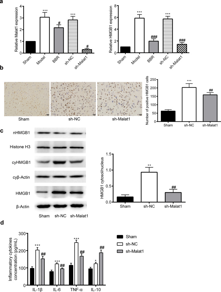
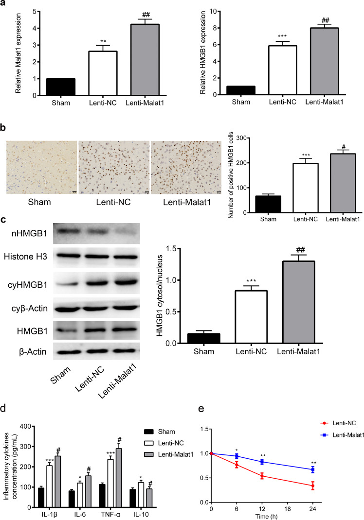
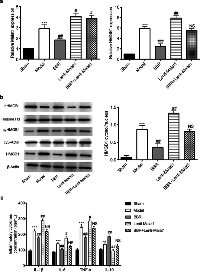
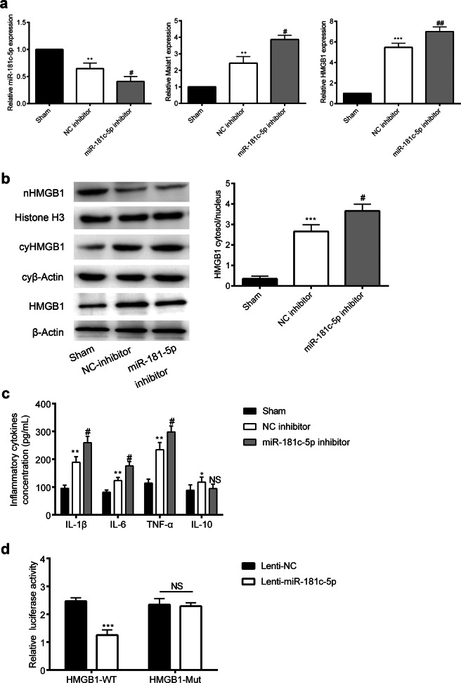
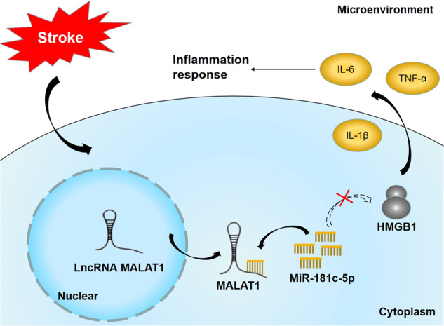
Long non-coding RNAs (lncRNAs) have been identified as essential mediators in neurological dysfunction. Our previous study shows that berberine (BBR) hampers the nuclear-to-cytosolic translocation of high-mobility group box 1 (HMGB1) in the process of poststroke inflammation. In this study, we explored the role of lncRNA metastasis-associated lung adenocarcinoma transcript 1 (Malat1) in the process of BBR-induced inhibition of HMGB1 in ischemic brain. Before the 60-min MCAO surgery, the mice were pretreated with BBR (50 mg· kg-1 per day, ig) for 14 days or ICV injected with specific lentiviral vector or shRNA. We showed that MCAO caused marked increase in the expression Malat1 and HMGB1 in the ipsilateral cortex, which was significantly attenuated by pretreatment with BBR. Knockdown of Malat1 attenuated the inflammatory injury after brain ischemia, whereas overexpression of Malat1 exacerbated ischemic brain inflammation. Overexpression of Malat1 also reversed BBR-induced reduction of HMGB1 and proinflammatory cytokines. The above results suggested a potential correlation between Malat1 and stroke inflammation. Based on informatics analysis we predicted that HMGB1 was a direct downstream target of miR-181c-5p, whereas Malat1 acted as a competitive endogenous RNA (ceRNA) for miR-181c-5p targeted the 3'-UTR of HMGB1 to promote inflammation after ischemic stroke. Knockdown of Malat1 significantly decreased HMGB1 level, which could be abrogated by transfection with miR-181c-5p inhibitors. Taken together, our results demonstrate for the first time that Malat1/miR-181c-5p/HMGB1 axis may be a key pathway of BBR-induced antiinflammation effects in stroke, and they may provide a novel avenue for targeted therapy.

Keywords: HMGB1; Malat1; berberine; inflammation; miR-181c-5p; stroke.


Conflict of interest statement

The authors declare that they have no conflict of interest.

Figures

Fig. 1
Fig. 1
Malat1 knockdown ameliorates ischemic brain inflammation in a mouse MCAO model (n = 3). a Malat1 and HMGB1 were detected by the real-time polymerase chain reaction after 24 h of MCAO. b Enlarged images from representative immunohistochemistry photographs of HMGB1 in different treatment groups and the number of cells with positive staining for HMGB1. Scale bar, 20 μm. Magnification of the microphotograph, ×400. c Representative photographs of Western blots for nHMGB1, cyHMGB1 and HMGB1, and the ratio of cyHMGB1 to nHMGB1. β-Actin and histone H3 were used as loading controls. d Concentrations of inflammatory cytokines (TNF-α, IL-1β, IL-6, and IL-10) were measured by ELISA. Data are presented as the means ± SD. *P < 0.05, ***P < 0.001 versus the Sham group, #P < 0.05, ##P < 0.01, ###P < 0.001 versus the Model group or sh-NC group. nHMGB1: nuclear HMGB1; cyHMGB1: cytosolic HMGB1; nβ-actin: nuclear β-actin; cyβ-actin: cytosolic β-actin
Fig. 2
Fig. 2
Malat1 overexpression exacerbates ischemic brain inflammation in mouse MCAO model (n = 3). a Malat1 and HMGB1 were detected by the real-time polymerase chain reaction after 24 h of MCAO. b Enlarged images from representative immunohistochemistry photographs of HMGB1 in different treatment groups and the number of cells with positive staining for HMGB1. Scale bar, 20 μm. Magnification of the microphotograph, ×400. c. Representative photographs of Western blots for nHMGB1, cyHMGB1 and HMGB1, and the ratio of cyHMGB1 to nHMGB1. β-actin and histone H3 were used as loading controls. d Concentrations of inflammatory cytokines (TNF-α, IL-1β, IL-6, and IL-10) were measured by ELISA. e The effect of Malat1 overexpression on HMGB1 mRNA stability was detected by the real-time polymerase chain reaction to determine the HMGB1 mRNA levels. Data are presented as the means ± SD. *P < 0.05, **P < 0.01, ***P < 0.001 versus the Sham group, #P < 0.05, ##P < 0.01 versus the Lenti-NC group. nHMGB1: nuclear HMGB1; cyHMGB1: cytosolic HMGB1; nβ-actin: nuclear β-actin; cyβ-actin: cytosolic β-actin
Fig. 3
Fig. 3
BBR exerts anti-inflammatory effects through Malat1 after MCAO (n = 3). a Malat1 and HMGB1 were detected by the real-time polymerase chain reaction after 24 h of MCAO. b Representative photographs of Western blots for nHMGB1, cyHMGB1 and HMGB1 and the ratio of cyHMGB1 to nHMGB1. β-actin and histone H3 were used as loading controls. c Concentrations of inflammatory cytokines (TNF-α, IL-1β, IL-6, and IL-10) were measured by ELISA. Data are presented as the means ± SD. **P < 0.01, ***P < 0.001 versus the Sham group, #P < 0.05, ##P < 0.01, ###P < 0.001 versus the Model group, NS versus the Model group. nHMGB1: nuclear HMGB1; cyHMGB1: cytosolic HMGB1; nβ-actin: nuclear β-actin; cyβ-actin: cytosolic β-actin
Fig. 4
Fig. 4
Malat1 binds to miR-181c-5p and functions as a ceRNA (n = 3). a Malat1 subcellular localization results indicated that it was expressed in the cytoplasm. b Predicted binding sites for miR-181c-5p in HMGB1 sequences. c Binding sites of Malat1 on miR-181c-5p predicted by bioinformatics software. d-e RT-PCR was performed on RNA collected from the ipsilateral brain after 24 h of MCAO and showed the expression of miR-181c-5p in the sh-Malat1 and Lenti-Malat1 groups, respectively. f Luciferase reporter assay revealed that Lenti-miR-181c-5p decreased the luciferase activity of Malat1-WT, but not of Malat1-Mut. Data are presented as the means ± SD. **P < 0.01, ***P < 0.001 versus the Sham group, #P < 0.05, ##P < 0.01 versus the sh-NC or Lenti-NC group
Fig. 5
Fig. 5
miR-181c-5p overexpression ameliorates ischemic brain inflammation in a mouse MCAO model (n = 3). a RT-PCR was performed on mRNA collected from the ipsilateral brain after 24 h of MCAO and showed the expression of miR-181c-5p, Malat1 and HMGB1 in the Lenti-miR-181c-5p group. b Representative photographs of Western blot for nHMGB1, cyHMGB1 and HMGB1 and the ratio of cyHMGB1 to nHMGB1. β-actin and histone H3 were used as loading controls. c Concentrations of inflammatory cytokines (TNF-α, IL-1β, IL-6, and IL-10) were measured by ELISA. Data are presented as the means ± SD. *P < 0.05, **P < 0.01, ***P < 0.001 versus the Sham group, #P < 0.05, ##P < 0.01, ###P < 0.001 versus the Lenti-NC group. nHMGB1: nuclear HMGB1; cyHMGB1: cytosolic HMGB1; nβ-actin: nuclear β-actin; cyβ-actin: cytosolic β-actin
Fig. 6
Fig. 6
miR-181c-5p knockdown exacerbates ischemic brain inflammation in a mouse MCAO model (n = 3). a RT-PCR was performed on RNA collected from the ipsilateral brain after 24 h of MCAO and showed the expression of miR-181c-5p, Malat1 and HMGB1 in the miR-181c-5p inhibitor group. b Representative photographs of Western blots for nHMGB1, cyHMGB1 and HMGB1 and the ratio of cyHMGB1 to nHMGB1. β-actin and histone H3 were used as loading controls. c Concentrations of inflammatory cytokines (TNF-α, IL-1β, IL-6, and IL-10) were measured by ELISA. d Luciferase reporter assay revealed that Lenti-miR-181c-5p decreased the luciferase activity of HMGB1-WT but not of HMGB1-Mut. Data are presented as the means ± SD. *P < 0.05, **P < 0.01, ***P < 0.001 versus the Sham group, #P < 0.05, ##P < 0.01 versus the NC inhibitor group. nHMGB1: nuclear HMGB1; cyHMGB1: cytosolic HMGB1; nβ-actin: nuclear β-actin; cyβ-actin: cytosolic β-actin
Fig. 7
Fig. 7
miR-181c-5p inhibitor rescues HMGB1 inhibition induced by Malat1 downregulation (n = 3). a RT-PCR was performed on mRNA collected from the ipsilateral brain after 24 h of MCAO and showed the expression of miR-181c-5p and HMGB1 in the Lenti-miR-181c-5p or miR-181c-5p inhibitor groups. b Representative photographs of Western blots for nHMGB1, cyHMGB1 and HMGB1 and the ratio of cyHMGB1 to nHMGB1. β-actin and histone H3 were used as loading controls. Data are presented as the means ± SD. *P < 0.05, **P < 0.01, ***P < 0.001 versus the Sh-NC + NC inhibitor group. nHMGB1: nuclear HMGB1; cyHMGB1: cytosolic HMGB1; nβ-actin: nuclear β-actin; cyβ-actin: cytosolic β-actin
Fig. 8
Fig. 8
The Malat1/miR-181c-5p/HMGB1 axis is a novel key pathway in stroke inflammation

References

    1. Pandian JD, Gall SL, Kate MP, Silva GS, Akinyemi RO, Ovbiagele BI, et al. Prevention of stroke: a global perspective. Lancet. 2018;392:1269–78. doi: 10.1016/S0140-6736(18)31269-8. - DOI - PubMed
    1. Della-Morte D, Guadagni F, Palmirotta R, Testa G, Caso V, Paciaroni M, et al. Genetics of ischemic stroke, stroke-related risk factors, stroke precursors and treatments. Pharmacogenomics. 2012;13:595–613. doi: 10.2217/pgs.12.14. - DOI - PubMed
    1. Zhang ZG, Chopp M. Neurorestorative therapies for stroke: underlying mechanisms and translation to the clinic. Lancet Neurol. 2009;8:491–500. doi: 10.1016/S1474-4422(09)70061-4. - DOI - PMC - PubMed
    1. Li D, Wang C, Yao Y, Chen L, Liu G, Zhang R, et al. mTORC1 pathway disruption ameliorates brain inflammation following stroke via a shift in microglia phenotype from M1 type to M2 type. FASEB J. 2016;30:3388–99. doi: 10.1096/fj.201600495R. - DOI - PubMed
    1. Brennan-Minnella AM, Won SJ, Swanson RA. NADPH oxidase-2: linking glucose, acidosis, and excitotoxicity in stroke. Antioxid Redox Signal. 2015;22:161–74. doi: 10.1089/ars.2013.5767. - DOI - PMC - PubMed

MeSH terms