. 2019 Oct;55(4):789-804.
doi: 10.3892/ijo.2019.4854. Epub 2019 Aug 5.

TFF3 and TFF1 expression levels are elevated in colorectal cancer and promote the malignant behavior of colon cancer by activating the EMT process

Affiliations

TFF3 and TFF1 expression levels are elevated in colorectal cancer and promote the malignant behavior of colon cancer by activating the EMT process

Aikeremu Yusufu et al. Int J Oncol. 2019 Oct.

Abstract

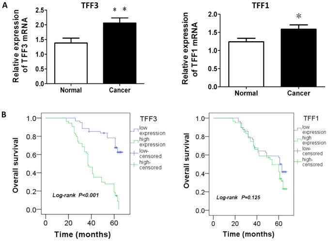
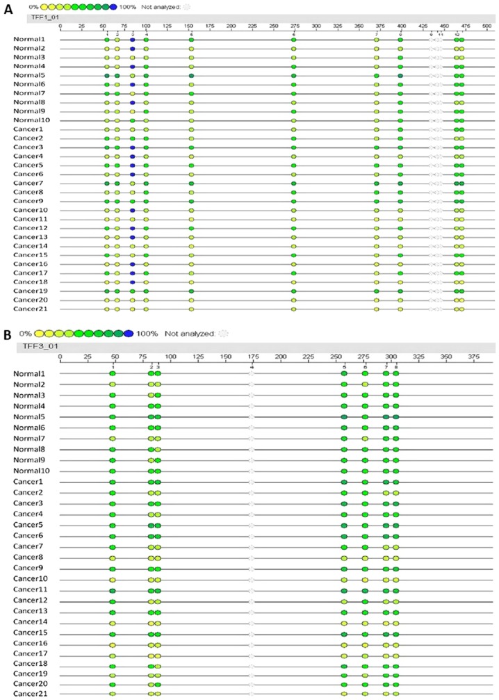
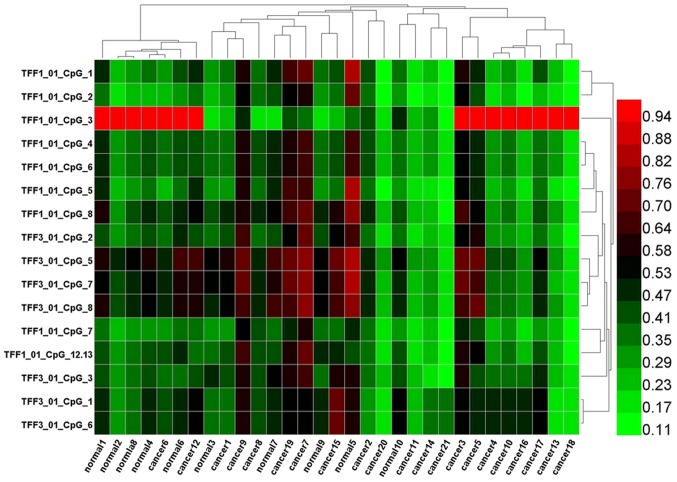
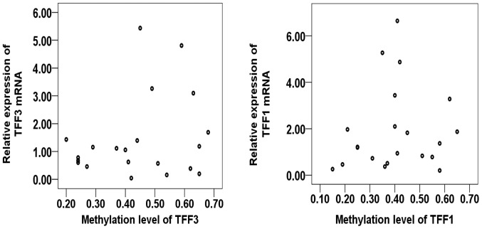
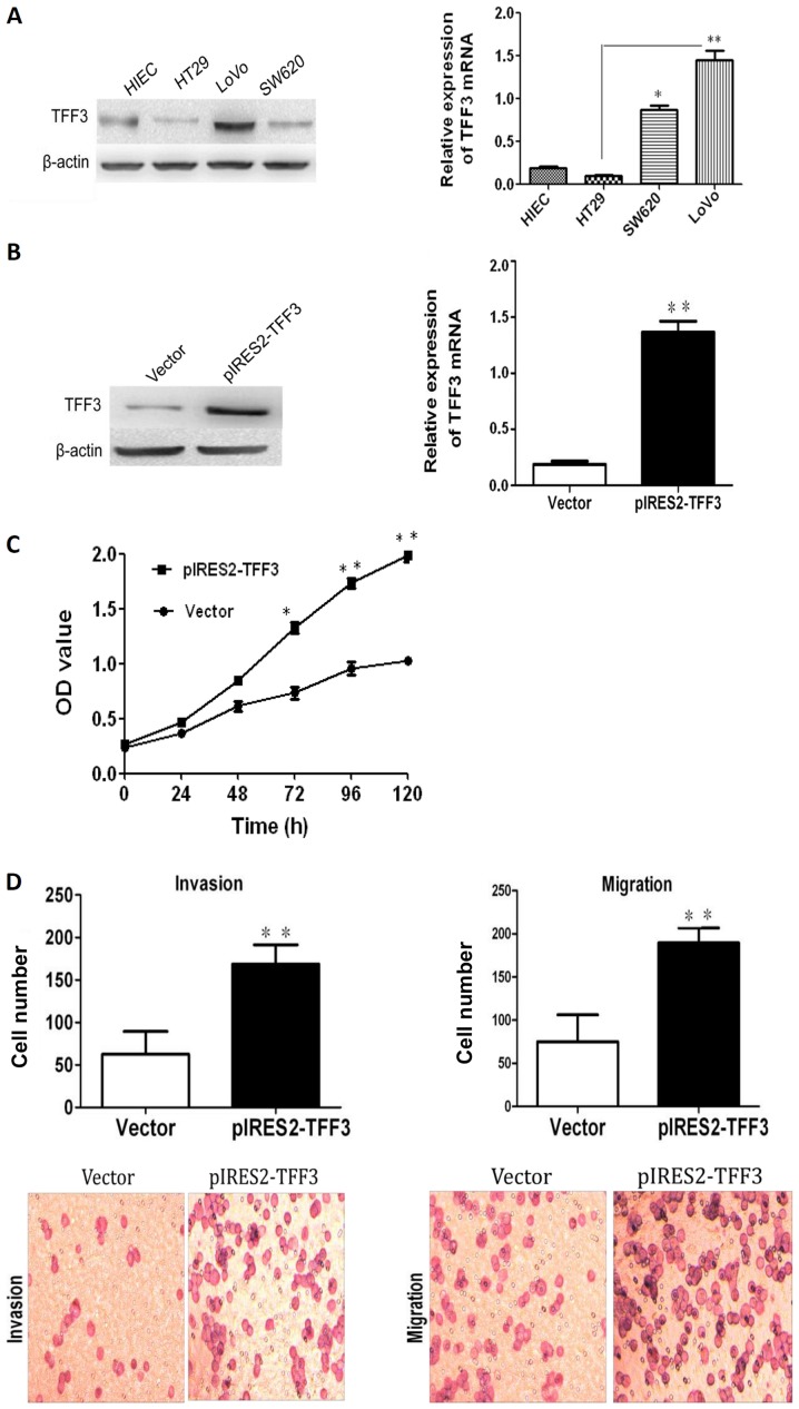
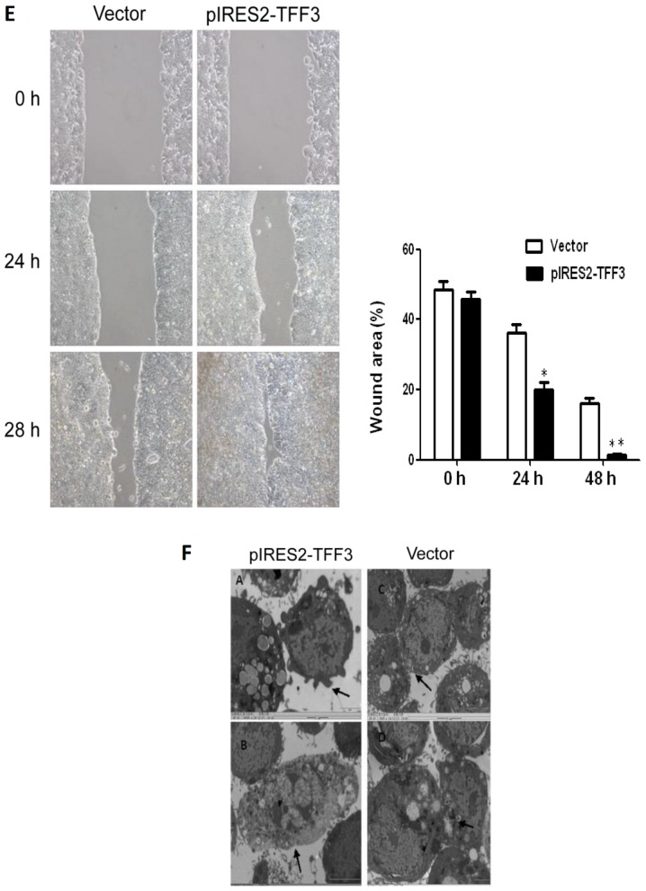
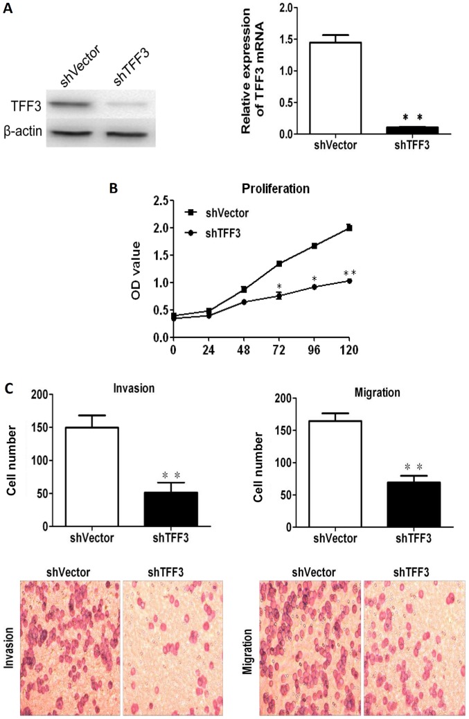
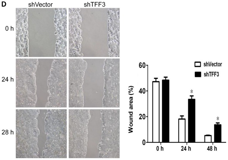
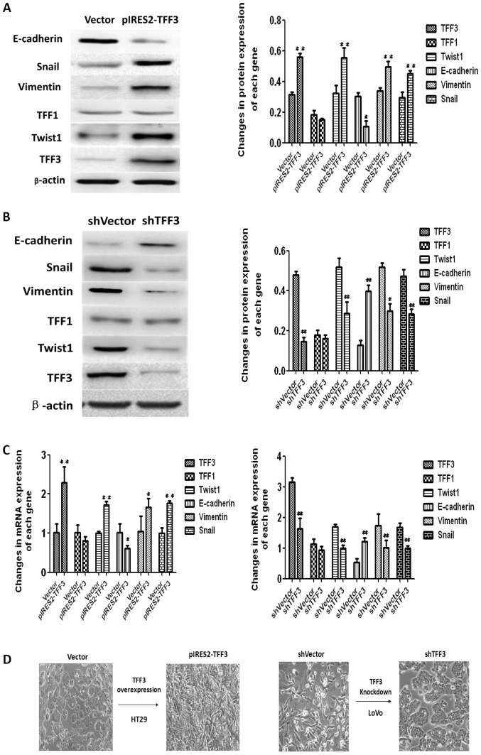
Reports on the roles of the secreted trefoil factor (TFF)1 and 3 in colorectal cancer (CRC) and their underlying mechanisms of action in tumorigenesis are not common and are controversial. In the present study, the mRNA expression and promoter methylation of TFF1 and TFF3 in cancer and adjacent normal tissues were investigated, and their association with other clinical factors and patient prognosis were evaluated. Moreover, the association between TFF3 and epithelial‑mesenchymal transition (EMT) was explored by overexpressing or inhibiting TFF3 expression. The results revealed that the mRNA level of TFF1 and TFF3 in the cancer tissues was significantly higher than that in the matched adjacent normal tissues (P=0.034 and P=0.007, respectively), and a higher expression of TFF3, but not TFF1, was predominantly associated with clinicopathological factors and a poorer prognosis. No correlation was observed between promoter methylation and the expression of TFF1 or TFF3. The overexpression of TFF3 promoted the proliferation, migration and invasiveness of HT29 cells, and induced an increase in the expression of Twist1, Snail and Vimentin, while causing a decrease in E‑cadherin expression. On the contrary, the knockdown of TFF3 resulted in opposite effects in the LoVo cells. On the whole, the findings of this study indicate that TFF3 may be a promising new factor for the estimation of the survival of patients with CRC, and may promote the malignant progression of CRC by activating the EMT process. Therefore, TFF3 may be a future potential therapeutic target for CRC.


Figures

Figure 1
Figure 1
mRNA expression of TFF1 and TFF3 in cancer and normal tissues and the association with survival. (A) mRNA expression of TFF1 or TFF3 in cancer tissues was higher than that in normal tissues (P=0.034 or P=0.007). (B) Survival analysis revealed that a higher expression of TFF3 was associated with a worse overall survival, while TFF1 expression demonstrated no association with patient survival (P<0.001 and P=0.125). *P<0.05 and **P<0.01 vs. normal group. TFF, trefoil factor.
Figure 2
Figure 2
Promoter methylation sites and levels of the TFF1 and TFF3 genes in cancer and normal tissues. (A) The length of promoter region of the TFF1 gene for detection was 489 bp, 13 units of CpG sites could be found and 10 units were measured successfully. (B) The length of 372 bp was detected for the TFF3 gene and 7 out of 8 units were analyzed favorably. The different colors represent the relative methylation changes in 10% increments (yellow, 0%; blue, 100% methylated). TFF, trefoil factor.
Figure 3
Figure 3
Two-way hierarchical cluster analysis of 21 cancer tissue and 10 normal tissue samples (columns) and DNA-methylation of CpG Units in promoter regions (rows). DNA-methylation levels are depicted in this heatmap on a continuous scale from light green (unmethylated) to light red (100% methylated). Poor quality data is shown in black. Less-dominant clustering was found in cancer or normal tissues. The hierarchical cluster algorithm could not separate colon cancer samples from normal tissue samples.
Figure 4
Figure 4
Correlation analysis between the promoter methylation level and mRNA expression of TFF1 or TFF3. No apparent variation trends were observed from the scatter diagrams for TFF1 or TFF3. TFF, trefoil factor.
Figure 5
Figure 5
The overexpression of TFF3 promotes the proliferation, migration and invasion capacities of the HT29 cells. (A) Both western blot analysis and RT-qPCR revealed that TFF3 expression in the HT29 and LoVo cells were the lowest and highest, respetively. (B) TFF3 expression was found to be enhanced significantly at the mRNA and protein level following transfection with the TFF3 expression plasmid. (C) CCK-8 assay revealed that the overexpression of TFF3 markedly increased the proliferation of the HT29 cells. (D) Transwell chamber assay displayed that the migration and invasion of the HT29 cells was markedly enhanced following the overexpression of TFF3. (E) The migration of the HT29 cells was also markedly increased according to wound healing assay. (F) Compared with the control group, the pIRES2-TFF3-transfected cells exhibited less pseudopodia (A), a reduced number of villi (B) and fewer intercellular junctions (C and D). An increased number of polygonal or spindle like shaped cells in the TFF3 overexpression group was also observed (B). *P<0.05 and **P <0.01 vs. control (HIEC) group or the empty vector (Vector). TFF, trefoil factor.
Figure 5
Figure 5
The overexpression of TFF3 promotes the proliferation, migration and invasion capacities of the HT29 cells. (A) Both western blot analysis and RT-qPCR revealed that TFF3 expression in the HT29 and LoVo cells were the lowest and highest, respetively. (B) TFF3 expression was found to be enhanced significantly at the mRNA and protein level following transfection with the TFF3 expression plasmid. (C) CCK-8 assay revealed that the overexpression of TFF3 markedly increased the proliferation of the HT29 cells. (D) Transwell chamber assay displayed that the migration and invasion of the HT29 cells was markedly enhanced following the overexpression of TFF3. (E) The migration of the HT29 cells was also markedly increased according to wound healing assay. (F) Compared with the control group, the pIRES2-TFF3-transfected cells exhibited less pseudopodia (A), a reduced number of villi (B) and fewer intercellular junctions (C and D). An increased number of polygonal or spindle like shaped cells in the TFF3 overexpression group was also observed (B). *P<0.05 and **P <0.01 vs. control (HIEC) group or the empty vector (Vector). TFF, trefoil factor.
Figure 6
Figure 6
Knockdown of TFF3 notably reduces the proliferation, migration and invasion of LoVo cells. (A) TFF3 expression was suppressed by shRNA transfection, as shown by both western blot analysis and RT-qPCR. (B) CCK-8 assay revealed that the silencing of TFF3 significantly decreased the proliferation of the LoVo cells. (C) The migration and invasion of Lovo cells were markedly suppressed following the silencing of TFF3, as shown by Transwell chamber assay. (D) Scratch wound healing assay demonstrated that the migration of the LoVo cells was markedly reduced by the silencing of TFF3. *P<0.05 and **P<0.01 vs. shvector. TFF, trefoil factor.
Figure 6
Figure 6
Knockdown of TFF3 notably reduces the proliferation, migration and invasion of LoVo cells. (A) TFF3 expression was suppressed by shRNA transfection, as shown by both western blot analysis and RT-qPCR. (B) CCK-8 assay revealed that the silencing of TFF3 significantly decreased the proliferation of the LoVo cells. (C) The migration and invasion of Lovo cells were markedly suppressed following the silencing of TFF3, as shown by Transwell chamber assay. (D) Scratch wound healing assay demonstrated that the migration of the LoVo cells was markedly reduced by the silencing of TFF3. *P<0.05 and **P<0.01 vs. shvector. TFF, trefoil factor.
Figure 7
Figure 7
TFF3 induces the EMT process in colorectal cancer cells. (A) Western blot analysis demonstrated that the enforced expression of TFF3 in HT29 cells increased the protein expression of Twist1, Snail and Vimentin, while it suppressed E-cadherin expression. (B) The downregulation of TFF3 expression in the Lovo cells decreased the protein expression of Twist1, Snail and Vimentin, while it enhanced E cadherin expression. (C) The same results were observed at the mRNA level when TFF3 was overexpressed or silenced. (D) EMT-like morphological changes in cells following the overexpression or silencing of TFF3. The morphology of the HT29 cells changed from a round or volmer-weber mode into a triangular or spindle form following the overexpression of TFF3. LoVo cells displayed a reversed morphological change from the scattered cord-like form to the round, rectangular or clustered pattern after the silencing of TFF3. #P<0.05 and ##P<0.01vs. empty vector (Vector) or shVector. TFF, trefoil factor.

References

    1. Cai SJ, Peng JJ. Colorectal cancer epidemiology and prevention strategies. Proc Academic Annual Conference of CSCO 2014; Peking, China. 2014. pp. 294–301.
    1. Li S, Wang J, Lu Y, Fan D. Screening and early diagnosis of colorectal cancer in China: A 12 year retrospect (1994 2006) J Cancer Res Clin Oncol. 2007;133:679–686. doi: 10.1007/s00432-007-0231-z. - DOI - PubMed
    1. Gespach C. Trefoil factors. In: Schwab M, editor. Encyclopedia of Cancer. Springer; Berlin, Heidelberg: 2011. pp. 4652–4658.
    1. Sands BE, Podolsky DK. The trefoil peptide family. Annu Rev Physiol. 1996;58:253–273. doi: 10.1146/annurev.ph.58.030196.001345. - DOI - PubMed
    1. Wright NA, Poulsom R, Stamp G, Van Noorden S, Sarraf C, Elia G, Ahnen D, Jeffery R, Longcroft J, Pike C, et al. Trefoil peptide gene expression in gastrointestinal epithelial cells in inflammatory bowel disease. Gastroenterology. 1993;104:12–20. doi: 10.1016/0016-5085(93)90830-6. - DOI - PubMed