Review
. 2020 Jan;77(2):305-321.
doi: 10.1007/s00018-019-03278-z. Epub 2019 Aug 20.

The enhancement of glycolysis regulates pancreatic cancer metastasis

Affiliations
Review

The enhancement of glycolysis regulates pancreatic cancer metastasis

Jinshou Yang et al. Cell Mol Life Sci. 2020 Jan.

Abstract

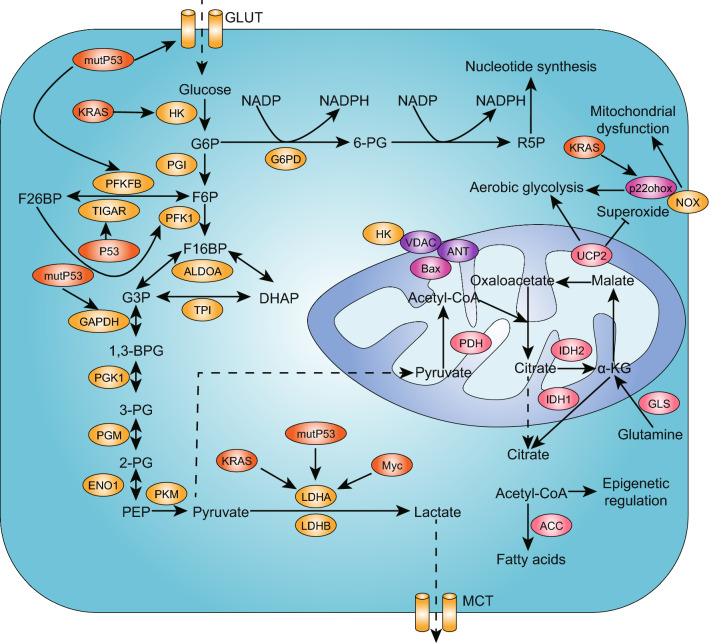
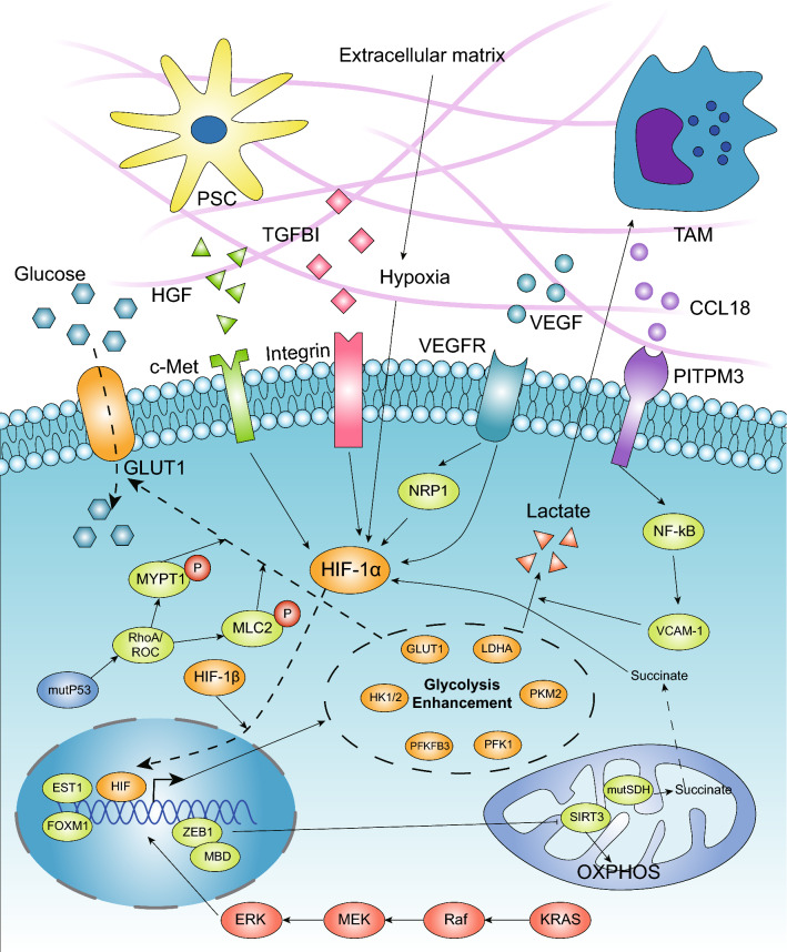
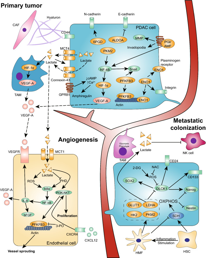
Pancreatic ductal adenocarcinoma is prone to distant metastasis and is expected to become the second leading cause of cancer-related death. In an extremely nutrient-deficient and hypoxic environment resulting from uncontrolled growth, vascular disturbances and desmoplastic reactions, pancreatic cancer cells utilize "metabolic reprogramming" to satisfy their energy demand and support malignant behaviors such as metastasis. Notably, pancreatic cancer cells show extensive enhancement of glycolysis, including glycolytic enzyme overexpression and increased lactate production, and this is caused by mitochondrial dysfunction, cancer driver genes, specific transcription factors, a hypoxic tumor microenvironment and stromal cells, such as cancer-associated fibroblasts and tumor-associated macrophages. The metabolic switch from oxidative phosphorylation to glycolysis in pancreatic cancer cells regulates the invasion-metastasis cascade by promoting epithelial-mesenchymal transition, tumor angiogenesis and the metastatic colonization of distant organs. In addition to aerobic glycolysis, oxidative phosphorylation also plays a critical role in pancreatic cancer metastasis in ways that remain unclear. In this review, we expound on the intracellular and extracellular causes of the enhancement of glycolysis in pancreatic cancer and the strong association between glycolysis and cancer metastasis, which we expect will yield new therapeutic approaches targeting cancer metabolism.

Keywords: Epithelial–mesenchymal transition; Hybrid metabolic phenotype; Metastatic niche; Mitochondrial respiration; Tumor microenvironment; Warburg effect.


Conflict of interest statement

The authors declare that they have no competing interests.

Figures

Fig. 1
Fig. 1
Warburg effect in PDAC. The “Warburg effect” exists in the majority of tumors with enhanced glycolysis and lactate production under aerobic conditions, which was initially considered to be the result of mitochondrial dysfunction. PDAC cells present a high-glycolysis phenotype in which glucose uptake is increased and the glycolysis rate is accelerated, which is regulated by Kras, mutp53 and c-Myc, as a result of respiratory injury and the overexpression of glycolytic enzymes, which are marked in the yellow ellipses in this figure. The enhancement of glycolysis shunts more glucose into the pentose phosphate pathway and results in the accumulation of lactate in the microenvironment
Fig. 2
Fig. 2
Driver genes and TME promote the enhancement of glycolysis in PDAC. The high-glycolysis phenotype is considered to be an emerging hallmark associated with activated oncogenes. Mutant Kras can upregulate the expression of glycolytic enzymes via the Raf/MEK/ERK pathway, which is dependent on Myc and functions transcriptionally. GOF mutp53 also promotes glycolysis mainly by regulating GLUT1 translocation to the cytoplasmic membrane. In the pancreatic microenvironment, hypoxia is considered the main regulator that transcriptionally upregulates the expression of multiple glycolytic enzymes. SDH mutations leading to succinate accumulation can promote glycolysis through stabilizing HIF-1α. Additionally, stroma cells, such as TAMs and pancreatic stellate cells, secrete cytokines that interact with membrane receptors in cancer cells, and soluble factors in the TME, such as VEGF and TGFBI, promote glycolysis mainly via the HIF-1α and NF-kB pathways. ZEB1, known as an EMT regulator, can also promote glycolysis via its interaction with MBD1, which transcriptionally suppresses the expression of SIRT3
Fig. 3
Fig. 3
Enhancement of glycolysis promotes PDAC metastasis. In the invasion–metastasis cascade in PDAC cells, glycolytic enzymes and lactate facilitate the process in three main ways. (1) Glycolysis promotes the EMT program by upregulating EMT-TFs, downregulating E-cadherin, and increasing MMP secretion and cytoskeleton remodeling. PFKFB3, ALDOA, ENO1, PKM2 and PGI/AMF can function in cytoplasmic, nuclear and extracellular forms. (2) Glycolysis promotes angiogenesis by increasing VEGF and amphiregulin secretion via a self-induced pathway and the regulation of stromal cells, which is dependent on the accumulation of lactate in the TME. Additionally, glycolytic enzymes such as PKM2 can be secreted into the blood stream to interact with receptors in the EC membrane to promote EC proliferation and migration. In addition, enhanced glycolysis in ECs also promotes vessel sprouting to facilitate angiogenesis. (3) Glycolysis promotes PDAC cell metastatic colonization. Glycolytic TAMs promote the extravasation of CTCs. In a distant microenvironment, the enhancement of glycolysis in metastatic PDAC cells maintains stemness through low ROS levels. In the hepatic microenvironment, many HSCs promote the elevated expression of SDH and enhance OXPHOS activity, reducing the self-renewal ability of pancreatic cancer metastatic cells via the reduced expression of Nanog and Nestin. In contrast, HMFs promote the downregulation of SDH and activate glycolysis, which promotes cancer cell proliferation and the formation of visible metastases. In addition, the secretion of lactate from PDAC cells reduces NK cell cytotoxicity, which promotes metastatic colonization

References

    1. Kamisawa T, Wood LD, Itoi T, Takaori K. Pancreatic cancer. Lancet. 2016;388:73–85. doi: 10.1016/S0140-6736(16)00141-0. - DOI - PubMed
    1. Siegel RL, Miller KD, Jemal A. Cancer statistics, 2019. CA Cancer J Clin. 2019;69:7–34. doi: 10.3322/caac.21551. - DOI - PubMed
    1. Rahib L., Smith B. D., Aizenberg R., Rosenzweig A. B., Fleshman J. M., Matrisian L. M. Projecting Cancer Incidence and Deaths to 2030: The Unexpected Burden of Thyroid, Liver, and Pancreas Cancers in the United States. Cancer Research. 2014;74(11):2913–2921. - PubMed
    1. Poruk KE, Firpo MA, Adler DG, Mulvihill SJ. Screening for pancreatic cancer: why, how, and who? Ann Surg. 2013;257:17–26. - PMC - PubMed
    1. Giovannetti E, et al. Never let it go: Stopping key mechanisms underlying metastasis to fight pancreatic cancer. Semin Cancer Biol. 2017;44:43–59. - PubMed

MeSH terms