. 2019 Oct;110(10):3068-3078.
doi: 10.1111/cas.14174. Epub 2019 Sep 24.

Interleukin-17 is associated with expression of programmed cell death 1 ligand 1 in ovarian carcinoma

Affiliations

Interleukin-17 is associated with expression of programmed cell death 1 ligand 1 in ovarian carcinoma

Aeri Aotsuka et al. Cancer Sci. 2019 Oct.

Abstract

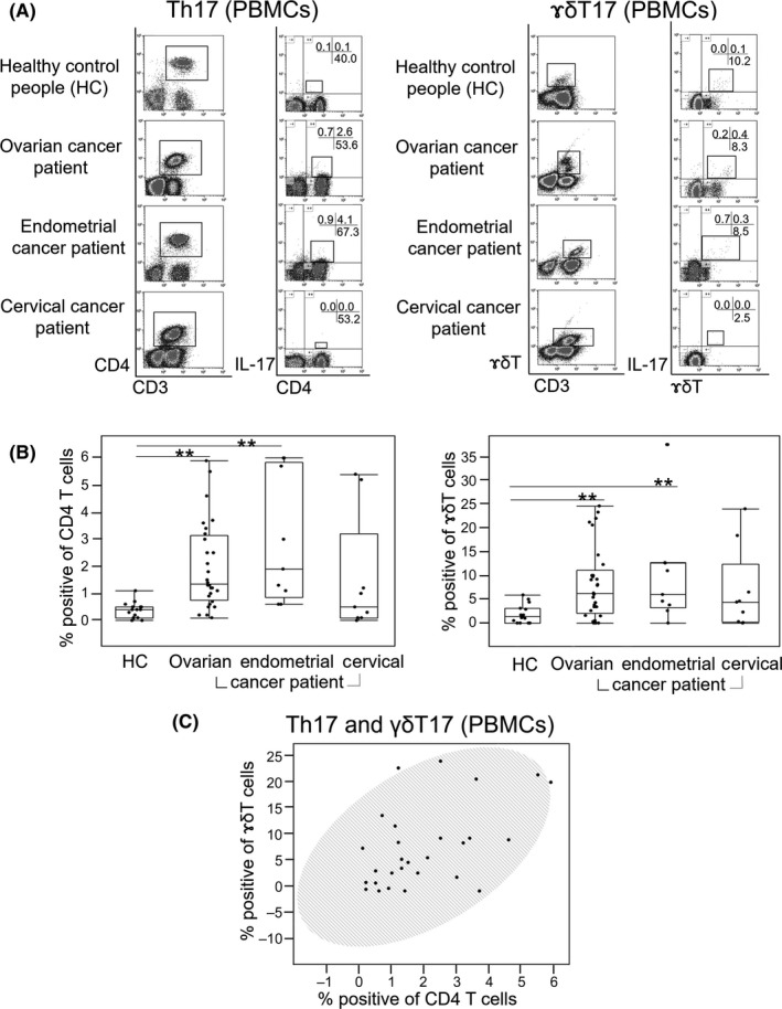
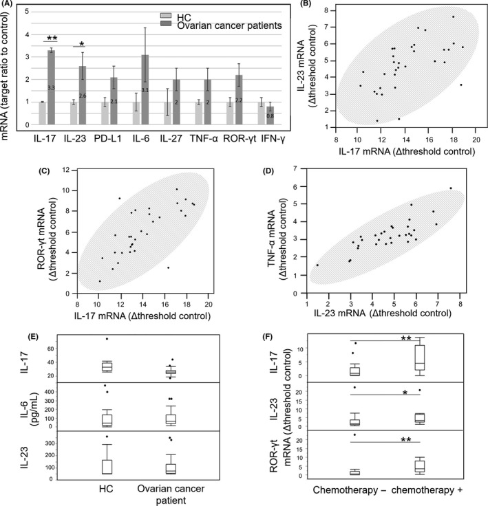
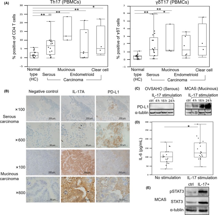
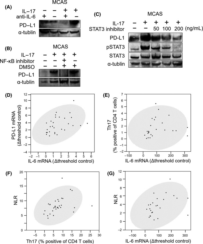
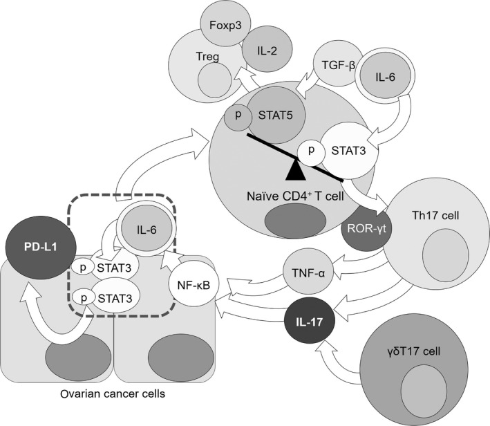
The programmed cell death 1/programmed cell death 1 ligand 1 pathway was successfully targeted in cancer immunotherapy. Elevated interleukin-17 (IL-17), which is known in autoimmune diseases, has recently been recognized in cancer patients. We investigated the role of IL-17 in the regulation of expression of programmed cell death 1 ligand 1 in ovarian cancer by evaluating changes in the number of IL-17-producing cluster of differentiation 4 helper T cells (Th17) and γδT cells (γδT17) in PBMC of 52 gynecological cancer patients (including 30 ovarian cancer patients) and 18 healthy controls. The occupancy ratio of Th17 and γδT17 was higher in ovarian cancer and endometrial cancer patients than in controls, determined by multi-color flow cytometry (Th17: P < 0.0001 and P = 0.0002, respectively; γδT17: P = 0.0020 and P = 0.0084, respectively). IL-17 mRNA level was elevated in PBMC of ovarian cancer patients (P = 0.0029), as measured by RT-PCR. The neutrophil-to-lymphocyte ratio, which is a prognostic biomarker of ovarian cancer, correlated with Th17 occupancy ratio in patients (P = 0.0068). We found that programmed cell death 1 ligand 1 expression and its associated factors (IL-6 and phospho-signal transducer and activator of transcription 3) were induced by IL-17 in an ovarian cancer cell line. These results suggest that increased Th17 counts and IL-17 level, which correlated with high neutrophil-to-lymphocyte ratio and programmed cell death 1 ligand 1 expression, are potential biomarkers for poor prognosis in ovarian cancer and likely indications for application of programmed cell death 1 ligand 1 pathway inhibitors.

Keywords: IL-17; PD-L1; Th17; neutrophil-to-lymphocyte ratio; ovarian cancer.


Figures

Figure 1
Figure 1
Ratio of Th17 and γδT17 cells in gynecological cancer patients. A, Squares in the left column indicate CD3+CD4+ and CD3+γδT+ cells; those in the right column indicate CD4+IL‐17+ and γδT+IL‐17+ cells. The proportion of CD3+CD4+IL‐17+ cells in CD3+CD4+ cells or the proportion of CD3+γδT+IL‐17+ cells in CD3+γδT+ cells was defined as the occupancy ratio of Th17 and γδT17, respectively. Numbers on cross bars in each column represent the percentage of the Th17 or γδT17 cells in CD3+ cell population. B, Occupancy ratio of Th17 or γδT17 for each carcinoma type (ovarian cancer [OC], n = 30; endometrial cancer, n = 12; and cervical cancer, n = 10) and 18 healthy control (HC) subjects. Lines in the middle of each box represent the median value and the lower and upper boundaries show the 5th and 95th percentiles, respectively. Results were analyzed using the Mann‐Whitney U test. *P < 0.05, **P < 0.01. C, Correlation between Th17 and γδT17 in PBMC of OC patients. Data were analyzed using the Spearman's test, and shaded ellipses represent 95% confidence level (r = 0.50, P = 0.0052)
Figure 2
Figure 2
mRNA and proteins levels of interleukin‐17 (IL‐17)‐related factors. A, Relative mRNA expression of IL‐17‐related genes in PBMC of ovarian cancer (OC) patients, as determined by RT‐PCR. Right and left bars represent OC patients and healthy control (HC) respectively. B‐D, Correlation between IL‐17 and IL‐23 mRNA levels in PBMC of OC patients. (C) and (D) show the correlation between IL‐17 and ROR‐γt mRNA and between IL‐23 and TNF‐α mRNA levels, respectively, in PBMC of OC patients (Spearman's test, b: r = 0.64, P < 0.0001; c: r = 0.69, P < .0001; and d: r = 0.84, P < .0001). Shaded ellipses represent 95% confidence level. E, Plasma IL‐17, IL‐23 and IL‐6 protein levels in HC and OC patients, as determined by ELISA. F, Comparative analysis of IL‐17, IL‐23 and ROR‐γt mRNA levels in PBMC of OC patients with or without chemotherapy. *P < 0.05, **P < 0.01 (Mann‐Whitney test). Black circles in (E) and (F) indicate outliers
Figure 3
Figure 3
Analysis of factors related to interleukin‐17 (IL‐17) according to ovarian cancer (OC) histopathological type. A, Occupancy ratio of Th17 and γδT17 cells in PBMC of OC patients or healthy controls (HC). B, Immunodetection of IL‐17A and programmed cell death 1 ligand 1 (PD‐L1) in surgical tumor specimens from OC patients. C, Effect of IL‐17 on PD‐L1 expression, as detected by western blotting in OVSAHO and MCAS cell lines. D, IL‐6 expression following stimulation with IL‐17A (right), as determined by ELISA; untreated cells served as control (left). In A‐D, lines in the middle of each box indicate the median value, and the lower and upper boundaries represent the 5th and 95th percentiles, respectively. E, STAT3 phosphorylation in MCAS cells evaluated 24 h after IL‐17A stimulation by ELISA. *P < .05, **P < .01 (Mann‐Whitney test). White circles in panel indicate outliers
Figure 4
Figure 4
Programmed cell death 1 ligand 1 (PD‐L1) expression induced by interleukin‐17A (IL‐17A) and its inhibition by IL‐6, phospho‐signal transducer and activator of transcription (pSTAT3) and nuclear factor‐κB (NF‐κB) inhibitors. A, Expression of IL‐17‐related genes in MCAS cells following IL‐17 stimulation in the presence of anti–IL‐6 Ab (mAbIL‐6), as determined by western blotting. B, Change in PD‐L1 expression upon IL‐17 stimulation in the presence of NF‐κB inhibitor (BAY11‐7082) and 0.1% DMSO, as determined by western blotting. C, Western blotting of changes in PD‐L1 and pSTAT3 expression following IL‐17 stimulation in the presence of STAT3 inhibitor (S31‐201: 0, 50, 10 and 200 μg/mL). Investigation of the relationship between PD‐L1, IL‐6, Th17 and neutrophil‐to‐lymphocyte ratio (NLR). D, Correlation between IL‐6 and PD‐L1 mRNA levels in PBMC of ovarian cancer (OC) patients. E, Relationship between IL‐6 mRNA and Th17 occupancy ratio in PBMC of OC patients. F,G, Relationship between NLR in preoperative blood collected from OC patients and Th17 occupancy ratio in PBMC (F) or plasma IL‐6 protein level (G). Results in (D) to (G) were analyzed using Spearman's test and shaded ellipses represent 95% confidence level ((D) ρ = 0.52, P = 0.0023; (D) ρ = 0.51, P = 0.0098; (F) ρ = 0.49, P = 0.0068; (G) ρ = 0.48, P = 0.0184)
Figure 5
Figure 5
Interleukin‐6 (IL‐6) and transforming growth factor beta (TGF‐β) phosphorylate STAT3 in naïve T cells. Naïve T cells are induced to differentiate into regulatory T cells by TGF‐β in the absence of IL‐6. ROR‐γt is activated to induce differentiation of the naïve T cells into Th17 cells, which produce IL‐17 and TNF‐α. Nuclear factor‐κB (NF‐κB) activated by IL‐17 or TNF‐α stimulation induces expression of IL‐6, which phosphorylates STAT3 in cancer cells. These steps result in programmed cell death 1 ligand 1 (PD‐L1) expression in ovarian cancer (OC) cells and reactivation of IL‐6 and STAT3 through a positive feedback loop

References

    1. Cancer Registry and Statistics . Cancer Information Service, National Cancer Center, Japan. 2015.
    1. Allemani C, Matsuda T, Di Carlo V, et al. Global surveillance of trends in cancer survival 2000–14 (CONCORD‐3): analysis of individual records for 37 513 025 patients diagnosed with one of 18 cancers from 322 population‐based registries in 71 countries. Lancet. 2018;391:1023‐1075. - PMC - PubMed
    1. Dong C. IL‐23/IL‐17 biology and therapeutic considerations. J Immunotoxicol. 2008;5:43‐46. - PubMed
    1. Kimura A, Kishimoto T. IL‐6: regulator of Treg/Th17 balance. Eur J Immunol. 2010;40:1830‐1835. - PubMed
    1. Ma J, Wang J, Wan J, et al. Morphine disrupts interleukin‐23 (IL‐23)/IL‐17‐mediated pulmonary mucosal host defense against Streptococcus pneumoniae infection. Infect Immun. 2010;78:830‐837. - PMC - PubMed

MeSH terms