Review
. 2019 Dec;18(6):e13031.
doi: 10.1111/acel.13031. Epub 2019 Aug 20.

Oxidative stress in the aging substantia nigra and the etiology of Parkinson's disease

Affiliations
Review

Oxidative stress in the aging substantia nigra and the etiology of Parkinson's disease

Benjamin G Trist et al. Aging Cell. 2019 Dec.

Abstract

Parkinson's disease prevalence is rapidly increasing in an aging global population. With this increase comes exponentially rising social and economic costs, emphasizing the immediate need for effective disease-modifying treatments. Motor dysfunction results from the loss of dopaminergic neurons in the substantia nigra pars compacta and depletion of dopamine in the nigrostriatal pathway. While a specific biochemical mechanism remains elusive, oxidative stress plays an undeniable role in a complex and progressive neurodegenerative cascade. This review will explore the molecular factors that contribute to the high steady-state of oxidative stress in the healthy substantia nigra during aging, and how this chemical environment renders neurons susceptible to oxidative damage in Parkinson's disease. Contributing factors to oxidative stress during aging and as a pathogenic mechanism for Parkinson's disease will be discussed within the context of how and why therapeutic approaches targeting cellular redox activity in this disorder have, to date, yielded little therapeutic benefit. We present a contemporary perspective on the central biochemical contribution of redox imbalance to Parkinson's disease etiology and argue that improving our ability to accurately measure oxidative stress, dopaminergic neurotransmission and cell death pathways in vivo is crucial for both the development of new therapies and the identification of novel disease biomarkers.

Keywords: Parkinson's disease; antioxidant; oxidative stress; reactive oxygen species.


Conflict of interest statement

None declared.

Figures
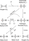
Figure 1
Figure 1
Common reactive oxygen species, their production, and clearance. Incomplete reduction of molecular oxygen (O2) produces superoxide radicals (O2 ), which may be converted to hydroxyl radicals (OH) via Haber–Weiss chemistry, or to hydrogen peroxide (H2O2) through the action of enzymes or molecules with superoxide dismutase (SOD) activity. Hydrogen peroxide is also a substrate for hydroxyl radical production via Fenton chemistry, catalyzed by labile ferrous iron. Hydrogen peroxide decomposition to water and oxygen is mediated by the enzymatic action of glutathione peroxidase (GPx) coupled to redox cycling of reduced (GSH) and oxidized (GSSG) glutathione, and also by catalase. Unpaired electrons are highlighted in red
Figure 2
Figure 2
Reactive oxygen species are an inherent by‐product of oxidative phosphorylation in the mitochondrial ETC. (a) Electrons generated by the tricarboxylic acid cycle in the mitochondrial matrix are shuttled to ETC complexes I and II by NADPH and FADH2, respectively. They are then transferred to complex IV of the ETC with the help of inner mitochondrial membrane (IMM) electron shuttles (Q, coenzyme Q; C, cytochrome c) where they reduce molecular oxygen to water, a process which simultaneously drives ATP production by ATP synthase (ETC complex V). A small amount of premature electron leakage occurs naturally during oxidative phosphorylation, whereby electrons bound within ETC complexes I and III diffuse into both the mitochondrial matrix and intermembrane space (IMS). Here, they may cause incomplete reduction of molecular oxygen (O2), generating superoxide radicals (O2 ) that may subsequently be converted to hydrogen peroxide (H2O2) through the action of superoxide dismutase 1 or 2 (SOD1/2). Electron leakage from the electron transport chain is worsened during healthy aging, or by pathogenic factors such as genetic mutations (SNCA, SOD1), environmental toxins (MPTP, rotenone), or misfolded proteins (α‐synuclein, SOD1). (b) Mitochondrial H2O2 levels are regulated by glutathione peroxidase (GPx) and peroxiredoxin (PRx), coupled to the redox cycling of glutathione (GSH/GSSG) and thioredoxin (TRxSH/TRxSS), respectively. While the oxidation component of each cycle is mediated by GPx and PRx, glutathione reductase (GR) and thioredoxin reductase (TRxR) drive NADPH‐dependent glutathione and thioredoxin reduction, respectively, to complete the redox loop
Figure 3
Figure 3
Dopamine metabolism and ROS production. Enzymatic decomposition of dopamine to homovanillic acid is mediated by monoamine oxidase (MAO), catechol‐o‐methyl transferase (COMT), and aldehyde dehydrogenase (ALDH). Conversely, dopamine may be oxidized to dopamine‐o‐quinone by tyrosinase (Tyr), cyclooxygenase (COX), or labile ferric iron (Fe3+). Dopamine‐o‐quinones are reactive intermediates for the generation of more damaging compounds, including 6‐hydroxydopamine and R‐Salsolinol. Endogenous detoxification of dopamine‐o‐quinones involves cyclization to produce aminochrome, and subsequent oxidation and polymerization to generate neuromelanin. Hydrogen peroxide (H2O2) produced by MAO, dopamine oxidation and dopamine‐o‐quinone oxidation, can participate in Fenton chemistry and react with labile ferrous iron (Fe2+) to generate damaging hydroxyl radicals (OH)
Figure 4
Figure 4
Alterations in dopamine, iron, and α‐synuclein promote oxidative stress selectively in the SNc. Under physiological conditions, α‐synuclein facilitates dynamin‐mediated endocytosis of transferrin receptor and iron‐bound holo‐transferrin. A facile cytoplasmic labile iron pool is tightly maintained by ferritin to enable ferroprotein function, including dopamine production by tyrosine hydroxylase (TH). α‐Synuclein facilitates multiple steps in synaptic dopamine release and repackaging, including VMAT2‐mediated dopamine packaging into synaptic vesicles, VAMP2 binding to tSNARE proteins in the presynaptic membrane, and dopamine transporter (DAT)‐mediated synaptic dopamine reuptake and repackaging into synaptic vesicles. In Parkinson's disease, oxidation and phosphorylation of α‐synuclein impair transferrin receptor‐mediated iron import, necessitating the utilization of divalent metal transporter 1 (DMT1), which is not regulated by intracellular iron levels. Combined with an age‐dependent diminution in the iron storage capacity of ferritin, this elevates the labile iron pool, which participates in Fenton chemistry and reacts with free dopamine to produce ROS. Free dopamine is also elevated due to impaired dopamine packaging into synaptic vesicles and reduced synaptic dopamine release, both of which are associated with atypical posttranslational modification of α‐synuclein. Oxidation and phosphorylation of α‐synuclein is associated with Lewy pathology deposition, exacerbating nigral oxidative stress. Figure adapted from Duce et al. (2017)
Figure 5
Figure 5
ROS‐dependent priming and activation of the intrinsic cell death pathway. In active mitochondria, cardiolipin (CL) anchors cytochrome c (Cyt C) in the inner mitochondrial membrane (IMM) between complexes III and IV of the ETC. The structure of inner mitochondrial membrane cristae junctions is maintained by OPA1 oligomers. Anti‐apoptotic Bcl‐2 family proteins (Bcl‐2, Bcl‐xL) dominate over pro‐apoptotic Bcl‐2 family proteins (Bak, Bax) preventing outer mitochondrial membrane (OMM) permeabilization and the induction of apoptosis. VDAC2 binding also inhibits Bak activation. Toxins such as MPTP and Rotenone (and possibly genetic mutations; Parkin, PINK‐1) induce significant mitochondrial oxidative stress, which triggers cristae remodeling via OPA1 oligomer dissociation, and catalyzes the disconnection of Cyt C from CL following oxidation of CL. Oxidized CL relocates to the OMM, where it binds cytoplasmic truncated (t)‐BID, and Cyt C accumulates in the intermembrane space (IMS). This does not trigger the intrinsic cell death pathway, but is thought to prime mitochondria for the release of Cyt C and other pro‐apoptotic factors (Smac, AIF; not shown) upon compounding cellular stress signals. Additional oxidative stress impairs Bcl‐2/Bcl‐xL and VDAC2 and activates p53/JNK and Bax/Bak, causing translocation of Bax/Bak to the OMM with the help of outer membrane CL‐BID complexes. This triggers OMM permeabilization and the release of Cyt C, Smac, and AIF. In the cytoplasm (Cyto), Cyt C binds to Apaf‐1 and procaspase‐9, which together activate executioner caspases that trigger apoptosis

References

    1. Abbruzzese, G. , Cossu, G. , Balocco, M. , Marchese, R. , Murgia, D. , Melis, M. , … Forni, G. L. (2011). A pilot trial of deferiprone for neurodegeneration with brain iron accumulation. Haematologica, 96(11), 1708–1711. 10.3324/haematol.2011.043018 - DOI - PMC - PubMed
    1. Ambani, L. M. , Van Woert, M. H. , & Murphy, S. (1975). Brain peroxidase and catalase in Parkinson disease. Archives of Neurology, 32(2), 114–118. 10.1001/archneur.1975.00490440064010 - DOI - PubMed
    1. Ayton, S. , Lei, P. , Duce, J. A. , Wong, B. X. W. , Sedjahtera, A. , Adlard, P. A. , … Finkelstein, D. I. (2013). Ceruloplasmin dysfunction and therapeutic potential for Parkinson disease. Annals of Neurology, 73(4), 554–559. 10.1002/ana.23817 - DOI - PubMed
    1. Ayton, S. , Lei, P. , Hare, D. J. , Duce, J. A. , George, J. L. , Adlard, P. A. , … Bush, A. I. (2015). Parkinson's disease iron deposition caused by nitric oxide‐induced loss of beta‐amyloid precursor protein. Journal of Neuroscience, 35(8), 3591–3597. 10.1523/JNEUROSCI.3439-14.2015 - DOI - PMC - PubMed
    1. Baksi, S. , Tripathi, A. K. , & Singh, N. (2016). Alpha‐synuclein modulates retinal iron homeostasis by facilitating the uptake of transferrin‐bound iron: Implications for visual manifestations of Parkinson's disease. Free Radical Biology and Medicine, 97, 292–306. 10.1016/j.freeradbiomed.2016.06.025 - DOI - PMC - PubMed