. 2020 Apr;16(4):724-734.
doi: 10.1080/15548627.2019.1656957. Epub 2019 Aug 26.

USP33 deubiquitinates PRKN/parkin and antagonizes its role in mitophagy

Affiliations

USP33 deubiquitinates PRKN/parkin and antagonizes its role in mitophagy

Kaifeng Niu et al. Autophagy. 2020 Apr.

Abstract

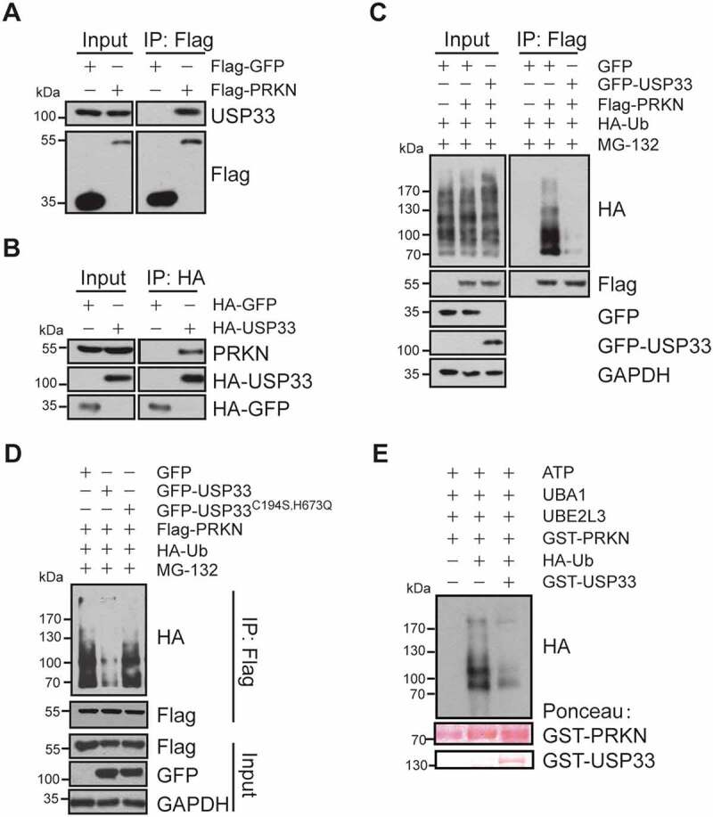
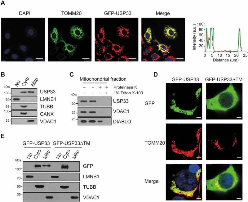
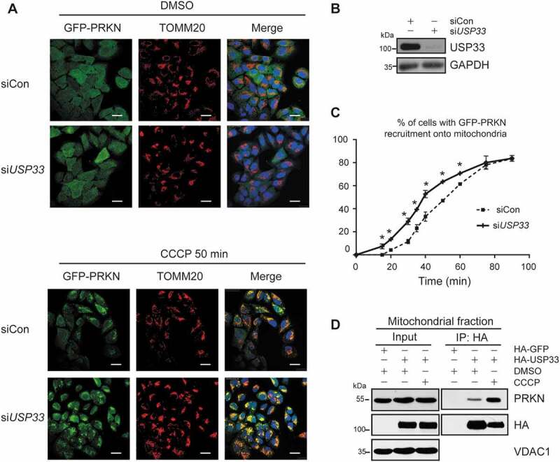
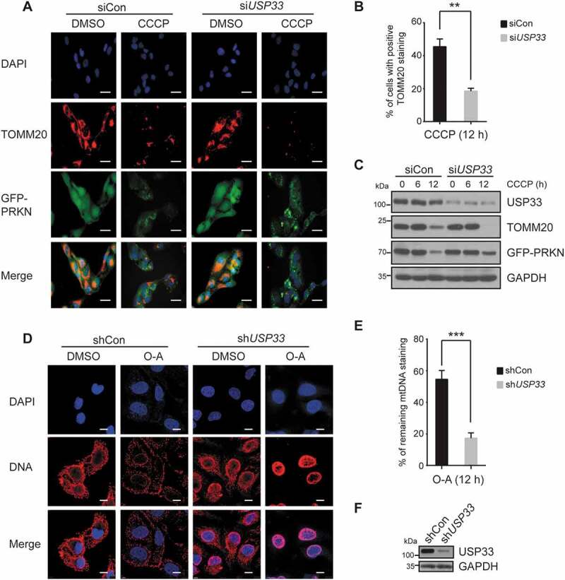
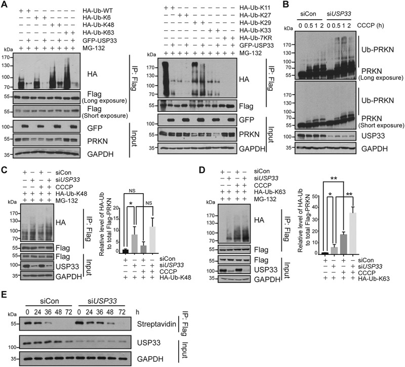
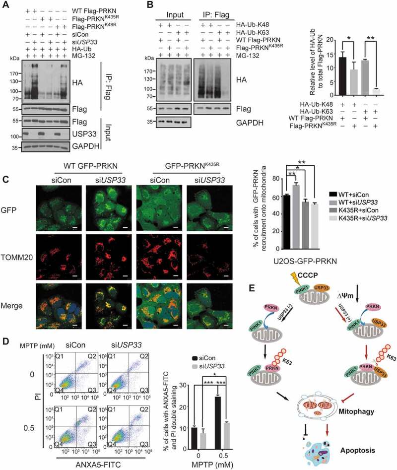
PRKN/parkin activation through phosphorylation of its ubiquitin and ubiquitin-like domain by PINK1 is critical in mitophagy induction for eliminating the damaged mitochondria. Deubiquitinating enzymes (DUBs) functionally reversing PRKN ubiquitination are critical in controlling the magnitude of PRKN-mediated mitophagy process. However, potential DUBs that directly target PRKN and antagonize its pro-mitophagy effect remains to be identified and characterized. Here, we demonstrated that USP33/VDU1 is localized at the outer membrane of mitochondria and serves as a PRKN DUB through their interaction. Cellular and in vitro assays illustrated that USP33 deubiquitinates PRKN in a DUB activity-dependent manner. USP33 prefers to remove K6, K11, K48 and K63-linked ubiquitin conjugates from PRKN, and deubiquitinates PRKN mainly at Lys435. Mutation of this site leads to a significantly decreased level of K63-, but not K48-linked PRKN ubiquitination. USP33 deficiency enhanced both K48- and K63-linked PRKN ubiquitination, but only K63-linked PRKN ubiquitination was significantly increased under mitochondrial depolarization. Further, USP33 knockdown increased both PRKN protein stabilization and its translocation to depolarized mitochondria leading to the enhancement of mitophagy. Moreover, USP33 silencing protects SH-SY5Y human neuroblastoma cells from the neurotoxin MPTP-induced apoptotic cell death. Our findings convincingly demonstrate that USP33 is a novel PRKN deubiquitinase antagonizing its regulatory roles in mitophagy and SH-SY5Y neuron-like cell survival. Thus, USP33 inhibition may represents an attractive new therapeutic strategy for PD patients.Abbreviations: CCCP: carbonyl cyanide 3-chlorophenylhydrazone; DUB: deubiquitinating enzymes; MPTP: 1-methyl-4-phenyl-1,2,3,6-tetrahydropyridine; OMM: outer mitochondrial membrane; PD: Parkinson disease; PINK1: PTEN induced kinase 1; PRKN/PARK2: parkin RBR E3 ubiquitin protein ligase; ROS: reactive oxygen species; TM: transmembrane; Ub: ubiquitin; UBA1: ubiquitin like modifier activating enzyme 1; UBE2L3/UbcH7: ubiquitin conjugating enzyme E2 L3; USP33: ubiquitin specific peptidase 33; WT: wild type.

Keywords: Apoptosis; PRKN/parkin; USP33 deubiquitinase; mitophagy; ubiquitination.


Figures

Figure 1.
Figure 1.
USP33 interacts with PRKN. (A) Endogenous USP33 protein was detected in anti-Flag immunoprecipitate from cell lysate of HEK293 cells overexpressing Flag-PRKN by western blotting. (B) Endogenous PRKN protein was verified in anti-HA immunoprecipitate from the lysate of HA-USP33 transfected HEK293 cells. HA-GFP was used for negative control. (C) Decreased ubiquitination level of PRKN in HEK293 cells overexpressing GFP-USP33. HEK293 cells were co-transfected with Flag-PRKN, HA-Ub and GFP-USP33, then treated with MG-132 for 3 h. Flag-PRKN was pulled-down with anti-Flag antibody and the PRKN ubiquitination level was detected with anti-HA antibody by western blotting. (D) Defective deubiquitination activity of USP33 mutant (USP33C194S,H673Q mutant) on PRKN. HEK293 cells were co-transfected with Flag-PRKN, HA-Ub and GFP-USP33 or GFP-USP33C194S,H673Q mutant, then treated with 10 μM MG-132 for 3 h. PRKN ubiquitination level was examined in Flag immunoprecipitates with anti-HA by western blotting. (E) USP33 removes PRKN Ub conjugates by in vitro ubiquitination assay. GST-PRKN and GST-USP33 levels stained with Ponceau were included.
Figure 2.
Figure 2.
USP33 is a novel OMM DUB. (A) GFP-USP33 colocalizes with mitochondrial marker TOMM20 by immunofluorescence staining. Images were captured using Leica TCS SP8 Confocal microscope. Line drawing graph was quantified by ImageJ software (http://rsbweb.nih.gov/ij/). a.u., arbitrary units. Scale bar: 25 μm. (B) USP33 level in different fractions (Nu: nucleus; Cyto: cytoplasm; Mito: mitochondria) of HEK293 cells by western blotting. LMNB1 (lamin B1): nuclear marker; TUBB/β-tubulin: cytoplasmic marker; CANX (calnexin): ER marker; VDAC1: mitochondrial marker. (C) Protease K assay showing localization of USP33 at OMM. VDAC1: mitochondrial outer membrane marker, DIABLO/SMAC: intermembrane space marker. (D) Colocalization of GFP-USP33, but not GFP-USP33ΔTM (residues 549-569 deletion), with mitochondrial protein TOMM20 by immunostaining analysis. Scale bar: 2.5 μm. (E) GFP-USP33 is present in both mitochondrial and cytosolic fractions, whereas GFP-USP33ΔTM only in cytosolic fraction of HEK293 cells transiently transfected with WT or mutant USP33.
Figure 3.
Figure 3.
USP33 depletion promotes PRKN recruitment to depolarized mitochondria. U2OS-GFP-PRKN cells were transfected with control siRNA (siCon) or USP33 siRNA1 + 2 (siUSP33) for 48 h and then treated with 20 μM CCCP. The cells were collected at indicated time-points for immunostaining with anti-TOMM20. (A) The appearance of GFP-PRKN puncta superposed on the mitochondrial TOMM20 was visualized. Scale bar: 25 μm. (B) Knockdown of USP33 validated by western blotting. (C) Percentage of cells with GFP-PRKN puncta colocalized with TOMM20 was quantified in siControl and USP33-depleted cells at 5-min interval. Over 150 cells were examined at each time-point, and data represents mean ± SD from three independent experiments. One-way ANOVA was used for statistical analysis. *P < 0.05. (D) Interaction between PRKN and USP33 in mitochondrial fraction of HEK293 cells with or without CCCP (20 μM) treatment for 2 h.
Figure 4.
Figure 4.
USP33 depletion enhances PRKN-mediated mitochondrial clearance. U2OS-GFP-PRKN cells were transfected with siCon or USP33 siRNA1 + 2 (siUSP33) for 48 h, treated with 20 μM CCCP for 6 or 12 h, and immunostained with anti-TOMM20. TOMM20 staining was visualized (A) and percentage cells with TOMM20 staining was quantified (B). A minimum of 200 cells at 12 h was analyzed. The data represents mean ± SD from three independent experiments. **P < 0.01, Student’s t test. Images were captured using Leica DM5000 microscope. Scale bar: 25 μm. (C) Expression levels of USP33, TOMM20 and PRKN determined by western blotting. (D) Images of mtDNA content and (E) quantification of mtDNA fluorescence density in U2OS-GFP-PRKN cells upon treatment of 10 μM oligomycin and 4 μM antimycin A for 12 h. Cells were infected with adenoviral scrambled control (shCon) or USP33 shRNA 1 + 2 (shUSP33) for 72 h before treatment. Images were captured using Leica TCS SP8 Confocal microscope. Scale bar: 10 μm. The mtDNAs from >300 cells per group were quantified by Image-Pro Plus software. The data represents mean ± SD from three independent experiments. ***P < 0.001, Student’s t test. (F) Knockdown of USP33 validated by western blotting.
Figure 5.
Figure 5.
USP33 preferentially removes K6, K11, K48 and K63-linked ubiquitin conjugates from PRKN. (A) HEK293 cells were co-transfected with a series of indicated expression plasmids and harvested after treated with 10 μM MG-132 for 3 h. The levels of different K-linked PRKN ubiquitination in Flag immunoprecipitates upon USP33 overexpression were determined by western blotting with anti-HA antibody. (B) U2OS-Flag-PRKN cells were transfected with siCon or USP33 siRNA1 + 2 (siUSP33) for 48 h, then treated with 20 μM CCCP for different times (0, 0.5, 1, 2 h). PRKN ubiquitination during mitophagy induced by CCCP was examined by western blotting. (C and D) K48 and K63-linked PRKN ubiquitination in U2OS cells after USP33 depletion with or without CCCP treatment for 45 min. The band density of PRKN ubiquitination level was quantified by Image J software. The data represent the mean ± SD from three independent experiments. A one-way ANOVA and Student’s t test were performed for statistical analysis. *P < 0.05, **P < 0.01. NS, not significant. (E) Stability of PRKN protein after USP33 depletion in U2OS-Flag-PRKN cells using metabolic labeling assay determined by immunoprecipitation followed by western blotting.
Figure 6.
Figure 6.
USP33 regulates PRKN ubiquitination at Lys435 and its silencing protects SH-SY5Y cells from the neurotoxin MPTP. (A) Lysine sites on PRKN deubiquitinated by USP33. U2OS cells were transfected with the indicated constructs for 24 h, then with siCon or USP33 siRNA1 + 2 (siUSP33) for 48 h. PRKN ubiquitination level in Flag immunoprecipitates was examined with anti-HA antibody by western blotting. (B) Levels of K48- and K63-linked Ub at Lys435. HEK293 cells were transfected with Flag-tagged WT PRKN or K435R mutant with HA-Ub-K48 or HA-Ub-K63 for 36 h, and harvested after 10 μM MG-132 treatment. The densities of PRKN ubiquitination were quantified using Image J software. The data represent the mean ± SD from three independent experiments. A one-way ANOVA and Student’s t test were performed for statistical analysis. *P < 0.05, **P < 0.01. (C) An enhanced recruitment of WT PRKN, but not K435R mutant, to depolarized mitochondria in USP33-depleted U2OS cells. U2OS cells stably expressing WT or K435-mutated PRKN were used. The images were captured using Leica TCS SP8 Confocal microscope. Scale bar: 10 μm. Percentage of cells with WT GFP-PRKN or GFP-PRKNK435R puncta colocalized with TOMM20 was quantified in siControl and USP33-depleted cells after 60 min of CCCP treatment. Over 100 cells were examined for each time-point, and data represents mean ± SD from three independent experiments. *P < 0.05, **P < 0.01, Student’s t test. (D) SH-SY5Y cells were transfected with siCon or USP33 siRNA1 + 2 (siUSP33) for 48 h, and then treated with 0.5 mM MPTP for 24 h. The cells were collected and stained with ANXA5-FITC and propidium iodide (PI) using the ANXA5/annexin V-FITC/PI Detection Kit (Beyotime, C1062M). The percentage of apoptotic cells were quantified by flow cytometry (BD FACSAria II). The data represents mean ± SD from three independent experiments. *P < 0.05 and ***P < 0.001, Student’s t test. (D) Model depicting the antagonizing effect of USP33 on PRKN-mediated mitophagy. USP33 as an OMM protein interacts with PRKN under mitochondrial depolarization and regulates the magnitude of PRKN-induced mitophagy mainly through removing K63-linked Ub chain from PRKN. USP33 deficiency leads a persistent elevation of K63-linked Ub conjugates on PRKN consequently stabilizing PRKN protein and augmenting mitophagy induction. Overall, USP33 depletion promotes an efficient clearance of damaged mitochondria and potently protects neuron-like cells from neurotoxin-induced apoptotic cell death.

References

    1. Gegg ME, Schapira AH.. PINK1-parkin-dependent mitophagy involves ubiquitination of mitofusins 1 and 2: implications for Parkinson disease pathogenesis. Autophagy. 2011;7(2):243–245. - PMC - PubMed
    1. Bingol B, Tea JS, Phu L, et al. The mitochondrial deubiquitinase USP30 opposes parkin-mediated mitophagy. Nature. 2014;510(7505):370–375. - PubMed
    1. Gladkova C, Maslen SL, Skehel JM, et al. Mechanism of parkin activation by PINK1. Nature. 2018;559(7714):410–414. - PMC - PubMed
    1. Cornelissen T, Haddad D, Wauters F, et al. The deubiquitinase USP15 antagonizes Parkin-mediated mitochondrial ubiquitination and mitophagy. Hum Mol Genet. 2014;23(19):5227–5242. - PMC - PubMed
    1. Durcan TM, Tang MY, Perusse JR, et al. USP8 regulates mitophagy by removing K6-linked ubiquitin conjugates from parkin. Embo J. 2014;33(21):2473–2491. - PMC - PubMed

Publication types

MeSH terms