Attenuation of Zika Virus by Passage in Human HeLa Cells
- PMID: 31434319
- PMCID: PMC6789458
- DOI: 10.3390/vaccines7030093
Attenuation of Zika Virus by Passage in Human HeLa Cells
Abstract
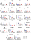
Zika virus (ZIKV) is a mosquito-borne Flavivirus. Previous studies have shown that mosquito-transmitted flaviviruses, including yellow fever, Japanese encephalitis, and West Nile viruses, could be attenuated by serial passaging in human HeLa cells. Therefore, it was hypothesized that wild-type ZIKV would also be attenuated after HeLa cell passaging. A human isolate from the recent ZIKV epidemic was subjected to serial HeLa cell passaging, resulting in attenuated in vitro replication in both Vero and A549 cells. Additionally, infection of AG129 mice with 10 plaque forming units (pfu) of wild-type ZIKV led to viremia and mortality at 12 days, whereas infection with 103 pfu of HeLa-passage 6 (P6) ZIKV led to lower viremia, significant delay in mortality (median survival: 23 days), and increased cytokine and chemokine responses. Genomic sequencing of HeLa-passaged virus identified two amino acid substitutions as early as HeLa-P3: pre-membrane E87K and nonstructural protein 1 R103K. Furthermore, both substitutions were present in virus harvested from HeLa-P6-infected animal tissue. Together, these data show that, similarly to other mosquito-borne flaviviruses, ZIKV is attenuated following passaging in HeLa cells. This strategy can be used to improve understanding of substitutions that contribute to attenuation of ZIKV and be applied to vaccine development across multiple platforms.
Keywords: HeLa cells; Zika virus; Zika virus NS1; Zika virus prM; attenuation; flavivirus.
Conflict of interest statement
The authors declare no conflict of interest.
Figures


Similar articles
-
Using Next Generation Sequencing to Study the Genetic Diversity of Candidate Live Attenuated Zika Vaccines.Vaccines (Basel). 2020 Apr 3;8(2):161. doi: 10.3390/vaccines8020161. Vaccines (Basel). 2020. PMID: 32260110 Free PMC article.
-
Zika Virus Attenuation by Codon Pair Deoptimization Induces Sterilizing Immunity in Mouse Models.J Virol. 2018 Aug 16;92(17):e00701-18. doi: 10.1128/JVI.00701-18. Print 2018 Sep 1. J Virol. 2018. PMID: 29925661 Free PMC article.
-
Chimeric yellow fever 17D-Zika virus (ChimeriVax-Zika) as a live-attenuated Zika virus vaccine.Sci Rep. 2018 Sep 4;8(1):13206. doi: 10.1038/s41598-018-31375-9. Sci Rep. 2018. PMID: 30181550 Free PMC article.
-
Zika virus structural biology and progress in vaccine development.Biotechnol Adv. 2018 Jan-Feb;36(1):47-53. doi: 10.1016/j.biotechadv.2017.09.004. Epub 2017 Sep 12. Biotechnol Adv. 2018. PMID: 28916391 Review.
-
Recent Perspectives on Genome, Transmission, Clinical Manifestation, Diagnosis, Therapeutic Strategies, Vaccine Developments, and Challenges of Zika Virus Research.Front Microbiol. 2017 Sep 14;8:1761. doi: 10.3389/fmicb.2017.01761. eCollection 2017. Front Microbiol. 2017. PMID: 28959246 Free PMC article. Review.
Cited by
-
Development of Langat virus infectious clones as a platform for live-attenuated tick-borne encephalitis vaccine.Npj Viruses. 2025 May 23;3(1):44. doi: 10.1038/s44298-025-00129-6. Npj Viruses. 2025. PMID: 40410304 Free PMC article.
-
Using Next Generation Sequencing to Study the Genetic Diversity of Candidate Live Attenuated Zika Vaccines.Vaccines (Basel). 2020 Apr 3;8(2):161. doi: 10.3390/vaccines8020161. Vaccines (Basel). 2020. PMID: 32260110 Free PMC article.
-
Zika virus infection in the genital tract of non-pregnant females: a systematic review.Rev Inst Med Trop Sao Paulo. 2020 Mar 2;62:e16. doi: 10.1590/S1678-9946202062016. eCollection 2020. Rev Inst Med Trop Sao Paulo. 2020. PMID: 32130356 Free PMC article.
-
Cell fusing agent virus isolated from Aag2 cells does not vertically transmit in Aedes aegypti via artificial infection.Parasit Vectors. 2023 Nov 6;16(1):402. doi: 10.1186/s13071-023-06033-3. Parasit Vectors. 2023. PMID: 37932781 Free PMC article.
-
IP-10 and CXCR3 signaling inhibit Zika virus replication in human prostate cells.PLoS One. 2020 Dec 30;15(12):e0244587. doi: 10.1371/journal.pone.0244587. eCollection 2020. PLoS One. 2020. PMID: 33378361 Free PMC article.
References
-
- Miller B.R., Adkins D. Biological Characterization of Plaque-Size Variants of Yellow Fever Virus in Mosquitoes and Mice. Acta Virol. 1988;32:227–234. - PubMed
LinkOut - more resources
Full Text Sources
Molecular Biology Databases
Research Materials

