Review
. 2019 Aug 21;17(1):90.
doi: 10.1186/s12951-019-0524-9.

Current outlook on radionuclide delivery systems: from design consideration to translation into clinics

Affiliations
Review

Current outlook on radionuclide delivery systems: from design consideration to translation into clinics

Oleksii O Peltek et al. J Nanobiotechnology. .

Erratum in

Abstract

Radiopharmaceuticals have proven to be effective agents, since they can be successfully applied for both diagnostics and therapy. Effective application of relevant radionuclides in pre-clinical and clinical studies depends on the choice of a sufficient delivery platform. Herein, we provide a comprehensive review on the most relevant aspects in radionuclide delivery using the most employed carrier systems, including, (i) monoclonal antibodies and their fragments, (ii) organic and (iii) inorganic nanoparticles, and (iv) microspheres. This review offers an extensive analysis of radionuclide delivery systems, the approaches of their modification and radiolabeling strategies with the further prospects of their implementation in multimodal imaging and disease curing. Finally, the comparative outlook on the carriers and radionuclide choice, as well as on the targeting efficiency of the developed systems is discussed.

Keywords: Diagnostic and therapeutic radionuclides; Drug carrier systems; Microspheres; Monoclonal antibodies; Multimodal imaging; Nanoparticles; Nuclear medicine; Radiopharmaceuticals; Theranostics.


Conflict of interest statement

The authors declare that they have no competing interests.

Figures

Fig. 1
Fig. 1
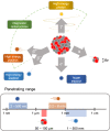
Administration of radionuclide carriers in the tumor site and their further accumulation via active targeting approach
Fig. 2
Fig. 2
Types of radioactive decay with the demonstration of the soft tissue penetration range
Fig. 3
Fig. 3
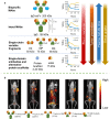
a Correlation between blood clearance, tumor penetration, plasma half-life and renal uptake and the previously discussed antibodies and antibody fragments of different size: bsAbs, intact MAbs, scFv, sdAbs and alternative protein scaffolds. Uptake is expressed as percentage of injected dose per gram (ID/g). b PET/computed tomography (CT) images of mice bearing prostate stem-cell antigen (PSCA)–expressing LAPC-9 prostate cancer xenografts. PSCA expression was visualized with intact mAbs, single-chain Fv–Fc (scFV-Fc) wild type (WT) mAbs, scFV-Fc double mutant (DM) mAbs, Minibodies and Diabody labeled with N-succinimidyl-4-[18F]-fluorobenzoate (18F-SFB). All microPET images were scaled individually to best show tumor targeting (this figure was reproduced from Scott et al. [29] with the required copyright permission)
Fig. 4
Fig. 4
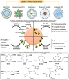
The main categories of organic NPs used for radiopharmaceutical formulations with schematic representation of their radiolabeling strategies. Below the commonly used macrocyclic and acylic chelators for radiolabeling are depicted
Fig. 5
Fig. 5
Schematic representation of various radiolabeling approaches of liposomes, targeting ligand modifications with further application in different field of nuclear medicine
Fig. 6
Fig. 6
PET and PET/CT images of [64Cu]MM-302 in lesions at different anatomic locations. The regions of interest used to measure tumor deposition of [64Cu]-MM-302 are shown in blue or turquoise outlines (this figure was reproduced from Lee et al. [127] with the required copyright permission)
Fig. 7
Fig. 7
Schematic illustration of multimodal imaging of radiolabeled inorganic NPs: plasmonic NPs for X-ray visualization, magnetic NPs for MRI visualization, C-based for optical imaging. Si-based NPs requires additional functionalization by contrast agents to be visualized (this figure is adopted from Yu et al. [189], Wang et al. [190], Liu et al. [191], Phillips et al. [192], Hoffman et al. [193] with required copyright permission)
Fig. 8
Fig. 8
Schematic illustration of SPION-based NPs fabrication (top), sagittal MR images of mouse animal model (bottom). The blue arrows indicate the brain, the red arrows indicate the liver and the yellow arrows indicate the bladder (this figure is reproduced from Hoffman et al. [193] with required copyright permission)
Fig. 9
Fig. 9
In vivo HER2-targeted PET imaging in xenograft breast cancer models. Serial coronal and axial tomographic PET images acquired at 2, 24, 48, and 72 h post i.v. injection of radiolabeled particle immunoconjugates in groups of tumor-bearing mice as follows: a Targeted group: 89ZrDFO-scFv-PEG-Cy5-C’ dots in BT-474 mice. b Non-targeted group: 89Zr-DFO-Ctr/scFv-PEG-Cy5-C’ dots in BT-474 mice. For each group, maximum intensity projection (MIP) images were also acquired at 48 h p.i. H: heart, B: bladder, L: liver. c Representative MIP PET, CT, and PET/CT fusion images of 89Zr-DFO-scFv-PEG-Cy5-C’ dots in a BT-474 tumor-bearing mouse. BT474 tumors are marked with yellow arrows (this figure was reproduced from Chen et al. [212] with the required copyright permission)
Fig. 10
Fig. 10
Schematic illustration of five major strategies for radiolabeling inorganic NPs. a Chelator-mediated complexation, b Specific trapping (b1) and ion-exchange (b2). c Hot-plus-cold precursor synthesis, d Proton beam activation. NP: nanoparticle (this figure was reproduced from Goel et al. [234] with the required copyright permission)
Fig. 11
Fig. 11
Superselective segmental radioembolization. a CT scan before treatment showing multinodular tumor. b PET scan showing intense radiation after administration of 90Y resin microspheres. c Significant atrophy of liver segment and lack of tumor activity 1 year after treatment (this figure is reproduced from Sangro et al. [253] with required copyright permission)

Similar articles

Cited by

References

    1. Yeong C-H, Cheng M, Ng K-H. Therapeutic radionuclides in nuclear medicine: current and future prospects. J Zhejiang Univ Sci B. 2014;15(10):845–863. doi: 10.1631/jzus.B1400131. - DOI - PMC - PubMed
    1. Srivastava SC, Mausner LF. Therapeutic radionuclides: production, physical characteristics, and applications. In: Baum RP, editor. Therapeutic nuclear medicine. Berlin: Springer; 2014. pp. 11–50.
    1. Oh J-R, Ahn B-C. False-positive uptake on radioiodine whole-body scintigraphy: physiologic and pathologic variants unrelated to thyroid cancer. Am J Nucl Med Mol Imaging. 2012;2(3):362–385. - PMC - PubMed
    1. Liu Z, Xing M. Induction of sodium/iodide symporter (NIS) expression and radioiodine uptake in non-thyroid cancer cells. PLoS ONE. 2012;7(2):e31729. doi: 10.1371/journal.pone.0031729. - DOI - PMC - PubMed
    1. Huang R, Zhao Z, Ma X, Li S, Gong R, Kuang A. Targeting of tumor radioiodine therapy by expression of the sodium iodide symporter under control of the survivin promoter. Cancer Gene Ther. 2011;18(2):144–152. doi: 10.1038/cgt.2010.66. - DOI - PMC - PubMed

LinkOut - more resources