. 2019 Oct 15;93(21):e00574-19.
doi: 10.1128/JVI.00574-19. Print 2019 Nov 1.

The HIV-1 Antisense Protein ASP Is a Transmembrane Protein of the Cell Surface and an Integral Protein of the Viral Envelope

Affiliations

The HIV-1 Antisense Protein ASP Is a Transmembrane Protein of the Cell Surface and an Integral Protein of the Viral Envelope

Yvonne Affram et al. J Virol. .

Abstract

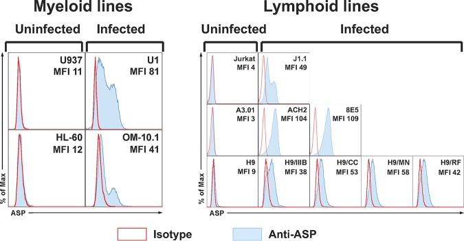
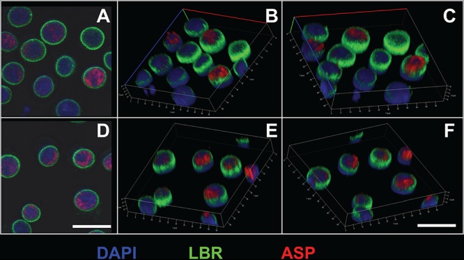
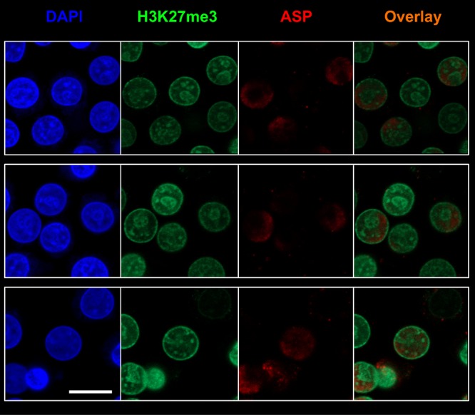
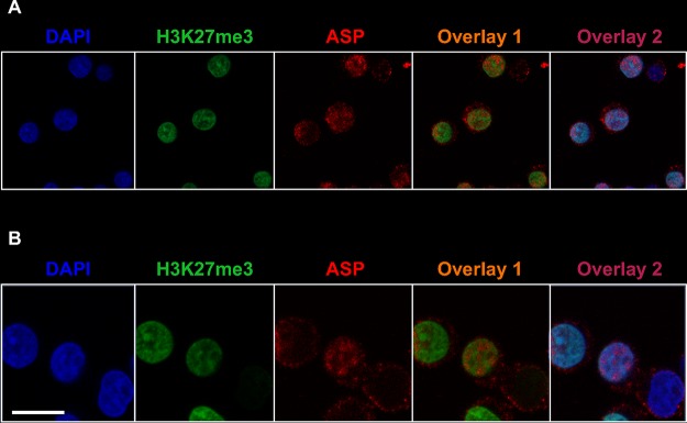
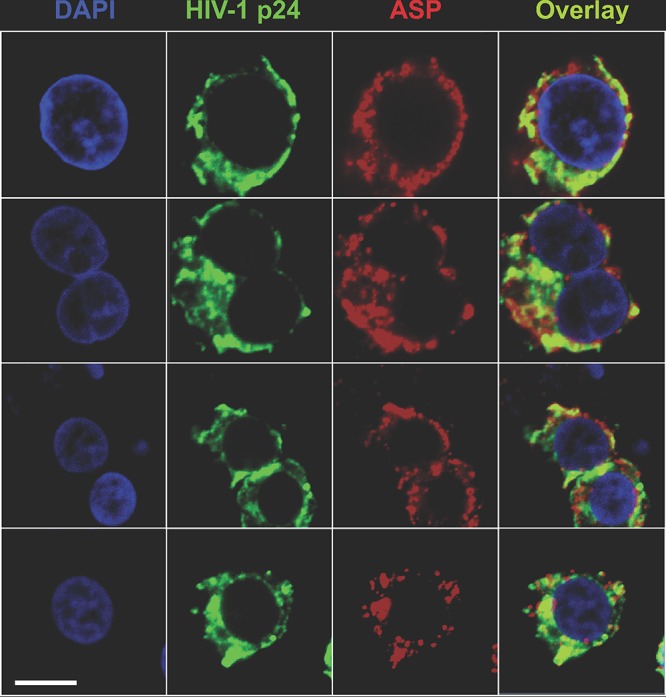
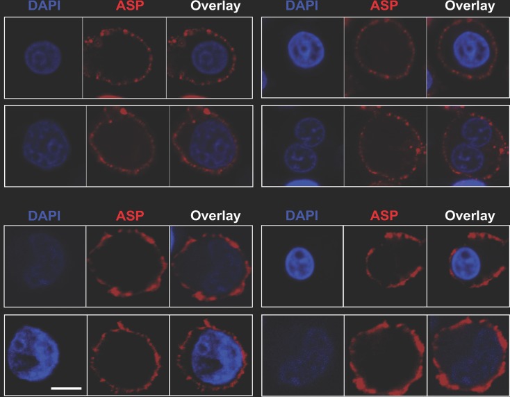
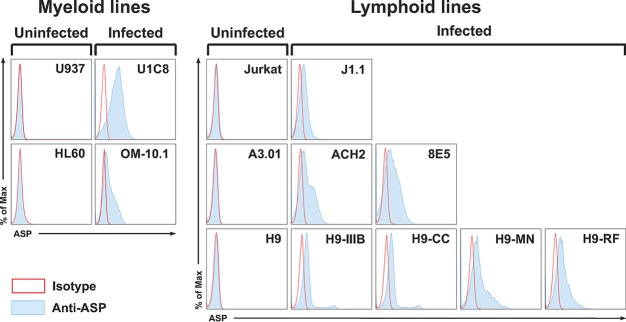
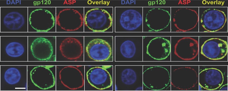
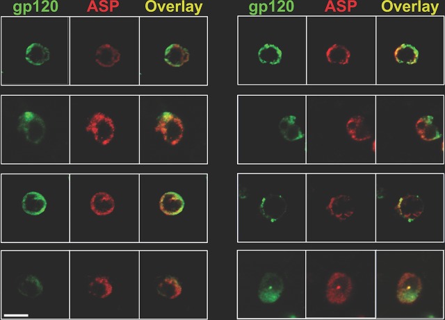
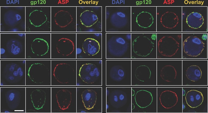
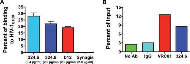
The negative strand of HIV-1 encodes a highly hydrophobic antisense protein (ASP) with no known homologs. The presence of humoral and cellular immune responses to ASP in HIV-1 patients indicates that ASP is expressed in vivo, but its role in HIV-1 replication remains unknown. We investigated ASP expression in multiple chronically infected myeloid and lymphoid cell lines using an anti-ASP monoclonal antibody (324.6) in combination with flow cytometry and microscopy approaches. At baseline and in the absence of stimuli, ASP shows polarized subnuclear distribution, preferentially in areas with low content of suppressive epigenetic marks. However, following treatment with phorbol 12-myristate 13-acetate (PMA), ASP translocates to the cytoplasm and is detectable on the cell surface, even in the absence of membrane permeabilization, indicating that 324.6 recognizes an ASP epitope that is exposed extracellularly. Further, surface staining with 324.6 and anti-gp120 antibodies showed that ASP and gp120 colocalize, suggesting that ASP might become incorporated in the membranes of budding virions. Indeed, fluorescence correlation spectroscopy studies showed binding of 324.6 to cell-free HIV-1 particles. Moreover, 324.6 was able to capture and retain HIV-1 virions with efficiency similar to that of the anti-gp120 antibody VRC01. Our studies indicate that ASP is an integral protein of the plasma membranes of chronically infected cells stimulated with PMA, and upon viral budding, ASP becomes a structural protein of the HIV-1 envelope. These results may provide leads to investigate the possible role of ASP in the virus replication cycle and suggest that ASP may represent a new therapeutic or vaccine target.IMPORTANCE The HIV-1 genome contains a gene expressed in the opposite, or antisense, direction to all other genes. The protein product of this antisense gene, called ASP, is poorly characterized, and its role in viral replication remains unknown. We provide evidence that the antisense protein, ASP, of HIV-1 is found within the cell nucleus in unstimulated cells. In addition, we show that after PMA treatment, ASP exits the nucleus and localizes on the cell membrane. Moreover, we demonstrate that ASP is present on the surfaces of viral particles. Altogether, our studies identify ASP as a new structural component of HIV-1 and show that ASP is an accessory protein that promotes viral replication. The presence of ASP on the surfaces of both infected cells and viral particles might be exploited therapeutically.

Keywords: HIV-1; antisense protein ASP; cell surface protein; viral envelope protein.


Figures

FIG 1
FIG 1
Intranuclear flow cytometry analysis of ASP expression in multiple cell lines. Chronically infected myeloid (U1 and OM-10.1) and lymphoid (J1.1, ACH2, 8E5, H9/IIIB, H9/CC, H9/MN, and H9/RF) cell lines and their uninfected parental myeloid (U937 and HL-60) and lymphoid (Jurkat, A3.01, and H9) cell lines were fixed and permeabilized with a FoxP3 staining kit to allow detection of nuclear proteins. Then, the cells were stained with the anti-ASP 324.6 (light-blue curve and line) or a control mouse IgG (open curve with red line) labeled with Alexa Fluor 647. Finally, the cells were washed, fixed, and analyzed by flow cytometry. All the chronically infected cell lines analyzed showed nuclear expression of ASP to varying degrees. The results shown are representative of several independent experiments. The mean fluorescence intensity (MFI) of samples stained with the 324.6 MAb is reported in each panel.
FIG 2
FIG 2
Intranuclear confocal microscopy analysis of ASP expression in unstimulated U1C8 cells. Cells were fixed and permeabilized with a FoxP3 staining kit to allow detection of nuclear proteins. Then, the cells were stained with Alexa Fluor 647-labeled anti-ASP 324.6 (red), FITC-labeled anti-LBR (lamin B receptor) (green), and DAPI (blue). Finally, the cells were washed, fixed, and analyzed by confocal microscopy as described in Materials and Methods. The cells display a nonuniform, polarized nuclear distribution of ASP. Bars, 5 μm. Panels B, C, E, and F are three-dimensional representations reconstructed from z-stack images 0.7 μm thick. The results shown are representative of at least three independent experiments.
FIG 3
FIG 3
ASP accumulates in nuclear areas lacking suppressive epigenetic marks in unstimulated infected U1C8 cells. Chronically infected U1C8 cells were fixed and permeabilized with a FoxP3 staining kit to allow detection of nuclear proteins. Then, the cells were stained with Alexa Fluor 647-labeled anti-ASP 324.6 (red), Alexa Fluor 488-labeled anti-H3K27me3 (green), and DAPI (blue). Finally, the cells were washed, fixed, and analyzed by confocal microscopy. The overlay of the images taken in the green (H3K27me3) and red (ASP) channels shows that the subnuclear distribution of the two markers is mutually exclusive, indicating that ASP is enriched in areas of the nucleus that contain actively transcribed chromatin. Bar, 5 μm. The results shown are representative of at least three independent experiments.
FIG 4
FIG 4
ASP exits the nucleus and localizes in the cytoplasm of PMA-treated U1C8 cells. Chronically infected U1C8 cells were treated with PMA to reactivate HIV-1 expression, as described in Materials and Methods. After fixation and permeabilization with a BD Cytofix/Cytoperm kit to allow detection of cytoplasmic proteins, the cells were stained with Alexa Fluor 647-labeled anti-ASP 324.6 (red), Alexa Fluor 488-labeled anti-H3K27me3 (green), and DAPI (blue). Finally, the cells were washed, fixed, and analyzed by confocal microscopy. The overlay of the images taken in the green (H3K27me3) and red (ASP) channels shows that PMA stimulation caused ASP to exit the nucleus and to translocate into the cytoplasm. Bar, 10 μm (panel A, low magnification) and 5 μm (panel B, high magnification). The results shown are representative of at least three independent experiments.
FIG 5
FIG 5
ASP and HIV-1 p24 colocalize in the cytoplasm of PMA-treated U1C8 cells. Chronically infected U1C8 cells were treated with PMA to reactivate HIV-1 expression as described in Materials and Methods. After fixation and permeabilization with a BD Cytofix/Cytoperm kit to allow detection of cytoplasmic proteins, the cells were stained with Alexa Fluor 647-labeled anti-ASP 324.6 (red), Alexa Fluor 488-labeled anti-HIV-1 p24 (green), and DAPI (blue). Finally, the cells were washed, fixed, and analyzed by confocal microscopy. The overlay of the images taken in the green (HIV-1 p24) and red (ASP) channels shows that PMA treatment induced HIV-1 expression, as shown by HIV-1 p24 presence in the cytoplasm. In addition, ASP and HIV-1 p24 present a significant degree of colocalization within the cytoplasm. Bar, 2 μm. The results shown are representative of at least three independent experiments.
FIG 6
FIG 6
ASP is an integral protein of the plasma membrane in PMA-treated U1C8 and OM-10.1 cells. Chronically infected U1C8 (top) and OM-10.1 (bottom) cells were treated with PMA to reactivate HIV-1 expression, as described in Materials and Methods. The cells were stained with Alexa Fluor 647-labeled anti-ASP 324.6 (red) and DAPI (blue) without prior fixation and permeabilization. Finally, the cells were washed, fixed, and analyzed by confocal microscopy. The overlay of the images taken in the red (ASP) and blue (DAPI) channels shows that PMA treatment induced ASP translocation to the cell membrane. Detection of ASP on the cell surface without prior permeabilization of the plasma membrane indicates that the 324.6 mAb recognizes an ASP epitope exposed in the extracellular milieu. Bar, 2 μm. The results shown are representative of at least three independent experiments.
FIG 7
FIG 7
Flow cytometry analysis of ASP expression on the surface in multiple cell lines. Chronically infected myeloid (U1C8 and OM-10.1) and lymphoid (J1.1, ACH2, 8E5, H9/IIIB, H9/CC, H9/MN, and H9/RF) cell lines and their uninfected parental myeloid (U937 and HL-60) and lymphoid (Jurkat, A3.01, and H9) cell lines were treated with PMA as indicated in Materials and Methods to reactivate HIV-1 expression. Then, the cells were surface stained with anti-ASP 324.6 (light-blue curve and line) or a control mouse IgG (open curve with red line) labeled with Alexa Fluor 647. After washing, the cells were fixed and analyzed by flow cytometry. All the chronically infected cell lines analyzed showed cell surface expression of ASP to varying degrees. The results shown are representative of several independent experiments.
FIG 8
FIG 8
ASP and HIV-1 gp120 colocalize on the surfaces of PMA-treated cells. U1C8 cells were treated with PMA as described in Materials and Methods to reactivate HIV-1 expression. Then, the cells were surface stained with Alexa Fluor 488-labeled anti-gp120 (green) and Alexa Fluor 647-labeled anti-ASP 324.6 (red). In addition, the cells were stained with DAPI to identify the nuclei. After washing, the cells were fixed and analyzed by confocal microscopy. The overlay of the images taken in the green (HIV-1 gp120), red (ASP), and blue (DAPI) channels shows that PMA treatment induced HIV-1 expression, as shown by the presence of both HIV-1 gp120 and ASP on the cell surface. In addition, ASP and HIV-1 gp120 present a significant degree of colocalization. Bar, 2 μm for all panels. The results shown are representative of several independent experiments.
FIG 9
FIG 9
ASP and HIV-1 gp120 colocalize on the surfaces of primary human CD4+ T cells infected with HIV-1Ada-M. CD4+ T cells were isolated, activated, and infected as described in Materials and Methods. At day 7 postinfection, the cells were surface stained with Alexa Fluor 488-labeled anti-gp120 (green) and Alexa Fluor 647-labeled anti-ASP 324.6 (red). After washing, the cells were fixed and analyzed by confocal microscopy. The overlay of the images taken in the green (HIV-1 gp120) and red (ASP) channels shows the presence of both HIV-1 gp120 and ASP on the cell surface. In addition, ASP and HIV-1 gp120 present a significant degree of colocalization. Bar, 5 μm for all panels. The results shown are representative of three independent experiments.
FIG 10
FIG 10
ASP and HIV-1 gp120 colocalize on the surfaces of primary human monocyte-derived macrophages infected with HIV-1Ada-M. Macrophages were generated from freshly isolated PBMC and infected as described in Materials and Methods. At day 15 postinfection, the cells were surface stained with Alexa Fluor 488-labeled anti-gp120 (green) and Alexa Fluor 647-labeled anti-ASP 324.6 (red). In addition, the cells were stained with DAPI to identify the nuclei. After washing, the cells were fixed and analyzed by confocal microscopy. The overlay of the images taken in the green (HIV-1 gp120), red (ASP), and blue (DAPI) channels shows the presence of both HIV-1 gp120 and ASP on the cell surface. In addition, ASP and HIV-1 gp120 present a significant degree of colocalization. Bar, 5 μm for all panels. The results shown are representative of three independent experiments.
FIG 11
FIG 11
ASP is an integral protein of the viral envelope. (A) HIV-1 viral particles concentrated from culture supernatants of U1C8 cells (HIV-1U1C8) treated with PMA were incubated with Alexa Fluor 647-labeled anti-gp120 (b12), anti-ASP (324.6), or anti-protein F of RSV (palivizumab). The samples were then analyzed by fluorescence correlation spectroscopy as described in Materials and Methods. (B) HIV-1 viral particles were captured from culture supernatants of PMA-treated U1C8 cells using anti-gp120 (VRC01), anti-ASP (324.6), or control mouse IgG conjugated to protein G Dynabeads. After extensive washes, the samples were analyzed by RT-PCR for the presence of virion-associated HIV-1 RNA. Both assays showed that ASP is expressed on the surfaces of HIV-1 particles as an integral protein of the viral envelope. The results shown are representative of three independent experiments.

References

    1. Barbeau B, Peloponese JM, Mesnard JM. 2013. Functional comparison of antisense proteins of HTLV-1 and HTLV-2 in viral pathogenesis. Front Microbiol 4:226. doi:10.3389/fmicb.2013.00226. - DOI - PMC - PubMed
    1. Ma G, Yasunaga J, Matsuoka M. 2016. Multifaceted functions and roles of HBZ in HTLV-1 pathogenesis. Retrovirology 13:16. doi:10.1186/s12977-016-0249-x. - DOI - PMC - PubMed
    1. Gaudray G, Gachon F, Basbous J, Biard-Piechaczyk M, Devaux C, Mesnard JM. 2002. The complementary strand of the human T-cell leukemia virus type 1 RNA genome encodes a bZIP transcription factor that down-regulates viral transcription. J Virol 76:12813–12822. doi:10.1128/jvi.76.24.12813-12822.2002. - DOI - PMC - PubMed
    1. Hivin P, Frédéric M, Arpin-André C, Basbous J, Gay B, Thébault S, Mesnard J-M. 2005. Nuclear localization of HTLV-I bZIP factor (HBZ) is mediated by three distinct motifs. J Cell Sci 118:1355–1362. doi:10.1242/jcs.01727. - DOI - PubMed
    1. Hivin P, Arpin-Andre C, Clerc I, Barbeau B, Mesnard JM. 2006. A modified version of a Fos-associated cluster in HBZ affects Jun transcriptional potency. Nucleic Acids Res 34:2761–2772. doi:10.1093/nar/gkl375. - DOI - PMC - PubMed

Publication types

MeSH terms

Substances