. 2019 Aug;572(7771):665-669.
doi: 10.1038/s41586-019-1501-z. Epub 2019 Aug 21.

Microbiota-derived lantibiotic restores resistance against vancomycin-resistant Enterococcus

Affiliations

Microbiota-derived lantibiotic restores resistance against vancomycin-resistant Enterococcus

Sohn G Kim et al. Nature. 2019 Aug.

Abstract

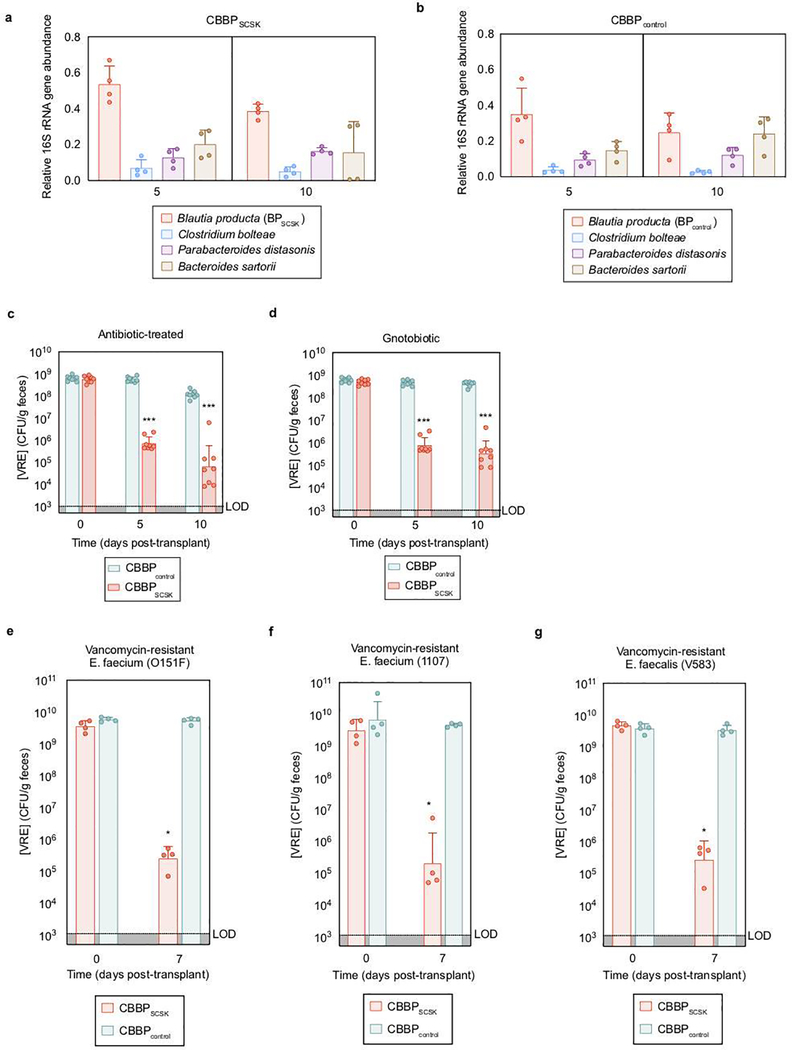
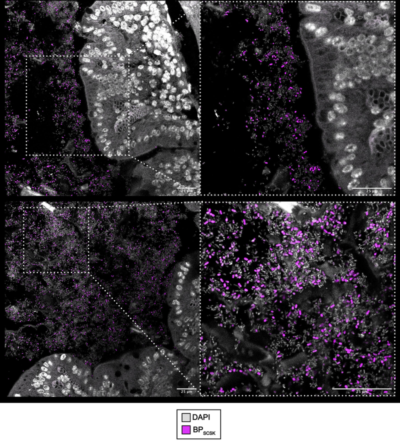
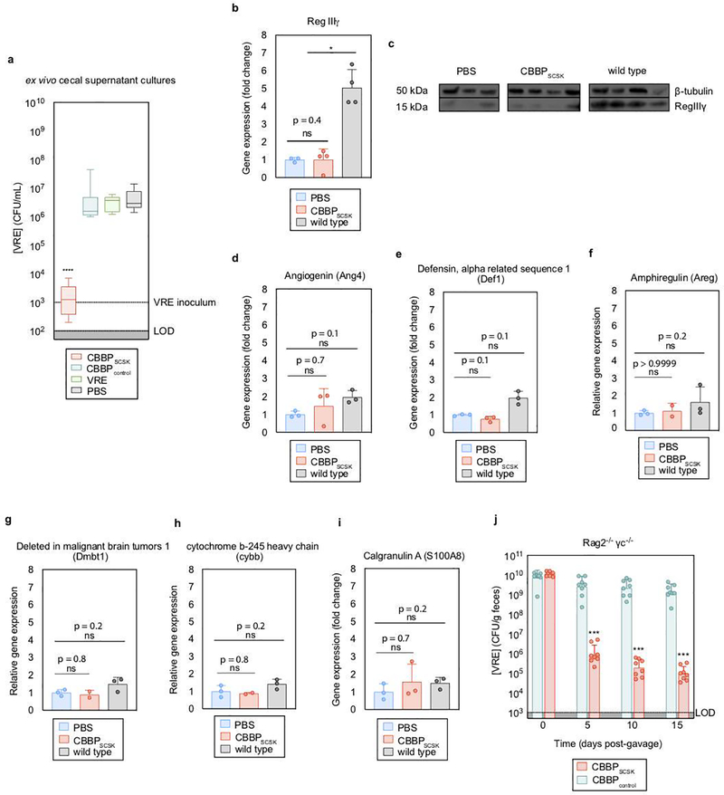
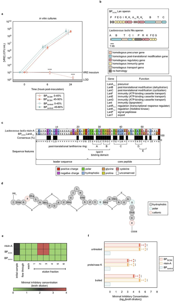
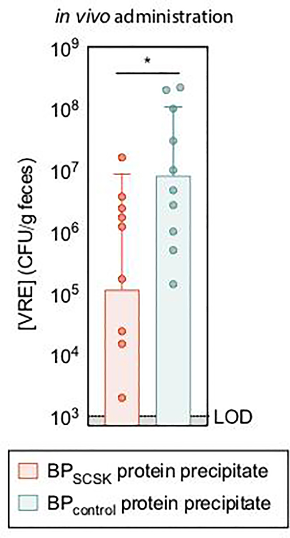
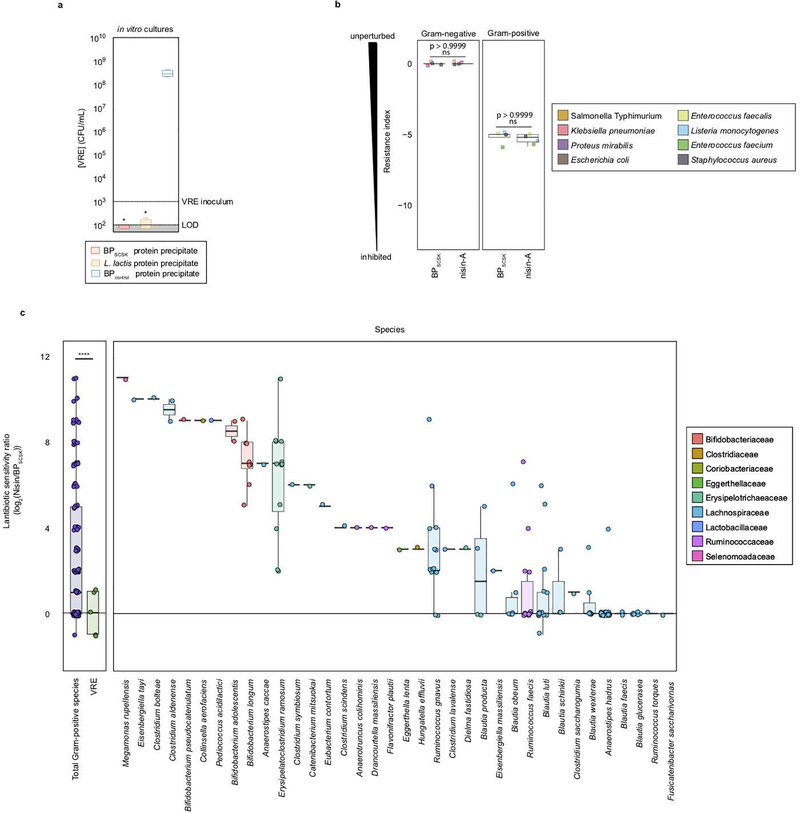
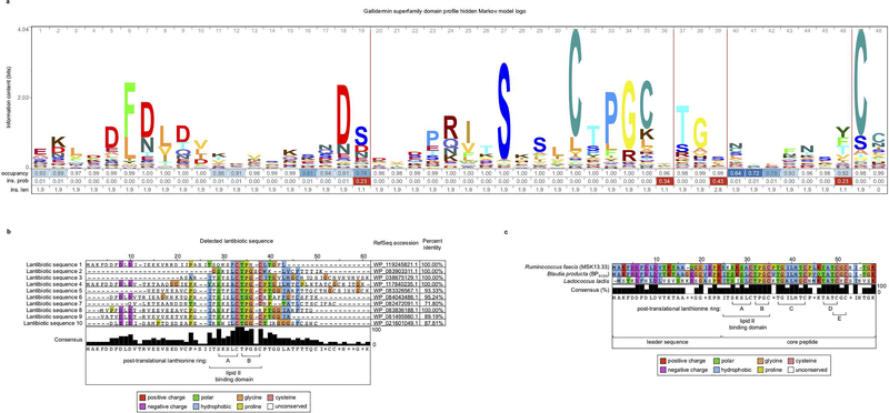
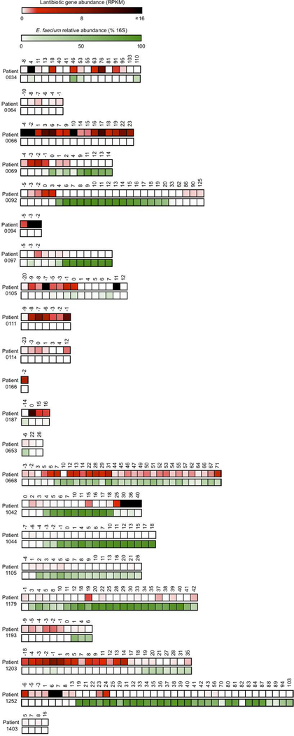
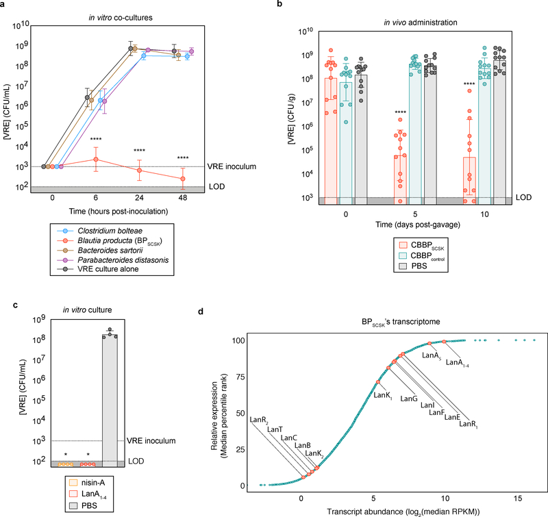
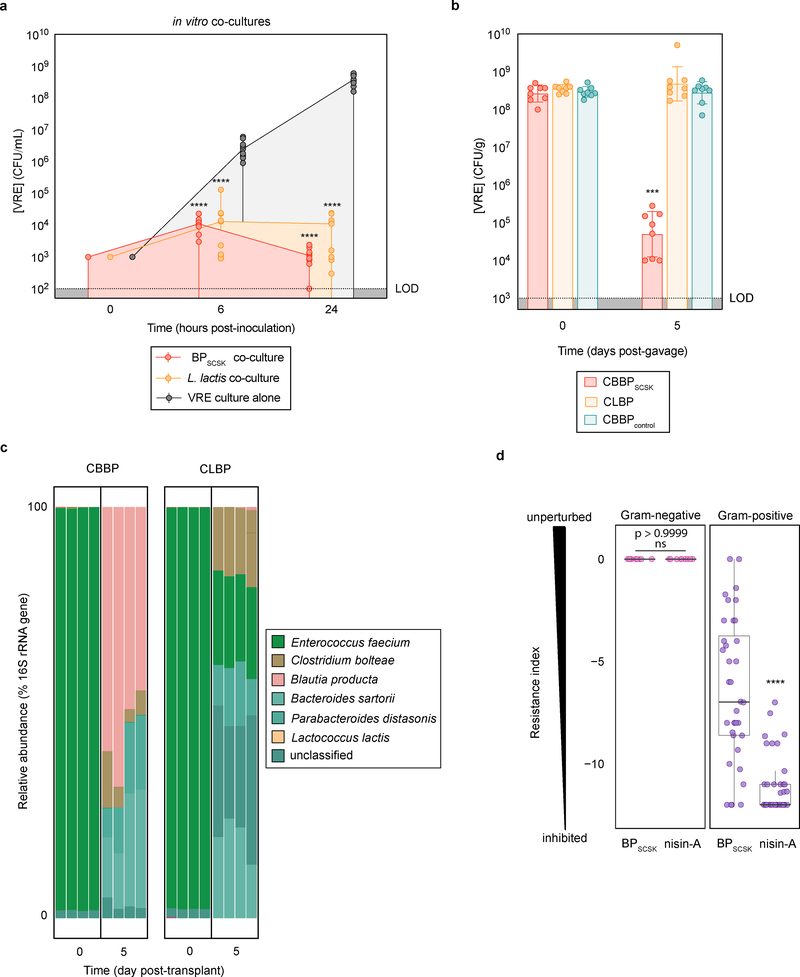
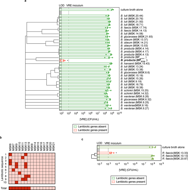
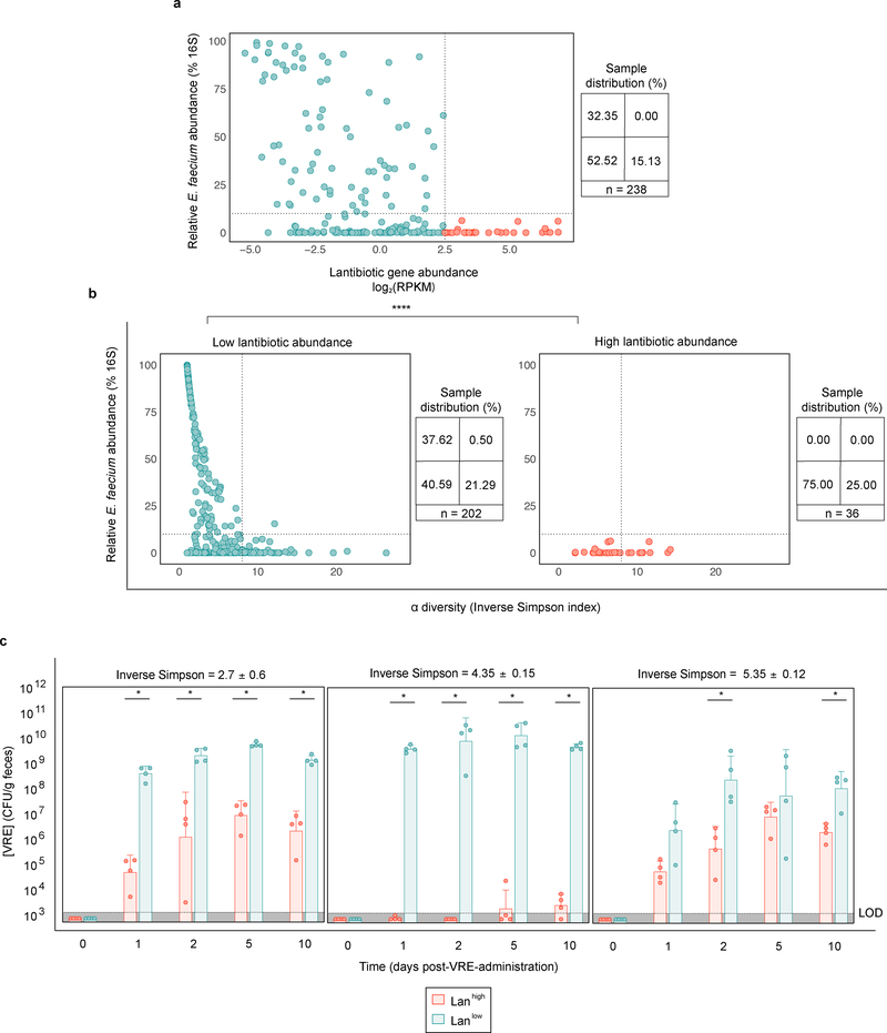
Intestinal commensal bacteria can inhibit dense colonization of the gut by vancomycin-resistant Enterococcus faecium (VRE), a leading cause of hospital-acquired infections1,2. A four-strained consortium of commensal bacteria that contains Blautia producta BPSCSK can reverse antibiotic-induced susceptibility to VRE infection3. Here we show that BPSCSK reduces growth of VRE by secreting a lantibiotic that is similar to the nisin-A produced by Lactococcus lactis. Although the growth of VRE is inhibited by BPSCSK and L. lactis in vitro, only BPSCSK colonizes the colon and reduces VRE density in vivo. In comparison to nisin-A, the BPSCSK lantibiotic has reduced activity against intestinal commensal bacteria. In patients at high risk of VRE infection, high abundance of the lantibiotic gene is associated with reduced density of E. faecium. In germ-free mice transplanted with patient-derived faeces, resistance to VRE colonization correlates with abundance of the lantibiotic gene. Lantibiotic-producing commensal strains of the gastrointestinal tract reduce colonization by VRE and represent potential probiotic agents to re-establish resistance to VRE.


Figures

Extended Data Figure 1 |
Extended Data Figure 1 |. BPSCSK directly inhibits VRE through a contact-independent mechanism.
a-d, VRE was co-cultured with each CBBP isolate (n = 15 biologically independent samples/3 independent experiments) and growth was monitored. e, VRE was inoculated in conditioned-media from each CBBP isolate culture (n = 15 biologically independent samples/3 independent experiments). f-i, VRE was inoculated in conditioned-media from each CBBP isolate culture (−VRE), or each CBBP isolate co-cultured with VRE (+VRE) (n = 5 biologically independent samples/5 independent experiments) and growth was monitored. j, VRE was inoculated in conditioned-media from Blautia species cultures (n = 6 strains/15 biologically independent samples/3 independent experiments). VRE (ATCC 700221) was used in all experiments shown. Median, error bars (range) (a-e, j); Data point (geometric mean), error bars (geometric s.d.) (f-i). All statistical analyses were performed using the Mann-Whitney rank sum test (two-tailed) comparing experimental conditions to a negative control. **** p-value < 0.0001.
Extended Data Figure 2 |
Extended Data Figure 2 |. BPSCSK, but not BPcontrol, reduces in vivo VRE colonization.
a,b, fecal samples collected from antibiotic- treated, VRE-dominated mice (n = 4 mice/1 independent experiment) orally gavaged with CBBP (a) or CBBPcontrol (b) were shotgun sequenced and the relative abundance of each species was determined by 16S rRNA. c,d, Antibiotic-treated (c) or germ- free (d) mice (n = 8 mice/2 independent experiments) were orally gavaged with VRE. Three days later, VRE-dominated mice received an oral gavage of CBBP or CBBPcontrol and VRE colonization was monitored by quantifying VRE in fecal samples. e-g, antibiotic-treated mice (n = 4 mice/1 independent experiment) were orally gavaged with different strains of clinical VRE isolates. Three days later, VRE-dominated mice received an oral gavage of CBBP or CBBPcontrol and VRE colonization was monitored by quantifying VRE in fecal samples. VRE strain 0151F is an E. faecium MLST type ST80 (e), VRE strain 1107 is an E. faceium MLST type ST412 (f), VRE strain V583 is an E. faecalis strain (g). VRE strains used were VRE (ATCC 700221) (a-d), VRE (0151F) (e), VRE (1107) (f), and VRE (V583) (g). *** p-value < 0.001. Center values (geometric mean), error bars (geometric s.d.). All statistical analyses were performed using the Mann-Whitney rank sum test (two-tailed) comparing two experimental conditions. *p- value < 0.05 (= 0.0286), ***p-value < 0.001.
Extended Data Figure 3 |
Extended Data Figure 3 |. BPSCSK colonizes the large intestine.
Antibiotic-treated mice (n = 5 mice/1 independent experiment) were orally administered CBBP. Two weeks later, BPSCSK localization around the mucosal epithelium (top) and lumen (bottom) of the cecum were visualized by fluorescent in situ hybridization. Entire cecum cross-sections were hybridized with a probe specific for BPSCSK. Sections were counterstained with Hoechst dye to visualize the nuclei. Representative images are shown.
Extended Data Figure 4 |
Extended Data Figure 4 |. CBBP mediates VRE colonization resistance by producing an inhibitor.
a, antibiotic-treated mice (n = 8 mice/2 independent experiments) received treatment by oral gavage containing CBBP, CBBPcontrol, PBS, or VRE. One week later, VRE was inoculated into the cecal content and growth was monitored 6 hours post-inoculation. b-i, antibiotic-treated mice received an oral gavage containing CBBP (n = 4 mice/1 independent experiment) or PBS (n = 3 mice/1 independent experiment). WT mice (n = 4 mice/1 independent experiment) were untreated and received no antibiotics. Four days later, RNA and proteins were extracted from the distal ileum, and RegIIIγ was measured by RT-qPCR (b) and western blot (c). Other genes involved in host- derived antimicrobial peptide production, including angiogenin-4 (Ang4) (d), defensin-1 (Def1) (e), amphiregulin (Areg) (f), and deleted in malignant brain tumors 1 (Dmbt1) (g); or inflammatory mediators including cytochrome b beta (cybb) (h) and calgranulin A (S100A8) (i) were measured by RT-qPCR. j, Rag2−/− γc−/− mice were treated with antibiotics, and orally gavaged with VRE. Three days later, VRE-dominated mice received CBBP or CBBPcontrol by oral gavage and VRE colonization was monitored by quantifying VRE in fecal samples. VRE (ATCC 700221) was used in experiments (a, j). All statistical analyses were performed using the Mann-Whitney rank sum test (two-tailed) comparing two experimental conditions. * p- value < 0.05 (= 0.0286), *** p-value < 0.001, ****p-value < 0.0001. Center values (median), error bars (range) (a); center values (mean), error bars (s.d.) (b, d-i); center values (geometric mean), error bars (geometric s.d.) (i).
Extended Data Figure 5 |
Extended Data Figure 5 |. BPSCSK encodes for a lantibiotic.
a, VRE was inoculated in media conditioned with BPSCSK or BPcontrol culture protein precipitate fractions (n = 8 biologically independent samples/2 independent experiments), and monitored for growth. b,c, BPSCSK was whole genome sequenced, assembled, and annotated. b, schematic comparing the lantibiotic operon discovered in BPSCSK’s genome to the nisin-A operon from Lactococcus lactis. Gene functions are based on the characterization of homologous genes in the nisin operon. c, amino acid sequence alignment comparing BPSCSK’s lantibiotic precursor (LanA1–4) and nisin-A precursor (NisA). Sequence features are based on the characterization of nisin. d, the molecular formula for the mature, post-translationally modified BPSCSK LanA1–4 lantibiotic with a predicted mass of 3152.45 Da. Dhb, dehydrobutyrine; Dha, dehydroalanine; Abu, alpha-aminobutyric acid. e, media conditioned with BPSCSK or BPcontrol culture protein precipitates, or commercial nisin-A, were incubated with proteinase K for 3 h at 37 °C, boiled at 100 °C, or left untreated. The treated protein precipitate (n = 8 biologically independent samples/4 independent experiments) was serially diluted and VRE was inoculated and cultured for 24 h. The minimal inhibitory concentration (MIC) was the highest mean dilution where VRE inhibition was observed. c, Proteins were precipitated from BPSCSK or BPcontrol, or nisin-A spiked cultures and applied to a SP sepharose column. Each fraction was serially diluted and VRE was inoculated and cultured for 24 h to determine the MIC (n = 4 biologically independent samples/4 independent experiments). VRE (ATCC 700221) was used in experiments (a,e,f). All statistical analyses were performed using the Mann-Whitney rank sum test (two-tailed) comparing experimental conditions to a negative control. *** p-value < 0.001, ****p-value < 0.0001. Data points (geometric mean), error bars (geometric s.d.) (a); mean (e); center values (geometric mean), error bars (geometric s.d.) (f).
Extended Data Figure 6 |
Extended Data Figure 6 |. Heterologous expression of BPSCSK LanA1–4 lantibiotic.
a, Genes involved in BPSCSK’s lantibiotic biosynthesis (His-tagged-LanA, LanB, and LanC) were cloned into expression vectors (pRSFDuet-1/LanA+LanB, pCDFDuet- 1/LanC) and heterologously expressed in E. coli. a, a schematic map indicating where each lantibiotic gene was inserted into the respective expression vectors. b,c, the His-tagged-LanA1–4 lantibiotic was purified from E. coli lysates by HiTrap HP nickel affinity chromatography and subsequently purified to homogeneity by reversed-phase high-performance liquid chromatography (RP-HPLC). The leader sequence and His-tag were removed by trypsin digestion to yield the mature lantibiotic. The purified His-tag product (b) and the purified mature lantibiotic (c) were analyzed by ESI-MS and the spectrum was deisotoped and deconvoluted using the Xtract algorithm in Xcalibur. The signals with labels correspond to the predicted mass of the His-tagged lantibiotic (M) and its incomplete forms that did not dehydrate all 9 residues (M + 1•H2O, M + 2•H2O, etc.).
Extended Data Figure 7 |
Extended Data Figure 7 |. Oral administrations of BPSCSK protein precipitate reduce VRE colonization in vivo.
Antibiotic-treated mice (n = 9 mice/3 independent experiments) were administered BPSCSK or BPcontrol protein precipitate. Three hours later VRE was orally gavaged, followed by oral administrations of BPSCSK or BPcontrol protein precipitate every three hours for twelve hours and VRE colonization was monitored by quantifying VRE in fecal samples. VRE (ATCC 700221) was used. All statistical analyses were performed using the Mann-Whitney rank sum test (two-tailed) comparing two experimental conditions. * p-value < 0.05 (= 0.0232). Center values (geometric mean), error bars (geometric s.d.).
Extended Data Figure 8 |
Extended Data Figure 8 |. BPSCSK’s lantibiotic has a narrower spectrum of activity against Gram-positive commensal strains.
a, VRE was inoculated in media conditioned with BPSCSK, Lactococcus lactis, or BPcontrol culture protein precipitate (n = 4 biologically independent samples/4 independent experiments) and growth was monitored 24 hours post-inoculation. b,c, Culture broth was conditioned with proteins precipitated from BPSCSK, BPcontrol, or commercial nisin-A and serially diluted. The minimal inhibitory concentration (MIC) was determined for common nosocomial pathogens (b) or 158 strains from a commensal biobank (n = 2 biologically independent samples/2 independent experiments) (c) by calculating the highest dilution factor that inhibited growth. The resistance index is a ratio between MIC of BPcontrol-conditioned media over the MIC of BPSCSK or nisin-A-conditioned media (b). The lantibiotic sensitivity ratio was calculated as the MIC of nisin-A over the MIC of BPSCSK’s lantibiotic for each strain (c). All statistical analyses were performed using the Mann-Whitney rank sum test (two-tailed) comparing experimental conditions to a negative control (a) or between two experimental conditions (b,c). *p-value < 0.05 (= 0.0286), **** p-value < 0.0001. Center values (median) center values (median), error bars (1.5 * interquartile range).
Extended Data Figure 9 |
Extended Data Figure 9 |. Identification of lantibiotic sequences from metagenomic sequencing of healthy human fecal samples.
a, the profile hidden Markov model used to identify the gallidermin superfamily domain, illustrated as a logo. b, Multiple sequence alignment of lantibiotic precursor sequences identified from shotgun sequencing of healthy-donor fecal samples. Detected lantibiotic sequences are the assembly of lantibiotic reads from shotgun metagenomic fecal samples. c, 421 species were individually isolated from healthy human fecal samples, whole genome sequenced, assembled, annotated, and mined for lantibiotic precursor sequences to identify a strain of Ruminococcus faecis encoding a homologous lantibiotic. The precursor lantibiotic sequence is compared to the sequences of BPSCSK LanA1–4 lantibiotic and nisin-A by multiple alignment.
Extended Data Figure 10 |
Extended Data Figure 10 |. Lantibiotic sequences identified from metagenomic sequencing of hospitalized patient fecal samples.
a, Stacked heatmap matrices represent a single patient. The top row illustrates lantibiotic gene abundance (RPKM). The bottom row illustrates relative Enterococcus faecium abundance (% 16S). Columns represent the sample collection day relative to transplant.
Figure 1 |
Figure 1 |. BPSCSK expresses a lantibiotic in vivo that inhibits VRE.
a, VRE was co-cultured in vitro with each CBBP isolate (n = 15 biologically independent samples/3 independent experiments) and monitored for growth. b, antibiotic-treated, VRE dominated mice (n = 12 mice/3 independent experiments) received treatment by oral gavage containing CBBP, CBBPcontrol, or PBS. VRE colonization was monitored by CFU quantification in fecal samples. c, VRE was inoculated in culture broth with commercial nisin-A (100 μM), purified BPSCSK’s LanA1–4 lantibiotic (100 μM), or PBS (n = 4 biologically independent samples/2 independent experiments). VRE CFUs were enumerated 8 hours post-inoculation. d, RNA-Seq analysis was performed on cecal content from mice treated with CBBP (n = 3 mice/1 independent experiment). VRE (ATCC 700221) was used in experiments shown in panels a-c. All statistical analyses were performed using the Mann-Whitney rank sum test (two-tailed) comparing experimental conditions to a negative control. **** p-value < 0.0001, * p-value < 0.05 (= 0.0286). Data points (geometric mean), error bars (geometric s.d.) (a); median, error bars (range) (b); center value (geometric mean), error bars (geometric s.d.) (c), data points (median) (d).
Figure 2 |
Figure 2 |. BPSCSK colonizes the GI tract and broadly inhibits Gram-positive pathogens while preserving some commensal species.
a, VRE was co-cultured in vitro with Lactococcus lactis or BPSCSK (n = 9 biologically independent samples/3 independent experiments) and growth was monitored. b, antibiotic-treated, VRE-dominated mice (n = 12 mice/3 independent experiments) received an oral gavage containing CBBP, CLBP, or CBBPcontrol. VRE colonization was monitored by CFU quantification in fecal samples. c, the microbiota composition determined by metagenomic sequencing of 16S rRNA genes from fecal samples collected from mice treated with CBBP or CLBP. d, Culture broth was conditioned with proteins precipitated from BPSCSK, BPcontrol, or commercial nisin-A and serially diluted. The minimal inhibitory concentration (MIC) was determined for 158 strains from a commensal biobank by calculating the highest dilution factor that inhibited growth (n = 2 biologically independent samples/2 independent experiments). The resistance index is a ratio between MIC of BPcontrol-conditioned media over the MIC of BPSCSK or nisin-A-conditioned media. VRE (ATCC 700221) was used for experiments shown in panels a-c. All statistical analyses were performed using the Mann-Whitney rank sum test (two-tailed) comparing experimental conditions to a negative control (a,b) or between two experimental conditions (d). **** p-value < 0.0001, *** p-value < 0.001. Center values (median) (a); center values (geometric mean), error bars (geometric s.d.) (b); center values (median), error bars (1.5 * interquartile range) (d).
Figure 3 |
Figure 3 |. Lantibiotic genes are present in human microbiomes of healthy individuals and gut resident, lantibiotic-producing species inhibit VRE.
a, Microbiota-derived Blautia species were whole genome sequenced, assembled, annotated, and mined for lantibiotic precursor sequences. VRE was inoculated in conditioned-media from 39 strains (n = 4 biologically independent samples/4 independent experiments) and monitored for growth. b, Lantibiotic detection from shotgun sequencing of human fecal samples (n = 15 fecal samples). c, 421 commensal biobank isolates were whole genome sequenced, assembled, annotated, and mined for lantibiotic precursor sequences to identify a strain of Ruminococcus faecis encoding a homologous lantibiotic. VRE was inoculated in conditioned-media from 3 strains of R. faecis cultures (n = 4 biologically independent samples/4 independent experiments) with or without detected lantibiotic genes and VRE growth was monitored 8 hours post-inoculation. VRE (ATCC 700221) was used in experiments shown in panels a, c. All statistical analyses were performed using the Mann-Whitney rank sum test (two-tailed) comparing experimental conditions to a negative control. * p-value < 0.05 (= 0.0286). Center values (geometric mean), error bars (geometric s.d.) (a, c).
Figure 4 |
Figure 4 |. Enrichment of lantibiotic genes correlates with reduced Enterococcus faecium in patient fecal samples.
a,b Longitudinally collected fecal samples (n = 238 biologically independent samples) from twenty-two allo-HCT patients were shotgun sequenced. The relative Enterococccus faecium abundance determined by 16S rRNA was plotted against lantibiotic gene abundance (Spearman correlation coefficient = −0.43, p-value = 2.08e−10) (a). Samples were then stratified by lantibiotic abundance, and the relative E. faecium abundance was plotted against microbiota α diversity (b). c, fecal microbiota transplants were performed on germ-free mice using diversity-matched microbiomes containing either high or low lantibiotic gene abundance. One week following FMT administration, the ex-germ-free mice were orally gavaged with VRE and colonization was monitored by quantifying VRE from fecal samples. VRE (ATCC 700221) was used for experiments shown in panel c. Low lantibiotic abundance ≤ 22.5 < high lantibiotic abundance (RPKM); low E. faecium abundance ≤ 10 < high E. faecium abundance (% relative 16S); low α diversity ≤ 8 < high α diversity (inverse Simpson index). All statistical analyses were performed using the Mann-Whitney rank sum test (two-tailed) comparing two experimental conditions. *p- value < 0.05 (= 0.0286), **** p-value < 0.0001.

References

    1. Lebreton F et al. Tracing the Enterococci from Paleozoic Origins to the Hospital. Cell 169, 849–861 e813, doi: 10.1016/j.cell.2017.04.027 (2017). - DOI - PMC - PubMed
    1. Gilmore M, Clewell D, Ike Y & Shankar N Enterococci: From Commensals to Leading Causes of Drug Resistant Infection. (2014). - PubMed
    1. Caballero S et al. Cooperating Commensals Restore Colonization Resistance to Vancomycin-Resistant Enterococcus faecium. Cell Host Microbe 21, 592–602 e594, doi: 10.1016/j.chom.2017.04.002 (2017). - DOI - PMC - PubMed
    1. U.S. Centers for Disease Control and Prevention, U. S. D. o. H. a. H. S. Antibiotic resistance threats in the United States, 2013, (U.S. Centers for Disease Control and Prevention, 2013).
    1. Pamer EG Resurrecting the intestinal microbiota to combat antibiotic-resistant pathogens. Science 352, 535–538, doi: 10.1126/science.aad9382 (2016). - DOI - PMC - PubMed

Additional References

    1. Vaishnava S et al. The antibacterial lectin RegIIIgamma promotes the spatial segregation of microbiota and host in the intestine. Science 334, 255–258, doi: 10.1126/science.1209791 (2011). - DOI - PMC - PubMed
    1. Swidsinski A, Weber J, Loening-Baucke V, Hale LP & Lochs H Spatial organization and composition of the mucosal flora in patients with inflammatory bowel disease. J Clin Microbiol 43, 3380–3389, doi: 10.1128/JCM.43.7.3380-3389.2005 (2005). - DOI - PMC - PubMed
    1. Shi Y, Yang X, Garg N & van der Donk WA Production of lantipeptides in Escherichia coli. J Am Chem Soc 133, 2338–2341, doi: 10.1021/ja109044r (2011). - DOI - PMC - PubMed
    1. Montalban-Lopez M, Buivydas A & Kuipers OP in Hydrocarbon and Lipid Microbiology Protocols Springer Protocols Handbooks (eds McGenity T, Timmis K, & Fernandez B. Nogales) (Springer, Berlin, Heidelberg, 2015).
    1. Caporaso JG et al. Ultra-high-throughput microbial community analysis on the Illumina HiSeq and MiSeq platforms. ISME J 6, 1621–1624, doi: 10.1038/ismej.2012.8 (2012). - DOI - PMC - PubMed

Publication types

MeSH terms