

doi: 10.1038/s41375-019-0546-1.
Epub 2019 Aug 21.
Cryptic genomic lesions in adverse-risk acute myeloid leukemia identified by integrated whole genome and transcriptome sequencing
Affiliations
- PMID: 31435024
- PMCID: PMC7214252
- DOI: 10.1038/s41375-019-0546-1
Item in Clipboard
Cryptic genomic lesions in adverse-risk acute myeloid leukemia identified by integrated whole genome and transcriptome sequencing
Leukemia.
2020 Jan.
No abstract available
Conflict of interest statement
The authors declare that they have no conflict of interest.
Figures
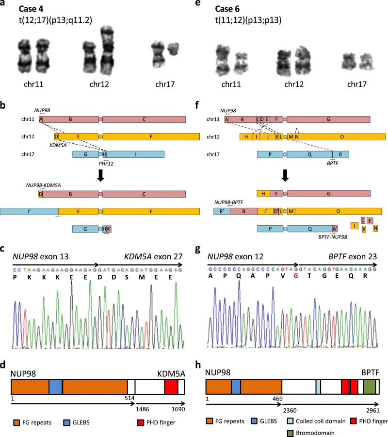
Cryptic NUP98-KDM5A and NUP98-BPTF fusion events in case 4 (left panels) and case 6 (right panels), respectively. a, e Partial karyograms of chromosomes 11, 12, and 17 and relevant cytogenetic findings. b, f Schematic representations of SVs in aforementioned chromosomes. Rearranged chromosomes, predicted from SVs and cytogenetics, are shown below. Arrows represent genes in 5′ to 3′ direction, alphabets represent genomic segments demarcated by case-specific breakpoints, apostrophes represent inverted segments, dashed lines represent SVs, and red dashed lines represent SVs leading to gene fusions. c, g Reverse transcription (RT)-PCR and Sanger sequencing of fusion transcripts. Arrows represent fused exons in 5′ to 3′ direction and letters below nucleotide codons represent corresponding amino acids. d, h Predicted fusion proteins and their domain structures adapted from UniProt. Arrows represent fused proteins and numbers represent amino acid positions
References
-
- King RL, Naghashpour M, Watt CD, Morrissette JJD, Bagg A. A comparative analysis of molecular genetic and conventional cytogenetic detection of diagnostically important translocations in more than 400 cases of acute leukemia, highlighting the frequency of false-negative conventional cytogenetics. Am J Clin Pathol. 2011;135:921–8. doi: 10.1309/AJCPJCW6BY0CNIHD. - DOI - PubMed
