. 2019 Jun 28:5:12.
doi: 10.1186/s42234-019-0023-1.

Spinal cord stimulation in chronic pain: evidence and theory for mechanisms of action

Affiliations

Spinal cord stimulation in chronic pain: evidence and theory for mechanisms of action

Jacob Caylor et al. Bioelectron Med. .

Abstract

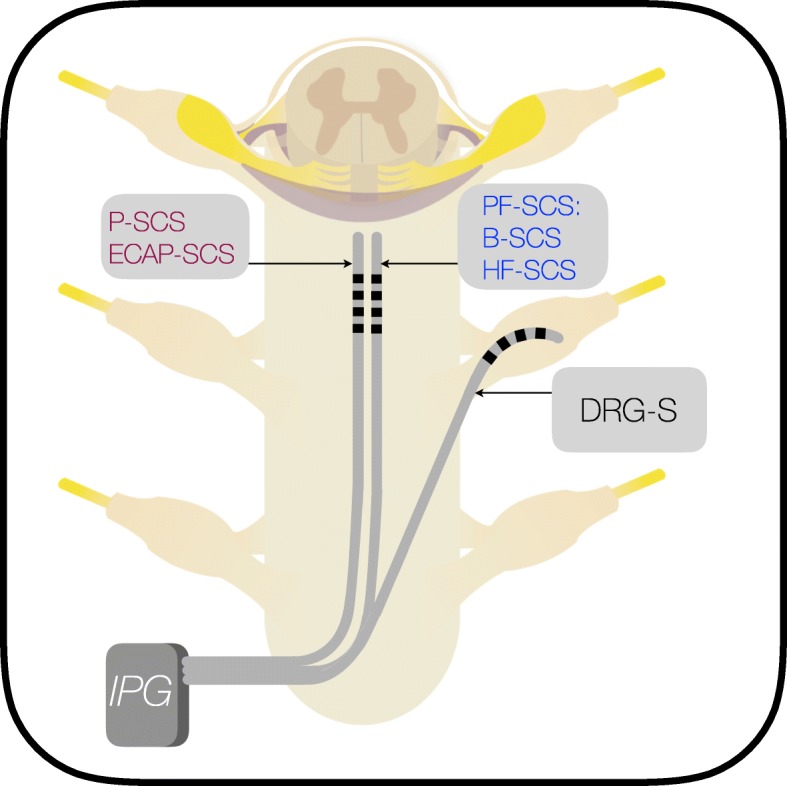
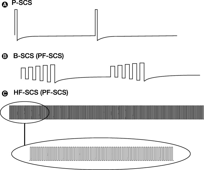
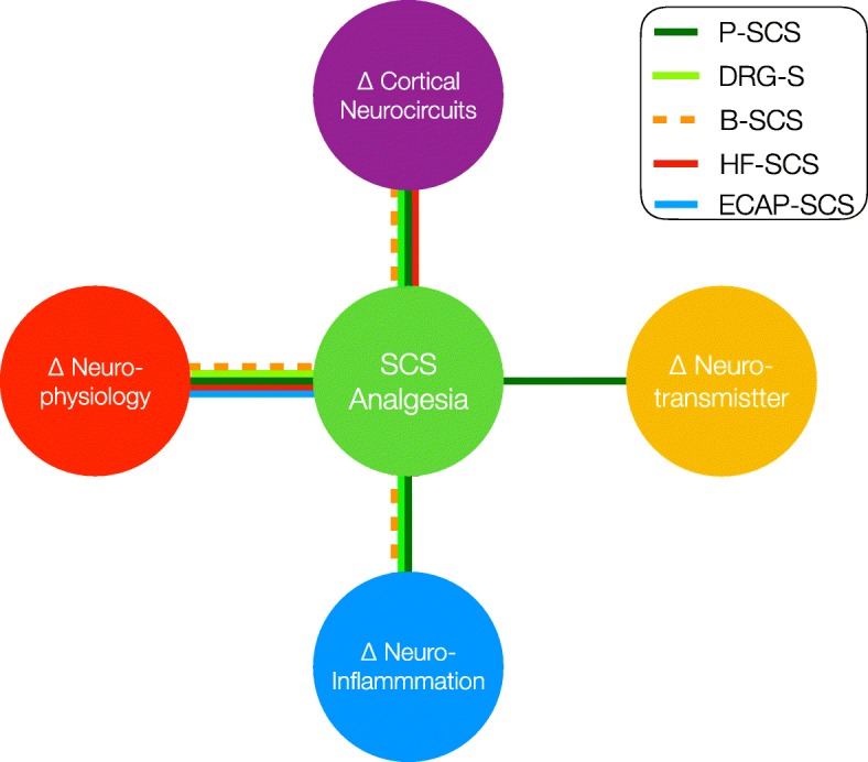
Well-established in the field of bioelectronic medicine, Spinal Cord Stimulation (SCS) offers an implantable, non-pharmacologic treatment for patients with intractable chronic pain conditions. Chronic pain is a widely heterogenous syndrome with regard to both pathophysiology and the resultant phenotype. Despite advances in our understanding of SCS-mediated antinociception, there still exists limited evidence clarifying the pathways recruited when patterned electric pulses are applied to the epidural space. The rapid clinical implementation of novel SCS methods including burst, high frequency and dorsal root ganglion SCS has provided the clinician with multiple options to treat refractory chronic pain. While compelling evidence for safety and efficacy exists in support of these novel paradigms, our understanding of their mechanisms of action (MOA) dramatically lags behind clinical data. In this review, we reconstruct the available basic science and clinical literature that offers support for mechanisms of both paresthesia spinal cord stimulation (P-SCS) and paresthesia-free spinal cord stimulation (PF-SCS). While P-SCS has been heavily examined since its inception, PF-SCS paradigms have recently been clinically approved with the support of limited preclinical research. Thus, wide knowledge gaps exist between their clinical efficacy and MOA. To close this gap, many rich investigative avenues for both P-SCS and PF-SCS are underway, which will further open the door for paradigm optimization, adjunctive therapies and new indications for SCS. As our understanding of these mechanisms evolves, clinicians will be empowered with the possibility of improving patient care using SCS to selectively target specific pathophysiological processes in chronic pain.

Keywords: Biomarker; Chronic pain; Complex regional pain syndrome; Failed back surgery syndrome; Mechanisms of action; Neuroinflammation; Neuropathic pain; Neurophysiology; Objective measures; Spinal cord stimulation.


Conflict of interest statement

Competing interests All other others claim no competing interests. Imanuel Lerman has been a paid consultant for Boston Scientific and received investigator initiated grants from Boston Scientific and Nevro Corporation. Ricardo Vallejo Ricardo Vallejo has been a paid consultant for and received grants from Boston Scientific and Nevro Corporation.

Figures

Fig. 1
Fig. 1
Lead Placement in P-SCS and PF-SCS: Dorsal column stimulation with traditional P-SCS, B-SCS and HF-SCS are anatomically placed over the dorsal columns. DRG-S is placed within the targeted foramina overlying the dorsal root ganglion. In all cases SCS can result in orthodromic activation or antidromic activation. Acronyms: IPG (Implantable Pulse Generator)
Fig. 2
Fig. 2
Parameters for Spinal Cord stimulation: Panel a: Amplitude: the peak current delivered, measured in milliamperes (mA). This impacts the number of fibers recruited and intensity of paresthesia. Amplitudes that are subthreshold do not generate an action potential and thus do not create paresthesia. Pulse Width (PW): the time over which the current is delivered, measured in microseconds (μs). The PW determines the amount of charge delivered for a given amplitude. Mathematical integration of the charge waveform yields the total charge delivered per pulse, measured in nanocoulombs. Increases in PW may recruit additional Aβ Fibers and broaden the area of paresthesia. Frequency: the number of pulses per second, measured in hertz (Hz). Panel b: Burst SCS parameters describing inter-burst frequency, or the number of bursts per second, and intraburst frequency, describing the number of pulses within a burst, measured in Hz. Panel c: The waveform or shape of the pulse can be divided into two segments: depolarization, or deflection above electroneutrality, and repolarization, the return to baseline. The depolarization waveform is determined by whether the system delivers the pulse in a Current-Controlled (CC) or Voltage-Controlled (VC) fashion. Current describes the flow of charge whereas voltage describes the potential difference between electrodes
Fig. 3
Fig. 3
Mechanisms of Hyperalgesia and Allodynia. Panel a) Non-pathologic nociception whereby C-fiber and Aβ inputs relay through an interneuron to modulate ascending signals via the projection neuron. Panel b) Nerve injury or repeated peripheral c-fiber stimuli leads to central sensitization through multiple mechanisms including changes in receptor kinetics, resting membrane potential and phenotypic transformation of Aβ afferent fibers. Through long term potentiation and altered receptor expression at the post receptor density zone, subthreshold stimulation evokes action potentials leading to classic hyperalgesia. Aβ axon sprouting after injury and secretion of substance P may offer further explanation for the development of tactile allodynia. Acronyms: Glu (Glutamate), Gly (Glycine), GABA (gamma amino butyric acid), SP (Substance P), BDNF (Brain Derived Neurotrophic Factor), CGRP (Calcitonin Gene Related Peptide), MGLUR (metabotropic glutamate receptor), TRKB (Tropomyosin receptor kinase), GABAR (GABA Receptor), AMPAR (AMPA Receptor), CGRPR (CGRP Receptor), NMDAR (NMDA Receptor), NK1R (Neurokinin-1 Receptor), LTP (Long Term Potentiation)
Fig. 4
Fig. 4
Changes in Dorsal Horn Circuitry to chronic pain and SCS. In the development of chronic pain, neural circuitry undergoes rewiring wherein abnormal enhancement of excitatory pathways and loss of inhibition facilitate nociceptive transmission to sub-threshold stimuli. Increased excitatory interneuron input, decreased inhibitory interneuron input and local factors contribute to pathologic destabilization of normal input balance to the projection neuron. SCS changes the balance of nociceptive and antinociceptive inputs through the activation of local segmental and descending supraspinal mechanisms to in-part restore balance to this network. Acronyms: DCN (Dorsal Column Nuclei), E (Excitatory Interneuron), I (Inhibitory Interneuron), PN (Projection Neuron), SP (Substance P), Glu (Glutamate), GABA (Gamma Aminobutyric Acid), 5-HT (5-Hydroxytryptamine, Serotonin)
Fig. 5
Fig. 5
Supraspinal Mechanisms of Spinal Cord Stimulation. A hallmark of chronic pain, abnormal enhancement of excitatory pathways and a loss of inhibition facilitate nociceptive transmission to sub-threshold stimuli. With SCS, orthodromic activation of supraspinal centers of pain control facilitates antinociception through activation of the DAS, largely through recruitment of the PAG, RVM and LC. Increases in spinal ACh, 5-HT and GABA as well as decreased spinal glutamate with SCS are in part thought to be a result of descending pathway recruitment. ‘Up’ arrows represent increased concentration or activity, whereas ‘down’ arrows represent opposite. ‘Sideways’ arrows represent no change. Acronyms: PAG (Periaqueductal Gray), RVM (Rostral Ventromedial Medulla), LC (Locus Coeruleus), A5 (Noradrenergic Cell Group A5), A7 (Noradrenergic Cell Group A7), DH (Dorsal Horn), DC (Dorsal Column), VLF (Ventrolateral Funiculus), DLF (Dorsolateral Funiculus), RVM ON (RVM On cells projecting from the RVM to the DH), RVM OFF (RVM OFF cells projecting from the RVM to the DH), RVM 5-HT Like (RVM 5-HT Like cells projecting from RVM to DH), cFOS (proto-oncogene), E (Excitatory Interneuron), I (Inhibitory Interneuron), PN (Projection Neuron), Glu (Glutamate), 5-HT (5-hydroxytryptamine), ACh (Acetylcholine), NE (Norepinephrine), GABA (Gamma Aminobutyric Acid)
Fig. 6
Fig. 6
Local Glia Modulate Synaptic Transmission After Nerve Injury. Glia are intimately associated with spinal excitatory and inhibitory synapses. The release of cytokines and glial mediators on pre and post-synaptic terminals modulates the activity of that synapse. Cytokines released at glutaminergic excitatory synapses augment transmission while cytokines released at GABAergic and Glycinergic synapses attenuates the signal. The net effect of gliosis and release of glial mediators is an increase in spinal cord pain transmission. Novel modes of SCS may modulate glial activity, thereby enacting their antinociceptive mechanisms through this pathway. Acronyms: Glu (Glutamate), GABA (Gamma Aminobutyric Acid), Gly (Glycine), TNF-α (Tumor Necrosis Factor Alpha), IL-1β (Interleukin 1β), IFN-γ (Interferon Gamma), NMDAR (NMDA Receptor), AMPAR (AMPA Receptor), BDNF (Brain Derived Neurotrophic Factor), GABAR (GABA Receptor), GlyR (Glycine Receptor), PGE2 (Prostaglandin E2)
Fig. 7
Fig. 7
Neurophysiological testing. Somatosensory evoked potentials (SSEP) waveforms are measured by applying an electrical stimuli to the tibial nerve. SSEP waveforms recorded with scalp electrodes at (CPz-Fz) are modulated with SCS. Panel a: During SCS ON P39-N50-P60 representative SSEP waveform decreases in amplitude. Flexor reflexes (RIII) are obtained with noxious electrical stimuli are applied to the sural nerve. The RIII waveform is recorded from the ipsilateral biceps femoris. Panel b: During SCS ON there is decrease the amplitude of the RIII waveform (adapted from (Sankarasubramanian et al., 2018b)). Quantitative sensory testing (QST) applies different sensory stimuli, such as vibratory, tactile or thermal stimuli to the subject extremity. Panels c, d: SCS results in variable effects on QST sensory thresholds and may increase increases pain threshold and tolerance. Advanced QST measures including temporal summation (TS) and conditioned pain modulation (CPM) can provide further insight into P-SCS, PF-SCS and DRG-S analgesic mechanisms
Fig. 8
Fig. 8
Cortical and Sub-cortical Pain Processing. Sensory-discriminative pain processing with thalamus, SI, SII and posterior IC, with sensory discriminative pain relayed through the ventroposterior-lateral and ventroposterior-medial thalamic nuclei and is also termed the neospinothalamic pathway. Affective-emotional pain processing with dorsal ACC and Anterior IC while affective sensory information to the AEN is thought to be relayed through the medio-dorsal thalamus, also termed the paleospinothalamic pathway. PFC (Prefrontal Cortex), OFC (orbitofrontal cortex) SI (Primary Somatosensory Cortex) SII (Secondary Somatosensory Cortex) dACC (Dorsal Anterior Cingulate Cortex) Amy (Amygdala), AI (Anterior Insula), RI (Rostral Insula) dPI (Dorsoposterior Insula), PAG (Periaqueductal Gray)
Fig. 9
Fig. 9
Chronic pain thalamocortical dysrhythmia is known to occur in distinct band, i.e. most commonly in theta, beta, and gamma bands. Work from De Ridder and Vannesste demonstrate p < .0001) with greatest differences found in theta-beta theta and gamma (Panel a), source localized to bilateral sensory discriminative pathways SI, which additional affective emotional pathways including dACC, sgACC, bilateral INS, bilateral PHC, and posterior cingulate cortex (Panel b). Pretrial and or implant characterization of chronic pain patient TCD could inform the practitioner of an optimal SCS paradigm, while post SCS TCD measures could track SCS efficacy (i.e., decrease in predominate θ theta, β beta, γ gamma dysrhythmias) as described by Schulman et al. (2005). dACC dorsal anterior cingulate cortex, sgACC subgenual anterior cingulate cortex, INS insula, PHC parahippocampus, SI primary somatosensory cortex, PCC posterior cingulate cortex, θ theta, α alpha, β beta, γ gamma. Figure adapted from Vanneste et al. (2018) Nature Communications (Vanneste et al., 2018)
Fig. 10
Fig. 10
Stimulation Patterns in Paresthesia and Paresthesia Free SCS Paradigms: Traditional SCS comprised of tonic or repetitive low frequency SCS usually in the range of 40–60 Hz produces paresthesia of P-SCS (a). Paresthesia free, burst spinal cord stimulation (B-SCS) employs incremental increase in amplitude with each burst. Inter-burst frequency is 40 Hz, while intra-burst frequency is 500 Hz (b). Between each burst there is a passive recharge phase. Paresthesia free high frequency SCS employs an ultra-high frequency of 10 kHz in continuous mode (c)
Fig. 11
Fig. 11
ECAP-SCS. Newly developed closed-loop SCS system measures evoked compound action potentials (ECAPs) from the spinal cord after each pulse. Greater ECAP amplitude represents more action potentials and is equivalent to increased fiber activation. Variable lead to cord contact occurs with change in position and over time. ECAP-SCS captures and calibrates SCS lead current to target desired ECAP waveform therefore minimizing variability in stimulation, which may improve clinical outcomes. ECAP-SCS is an objective measure of spinal cord activation that may help to predict responders to P-SCS
Fig. 12
Fig. 12
The Current literature supports multiple domains of MOA of SCS. Major P-SCS, PF-SCS and DRG-S analgesic MOA include modulation of 1) segmental and supraspinal neurotransmitters, 2) segmental and supraspinal neurophysiology/neuroplasticity, 3) central and peripheral neuroinflammation and 4) cortical and subcortical neurocircuits. P-SCS has accrued the largest literature in support of all four MOA, while only P-SCS preclinical studies consistently demonstrate modulation of neurotransmitters critical to analgesia. Emerging and published literature support the concept that all SCS paradigms (P-SCS, B-SCS, DRG-S, HF-SCS, ECAP-SCS) contribute to altered neuronal activity (i.e. neurophysiological). Pre-clinical and clinical work support cortical SCS paradigms (P-SCS, B-SCS, DRG-S, HF-SCS, ECAP-SCS) contribute to altered neurocircuit activity

References

    1. Abejon D, Feler CA. Is impedance a parameter to be taken into account in spinal cord stimulation? Pain Physician. 2007;10(4):533. - PubMed
    1. Abejón D, Rueda P, del Saz J, Arango S, Monzón E, Gilsanz F. Is the introduction of another variable to the strength–duration curve necessary in Neurostimulation? Neuromodulation. 2015;18(3):182–190. doi: 10.1111/ner.12223. - DOI - PubMed
    1. Ahmed SU, Zhang Y, Chen L, St. Hillary K, Cohen A, Vo T, Houghton M, Mao J. Effects of spinal cord stimulation on pain thresholds and sensory perceptions in chronic pain patients. Neuromodulation. 2015;18(5):355–360. doi: 10.1111/ner.12316. - DOI - PubMed
    1. Akil H, Liebeskind JC. Monoaminergic mechanisms of stimulation-produced analgesia. Brain Res. 1975;94(2):279–296. doi: 10.1016/0006-8993(75)90062-1. - DOI - PubMed
    1. Al-Kaisy A, Palmisani S, Smith T, Harris S, Pang D. The use of 10-kilohertz spinal cord stimulation in a cohort of patients with chronic neuropathic limb pain refractory to medical management. Neuromodulation. 2015;18(1):18–23. doi: 10.1111/ner.12237. - DOI - PubMed