. 2019 Oct 1;142(10):3028-3044.
doi: 10.1093/brain/awz250.

Synaptic clustering differences due to different GABRB3 mutations cause variable epilepsy syndromes

Affiliations

Synaptic clustering differences due to different GABRB3 mutations cause variable epilepsy syndromes

Yi-Wu Shi et al. Brain. .

Abstract

GABRB3 is highly expressed early in the developing brain, and its encoded β3 subunit is critical for GABAA receptor assembly and trafficking as well as stem cell differentiation in embryonic brain. To date, over 400 mutations or variants have been identified in GABRB3. Mutations in GABRB3 have been increasingly recognized as a major cause for severe paediatric epilepsy syndromes such as Lennox-Gastaut syndrome, Dravet syndrome and infantile spasms with intellectual disability as well as relatively mild epilepsy syndromes such as childhood absence epilepsy. There is no plausible molecular pathology for disease phenotypic heterogeneity. Here we used a very high-throughput flow cytometry assay to evaluate the impact of multiple human mutations in GABRB3 on receptor trafficking. In this study we found that surface expression of mutant β3 subunits is variable. However, it was consistent that surface expression of partnering γ2 subunits was lower when co-expressed with mutant than with wild-type subunits. Because γ2 subunits are critical for synaptic GABAA receptor clustering, this provides an important clue for understanding the pathophysiology of GABRB3 mutations. To validate our findings further, we obtained an in-depth comparison of two novel mutations [GABRB3 (N328D) and GABRB3 (E357K)] associated with epilepsy with different severities of epilepsy phenotype. GABRB3 (N328D) is associated with the relatively severe Lennox-Gastaut syndrome, and GABRB3 (E357K) is associated with the relatively mild juvenile absence epilepsy syndrome. With functional characterizations in both heterologous cells and rodent cortical neurons by patch-clamp recordings, confocal microscopy and immunoblotting, we found that both the GABRB3 (N328D) and GABRB3 (E357K) mutations reduced total subunit expression in neurons but not in HEK293T cells. Both mutant subunits, however, were reduced on the cell surface and in synapses, but the Lennox-Gastaut syndrome mutant β3 (N328D) subunit was more reduced than the juvenile absence epilepsy mutant β3 (E357K) subunit. Interestingly, both mutant β3 subunits impaired postsynaptic clustering of wild-type GABAA receptor γ2 subunits and prevented γ2 subunits from incorporating into GABAA receptors at synapses, although by different cellular mechanisms. Importantly, wild-type γ2 subunits were reduced and less clustered at inhibitory synapses in Gabrb3+/- knockout mice. This suggests that impaired receptor localization to synapses is a common pathophysiological mechanism for GABRB3 mutations, although the extent of impairment may be different among mutant subunits. The study thus identifies the novel mechanism of impaired targeting of receptors containing mutant β3 subunits and provides critical insights into understanding how GABRB3 mutations produce severe epilepsy syndromes and epilepsy phenotypic heterogeneity.

Keywords: GABRB3 mutation; GABAA receptor; Lennox-Gastaut syndrome; intellectual disability; juvenile absence epilepsy.


Figures

Figure 1
Figure 1
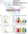
Reduced partnering GABAA receptor γ2 subunit at cell surface is a common phenomenon across GABRB3 epilepsy mutations. (A) Schematic presentation of β3 subunit protein topology and locations of mutations in human GABRB3 associated with various epilepsy syndromes and neurodevelopmental disorders. These mutations are distributed in various locations and domains of the β3 subunit protein peptide. The coloured dots represent the relative locations of the epilepsy related mutations. (BE) The flow cytometry histograms depict surface β3 (B) or HA-tagged γ2 subunit (γ2HA) (D) expression levels on HEK293T cells co-expressing the human wild-type or the mutant β3 subunit cDNAs with the α1 and HA-tagged γ2 subunit cDNAs for 48 h. The expression levels of the β3 or γ2HA subunits in the mutant α1β3(Y182F)γ2HA receptors was chosen as example. The HA-tagged γ2 subunit protein was immunostained with a mouse anti-HA antibody while β3 subunits were immunostained with the mouse anti-β2/3 antibody (BD17). The relative surface levels of each mutant β3 subunit were normalized to the wild-type β3 subunit when co-expressed with α1 and γ2HA cDNAs. The relative subunit expression level of β3 (C) or γ2HA (E) subunits were normalized to those obtained with co-expression of γ2HA subunit with α1 and the wild-type β3 subunit cDNAs. In C and E, n = 4 different transfections, *P < 0.05; **P < 0.01, ***P < 0.001, one-way ANOVA with post hoc Tukey test.
Figure 2
Figure 2
Clinical and molecular genetic findings in GABRB3 in patients with Lennox-Gastaut syndrome (LGS) or juvenile absence epilepsy (JAE). (A and B) Representative EEGs from each patient are presented. The top traces show EEG recordings from the patient carrying the GABRB3 (N328D) variation, showing a generalized poly-spike-waves discharge, accompanied by time-locked muscle activity on the EMG recording (A), and an interictal EEG of a fast rhythm that was superimposed on the following irregular slow waves during sleep (B). (C and D) EEG traces show an ictal EEG with paroxysmal 3.0–3.5 Hz generalized spike-and-wave discharges with behavioural arrest, lasting for ∼5 s (C) and an interictal EEG of a single generalized spike or spike-and-waves discharge in the patient carrying the GABRB3 (E357K) variation (D). (E and F) Two novel mutations were identified in patients with a severe epilepsy syndrome (Lennox-Gastaut syndrome) with intellectual disability or a mild epilepsy syndrome (juvenile absence epilepsy) without intellectual disability. (F) Cartoon illustrates that the location of mutations in GABRB3 in the subunit protein topology. (G) The amino acids coded by both variations are conserved across species as showing in the boxed regions.
Figure 3
Figure 3
Mutant α1β3γ2 receptors showed decreased GABA-evoked whole-cell currents and altered zinc inhibition. (AC) HEK 293 T cells were transfected with human α1 and γ2 subunits in combination with the wild-type or mutant β3(N328D) or β3(E357K) subunits for 48 h. EGFP cDNA was included as a marker for positive transfection. Representative GABA-evoked current traces were obtained following rapid application of 1 mM GABA for 6 s to lifted HEK293T cells voltage clamped at −50 mV (A). (B) Bar graphs showing average peak current amplitude from the wild-type or the mutant α1β3γ2 receptors. Values are expressed as mean ± standard error of the mean (SEM). (C) Per cent current loss was measured by subtracting the remaining current during GABA and zinc co-application from the peak current during GABA application. In B, n = 6 patches for wild-type, n = 8 for N328D mix, n = 9 for N328D, n = 11 for E357K mix and n = 7 for E357K. In C, n = 6 patches for wild-type, n = 5 for N328D mix, n = 5 for N328D, n = 6 for E357K mix and n = 5 for E357K. In B and C, ***P < 0.001 versus wild-type, ††P < 0.01 versus mixed in N328D or in E357K; §P < 0.05; §§P < 0.01 versus mutant in E357K. One-way ANOVA and Newman-Keuls test was used to determine significance compared to the wild-type condition and between mutations.
Figure 4
Figure 4
Both mutant α1β3(N328D)γ2 and α1β3(E357K)γ2 receptors had reduced subunit surface expression. (A) HEK293 T cells were transfected with human α1 and γ2 subunits in combination with the wild-type β3 subunit alone (WT), mixed wild-type and mutant β3 subunit for mixed, or the mutant β3(N328D)HA or β3(E357K)HA subunits alone for mutant (mut) for 48 h. Surface protein samples were collected through biotinylation and probed by rabbit polyclonal anti-β3 or anti-γ2 subunit or mouse monoclonal anti-α1 subunit antibodies. LC = loading control (ATPase). (B) Surface expression of each subunit protein from western blot was quantified. The integrated protein density values (IDVs) were normalized to the loading control first and then to the wild-type which is arbitrarily taken as 1. **P < 0.01, ***P < 0.001 versus wild-type, P < 0.05, ††P < 0.01, N328 heterozygous (het) versus E357K het, ns = non-significant, n = 4 different transfections, one-way ANOVA and Newman-Keuls test. Values are expressed as mean ± SEM.
Figure 5
Figure 5
Both mutant β3(N328D)HA and β3(E357K)HA subunits had reduced distribution in dendrites in neurons. (A) Rat cortical neurons were transfected with wild-type (β3HA) or mutant [β3(N328D)HA and β3(E357K)HA] subunits at Day 5–7 in culture. At 8–10 days after transfection, the neurons were permeabilized and stained with mouse monoclonal anti-HA antibody and visualized with fluorophore Alexa-488. (B) Total lysates of mouse cortical neurons expressing the empty vector pCDNA (control), the HA-tagged wild-type (WT) (β3HA) or the mutant β3 subunits β3 (N328D)HA and β3 (E357K)HA) in 100 mm2 dishes were collected at Day 8–10 after transfection. The lysates were separated by SDS-PAGE and immunoblotted with mouse monoclonal anti-HA antibody. ATPase was used for loading control. (C) The raw fluorescence values in somata were measured. The identification of somata as red circled area for each neuron was arbitrary because no neuronal nuclei marker was applied in this experiment. (D) β3HA subunit fluorescence puncta in neurons expressing the wild-type or the mutant β3HA subunits were quantified. The total fluorescence puncta per 100 µm were measured. In C and D, n = 20 neurons for each condition from four different transfections. At least three to five coverslips were surveyed from each transfection (E). The total expression of the wild-type or the mutant β3HA subunit in neurons was quantified. The IDVs were normalized to the loading control first and then to the wild-type which is arbitrarily taken as 1 (n = 4 different transfections). **P < 0.01, ***P < 0.001 versus wild-type, P < 0.05, †††P < 0.001 versus E357K. For C and D, one-way ANOVA and Newman-Keuls test. For E, one sample t-test and unpaired t-test were used for N328D versus E357K. Values are expressed as mean ± SEM.
Figure 6
Figure 6
Rat cortical neurons expressing the mutant β3 (N328D)HA or β3 (E357K)HA subunit had reduced γ2 subunits in synapses. (A) Rat cortical neurons were transfected with wild-type or mutant β3 subunits at Day 5 in culture. At 8 days after transfection, the neurons were permeabilized and co-stained with rabbit polyclonal anti-γ2 and mouse monoclonal anti-vGAT (vesicular GABA transporter) antibodies. Mouse polyclonal IgGs were visualized with the fluorophore Alexa-488 while the rabbit IgGs were visualized with rhodamine. Cell nuclei were stained with TO-PRO-3. White arrow indicates area enlarged in insets. (B) Total fluorescence intensity of vGAT in rat cortical neurons expressing the wild-type or the mutant β3 subunits was quantified by ImageJ. (C) Total γ2 subunit fluorescence puncta in neurons expressing the wild-type or the mutant β3 subunits in each visualized whole field were quantified. In B and C, n = 11 for wild-type, 12 for N328D and 12 for E357K non-overlapping visualized fields from four different transfections. ***P < 0.001 versus wild-type, One-way ANOVA and Newman-Keuls test. Values are expressed as mean ± SEM.
Figure 7
Figure 7
The mutant β3(N328D)HA and β3(E357K)HA subunits have reduced co-localization with the inhibitory postsynaptic marker gephyrin. (A) Rat cortical neurons were transfected with wild-type or mutant β3 subunits at Days 5–7 in culture. At 8–10 days after transfection, the neurons were permeabilized and co-stained with rabbit polyclonal anti-HA and mouse monoclonal anti-gephyrin antibodies. Mouse polyclonal IgGs were visualized with the fluorophore Alexa-488 while the rabbit IgGs were visualized with rhodamine. Cell nuclei were stained with TO-PRO-3. White arrow indicates area enlarged in insets. (B) Total fluorescence puncta of β3HA subunits in rat cortical neurons expressing wild-type or mutant β3 subunits in each whole visualized field were quantified. (C) The per cent β3HA subunit fluorescence signal overlapping gephyrin fluorescence signal was quantified in neurons expressing the wild-type or the mutant β3 subunits in each whole field. In B and C, n = 15 for wild-type, 11 for N328D, and 9 for E357K; non-overlapping visualized fields from four different transfections. (*P < 0.05, **P < 0.01, ***P < 0.001 versus wild-type, P < 0.05, ††P < 0.01 versus E357K, one-way ANOVA and Newman-Keuls test). Values are expressed as mean ± SEM.
Figure 8
Figure 8
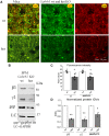
Loss of β3 subunit function impaired γ2 subunit clustering at inhibitory synapses in Gabrb3+/− mice. (A) Brain tissue from wild-type (wt) or heterozygous (het) Gabrb3+/− mice were briefly fixed, permeabilized and co-stained with rabbit polyclonal anti-γ2 and mouse monoclonal anti-gephyrin (gep) antibodies. Rabbit polyclonal IgGs were visualized with the fluorophore Alexa-488 while the mouse IgGs were visualized with rhodamine. Co = co-localization; KO = knockout. (B) Synaptosome protein samples were collected through subcellular fractionation and probed by rabbit polyclonal anti-β3 or anti-γ2 subunit or mouse monoclonal gephyrin antibodies. LC = control (GAPDH). (C) Fluorescence values of γ2 subunits or gephyrin were quantified (*P < 0.05 versus wild-type, unpaired t-test). Values were expressed as mean ± SEM, n = 12 sections for each genotype from four different pairs of mice for γ2, and n = 20 sections for wild-type and 23 sections for heterozygous from seven different pairs of mice for gephyrin. For each section, three representative regions were chosen, and the average value of the three regions was taken as n = 1). (D) The integrated protein density values (IDVs) from synaptosome preparations were normalized to the loading control first and then to the wild-type, which was arbitrarily taken as 1 (n = 4 pairs of 2–4-month-old mice, one-sample t-test).

References

    1. Alldred MJ, Mulder-Rosi J, Lingenfelter SE, Chen G, Luscher B. Distinct gamma2 subunit domains mediate clustering and synaptic function of postsynaptic GABAA receptors and gephyrin. J Neurosci 2005; 25: 594–603. - PMC - PubMed
    1. Allen AS, Berkovic SF, Cossette P, Delanty N, Dlugos D, Eichler EE et al. . De novo mutations in epileptic encephalopathies. Nature 2013; 501: 217–21. - PMC - PubMed
    1. Andäng M, Hjerling-Leffler J, Moliner A, Lundgren TK, Castelo-Branco G, Nanou E et al. . Histone H2AX-dependent GABA(A) receptor regulation of stem cell proliferation. Nature 2008; 451: 460–64. - PubMed
    1. Baulac S, Huberfeld G, Gourfinkel-An I, Mitropoulou G, Beranger A, Prud’homme JF et al. . First genetic evidence of GABA(A) receptor dysfunction in epilepsy: a mutation in the gamma2-subunit gene. Nat Genet 2001; 28: 46–8. - PubMed
    1. Brooks-Kayal A. Epilepsy and autism spectrum disorders: are there common developmental mechanisms? Brain Dev 2010; 32: 731–8. - PubMed

Publication types

MeSH terms