. 2019 Nov 1;129(11):4979-4991.
doi: 10.1172/JCI126402.

Airway epithelium-shifted mast cell infiltration regulates asthmatic inflammation via IL-33 signaling

Affiliations

Airway epithelium-shifted mast cell infiltration regulates asthmatic inflammation via IL-33 signaling

Matthew C Altman et al. J Clin Invest. .

Abstract

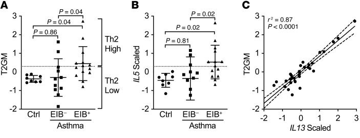
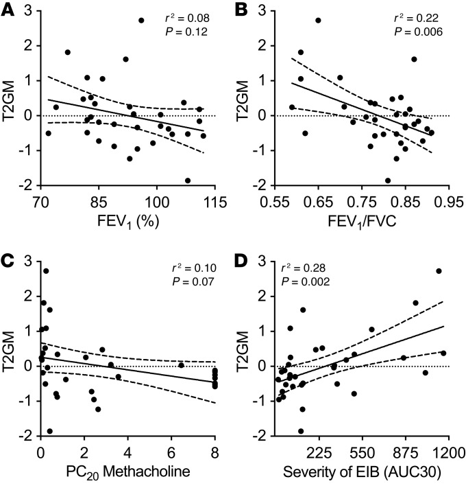
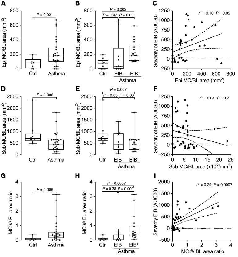
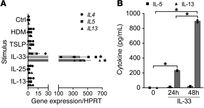
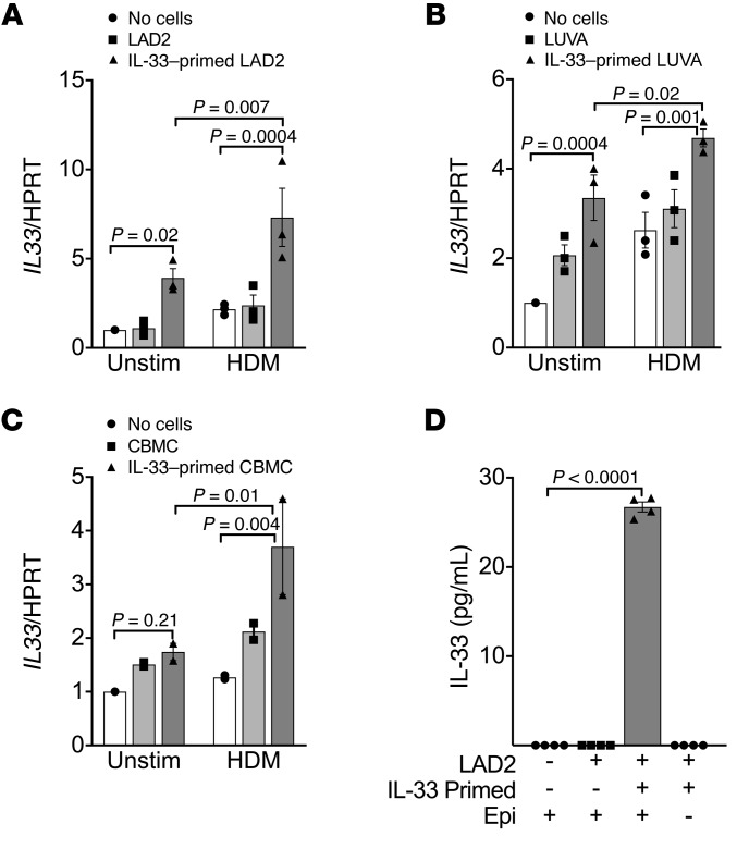
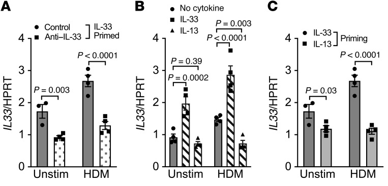
Asthma is a heterogeneous syndrome that has been subdivided into physiologic phenotypes and molecular endotypes. The most specific phenotypic manifestation of asthma is indirect airway hyperresponsiveness (AHR), and a prominent molecular endotype is the presence of type 2 inflammation. The underlying basis for type 2 inflammation and its relationship to AHR are incompletely understood. We assessed the expression of type 2 cytokines in the airways of subjects with and without asthma who were extensively characterized for AHR. Using quantitative morphometry of the airway wall, we identified a shift in mast cells from the submucosa to the airway epithelium specifically associated with both type 2 inflammation and indirect AHR. Using ex vivo modeling of primary airway epithelial cells in organotypic coculture with mast cells, we show that epithelial-derived IL-33 uniquely induced type 2 cytokines in mast cells, which regulated the expression of epithelial IL33 in a feed-forward loop. This feed-forward loop was accentuated in epithelial cells derived from subjects with asthma. These results demonstrate that type 2 inflammation and indirect AHR in asthma are related to a shift in mast cell infiltration to the airway epithelium, and that mast cells cooperate with epithelial cells through IL-33 signaling to regulate type 2 inflammation.

Keywords: Asthma; Immunology; Mast cells; Pulmonology; Th2 response.


Conflict of interest statement

Conflict of interest: The authors have declared that no conflict of interest exists.

Figures

Figure 1
Figure 1. Type 2 gene expression in induced sputum is specifically elevated in EIB+ asthma.
(A) The T2GM was elevated in EIB+ asthma subjects (n = 14) compared with EIB asthma subjects (n = 11) and healthy controls (n = 8). (B) Of the 3 type 2 genes, expression of IL5 showed the most significant difference among the groups. Shown are means and SDs. Analyses are by 1-way ANOVA with correction for multiple comparisons. (C) IL13 expression was most highly associated with the T2GM by linear regression (95% confidence intervals are shown).
Figure 2
Figure 2. Sputum type 2 gene expression correlates with airway obstruction and the severity of indirect AHR.
(A and B) The T2GM did not correlate with FEV1 % predicted (A) but did correlate with the FEV1/FVC ratio (B). (C and D) The T2GM trended toward an association with severity of direct AHR (C) and was significant in association with indirect AHR (D). Associations were assessed by linear regression; shown are regression lines and 95% confidence intervals.
Figure 3
Figure 3. Sputum type 2 gene expression is associated with mast cell gene expression in sputum and bronchial brushings.
The T2GM expression in sputum was significantly correlated with the expression of the mast cell genes TPSAB1 and CPA3, but not CMA1, in induced sputum cells (AC) and also in bronchial epithelial brushing (DF). All associations are by linear regression; shown are regression lines and 95% confidence bounds.
Figure 4
Figure 4. A shift in mast cells from the submucosa to the epithelium is associated with indirect AHR.
(A) The number of intraepithelial mast cells relative to the area of the basal lamina (Epi MC/BL area) is increased in asthma compared with healthy controls. (B) It is highest in EIB+ asthma subjects compared with EIB subjects and healthy controls. (C) Additionally, it is significantly correlated with severity of indirect AHR measured by AUC30. (DF) In contrast, the number of submucosal mast cells relative to the area of the basal lamina (Sub MC/BL area) is lower in asthma compared with healthy controls (D), is similar in EIB+ and EIB asthma (E), and is not correlated with indirect AHR (F). (G) The ratio of the number of mast cells per area of the basal lamina in the epithelium relative to the number in the submucosa is increased in asthma. (H) It is highest in the EIB+ asthma subjects relative to the EIB subjects and healthy controls. (I) Additionally, it is significantly associated with the severity of indirect AHR measured by AUC30. Group comparisons are shown as box plots with median, interquartile range, minimum, and maximum. Significance was assessed by the Mann-Whitney U test (2-group) or the Kruskal-Wallis test with Dunn’s post hoc test for multiple comparisons (3-group). Associations are by linear regression; shown are regression lines and 95% confidence bounds.
Figure 5
Figure 5. Sputum type 2 gene expression is correlated with a shift in mast cells from the submucosa to the airway epithelium.
(A and B) The sputum T2GM is correlated with the number of intraepithelial mast cells relative to the area of the basal lamina (Epi MC/BL area) (A), and not with the number of submucosal mast cells relative to the area of the basal lamina (Sub MC/BL area) (B). (C) The sputum T2GM is correlated with the volume density ratio of mast cells in the epithelium relative to the submucosa. (D) Sputum T2GM is also weakly correlated with sputum eosinophil concentration. All associations are by linear regression; shown are regression lines and 95% confidence bounds.
Figure 6
Figure 6. IL-33 uniquely induces production of type 2 cytokines in mast cells.
(A) IL-33 induced gene expression of the type 2 cytokines IL5 and IL13, and to a lesser extent IL4, in primary human CBMCs compared with unstimulated control (Ctrl), whereas IL-25, TSLP, HDM extract, or IL-13 did not (n = 4 per condition). *P < 0.0001 vs. Ctrl. (B) IL-33 induced production of IL-13 protein and to a lesser extent IL-5 protein in LUVA mast cells (n = 2 per condition at 3 time points). Differences between multiple conditions were assessed by 1-way ANOVA with correction for multiple comparisons. Shown are mean values and SEM bars. *P < 0.0001.
Figure 7
Figure 7. Mast cells regulate the epithelial expression of IL33 in a feed-forward loop.
In a coculture system with differentiated epithelial cells cocultured with or without mast cells for 48 hours, priming of the mast cells with IL-33 significantly amplified the epithelial expression of IL33. Stimulation of the epithelial surface with HDM further amplified IL33 expression. Similar results were identified for LAD2 mast cells (n = 3 per condition) (A), LUVA mast cells (n = 3 per condition) (B), and primary CBMCs (n = 2 per condition) (C). (D) IL-33 protein was detected in the basolateral media only when IL-33–primed mast cells were added to the coculture system in which HDM was added to the apical surface (n = 4 per condition). IL-33 protein was not detectable when IL-33–primed mast cells that were similarly exposed to HDM were cultured in the absence of the epithelium. Differences between multiple conditions were assessed by 1-way ANOVA with correction for multiple comparisons. Shown are mean values and SEM bars.
Figure 8
Figure 8. Feed-forward amplification of epithelial IL33 gene expression is mediated by IL-33 and is not a consequence of IL-13 signaling.
(A) A blocking antibody against IL-33 during the 48 hours of coculture reduced the amplification of epithelial IL33 expression during coculture with IL-33–primed LAD2 mast cells, particularly when the apical surface of the epithelium was treated with HDM. (B) Direct treatment of differentiated primary epithelial cells alone (without mast cells) with IL-33 significantly increased the expression of IL33, while treatment with IL-13 did not induce the expression of epithelial IL33. HDM also induced expression of epithelial IL33 (P = 0.02), which was further augmented by treatment with IL-33 in the basolateral compartment and was diminished by the addition of IL-13 in the basolateral fluid. (C) Priming mast cells with IL-33 before coculture increased epithelial IL33 expression in comparison with priming with IL-13 (n = 4 per condition for all conditions, except one condition in which poor quantitative PCR amplification of the housekeeping gene occurred). Differences between multiple conditions were assessed by 1-way ANOVA with correction for multiple comparisons. Shown are mean values and SEM bars.
Figure 9
Figure 9. Feed-forward amplification of epithelial IL33 expression is higher in epithelial cells from children with asthma compared with healthy children.
Amplification of epithelial IL33 expression by coculture with IL-33–primed LUVA mast cells was greater in epithelial cells isolated from children with asthma compared with epithelial cells isolated from healthy nonatopic children (n = 3 per group). Significance was assessed by 2-way ANOVA. There was a notable trend toward an increased effect of IL-33–primed mast cells on epithelial cells derived from children with asthma. *P = 0.1 for the post hoc test.

References

    1. Coates AL, et al. ERS technical standard on bronchial challenge testing: general considerations and performance of methacholine challenge tests. Eur Respir J. 2017;49(5):1601526. doi: 10.1183/13993003.01526-2016. - DOI - PubMed
    1. Hallstrand TS, et al. ERS technical standard on bronchial challenge testing: pathophysiology and methodology of indirect airway challenge testing. Eur Respir J. 2018;52(5):1801033. doi: 10.1183/13993003.01033-2018. - DOI - PubMed
    1. Martinez FD. Links between pediatric and adult asthma. J Allergy Clin Immunol. 2001;107(5 suppl):S449–S455. - PubMed
    1. et al. T-helper type 2-driven inflammation defines major subphenotypes of asthma. Am J Respir Crit Care Med. 2009;180(5):388–395. doi: 10.1164/rccm.200903-0392OC. - DOI - PMC - PubMed
    1. Cabral AL, Conceição GM, Fonseca-Guedes CH, Martins MA. Exercise-induced bronchospasm in children: effects of asthma severity. Am J Respir Crit Care Med. 1999;159(6):1819–1823. doi: 10.1164/ajrccm.159.6.9805093. - DOI - PubMed

Publication types