. 2019 Aug 22;14(8):e0219765.
doi: 10.1371/journal.pone.0219765. eCollection 2019.

Differential tetraspanin genes expression and subcellular localization during mutualistic interactions in Phaseolus vulgaris

Affiliations

Differential tetraspanin genes expression and subcellular localization during mutualistic interactions in Phaseolus vulgaris

Saul Jimenez-Jimenez et al. PLoS One. .

Abstract

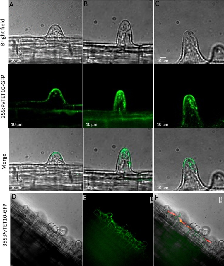
Arbuscular mycorrhizal fungi and rhizobia association with plants are two of the most successful plant-microbe associations that allow the assimilation of P and N by plants, respectively. These mutualistic interactions require a molecular dialogue, i.e., legume roots exude flavonoids or strigolactones which induce the Nod factors or Myc factors synthesis and secretion from the rhizobia or fungi, respectively. These Nod or Myc factors trigger several responses in the plant root, including calcium oscillations, and reactive oxygen species (ROS). Furthermore, superoxide and H2O2 have emerged as key components that regulate the transitions from proliferation to differentiation in the plant meristems. Similar to the root meristem, the nodule meristem accumulates superoxide and H2O2. Tetraspanins are transmembrane proteins that organize into tetraspanin web regions, where they recruit specific proteins into platforms required for signal transduction, membrane fusion, cell trafficking and ROS generation. Plant tetraspanins are scaffolding proteins associated with root radial patterning, biotic and abiotic stress responses, cell fate determination, and hormonal regulation and recently have been reported as a specific marker of exosomes in animal and plant cells and key players at the site of plant fungal infection. In this study, we conducted transcriptional profiling of the tetraspanin family in common bean (Phaseolus vulgaris L. var. Negro Jamapa) to determine the specific expression patterns and subcellular localization of tetraspanins during nodulation or under mycorrhizal association. Our results demonstrate that the tetraspanins are transcriptionally modulated during the mycorrhizal association, but are also expressed in the infection thread and nodule meristem development. Subcellular localization indicates that tetraspanins have a key role in vesicular trafficking, cell division, and root hair polar growth.


Conflict of interest statement

The authors have declared that no competing interests exist.

Figures

Fig 1
Fig 1. A rooted neighbor-joining phylogenetic tree of common bean tetraspanin.
(A) On the left shown is the phylogenetic tree constructed using MEGA6.06, with amino acidic sequences from phytozome.org. Numbers above branches indicate bootstrap percentage values. PvTET proteins were clustered based on a significant bootstrap value of ≥50%. (B) On the right using an online tool, Gene Structure Display Server (GSDS; http://gsds.cbi.pku.edu.cn/), was used to draw the tetraspanin gene structure. Red boxes indicate exons, black lines depict introns, upstream/downstream sequences are shown as oranges boxes. Intron phases are indicated at exon–intron junctions.
Fig 2
Fig 2. Tetraspanin gene expression profile in P. vulgaris root or root hairs from different developmental stages.
The relative expression of each PvTET gene was evaluated by qRT–PCR in three different sections of root at 48 h post germination (hpg). Mature zone or Zone III (blue), elongation zone or Zone II (red), and meristematic, elongating and differentiating region or Zone I (green). Transcript accumulation was normalized to the expression of EF1a, which was used as a reference gene. Bars represent means ± SEM from at least three independent biological replicates with three technical repeats. (A) Tetraspanin (TET) gene expression in shaved root and (B) root hairs at different development stages at 48 hpg. Tissues enriched with emerging or bulging root hairs from Zone I (green bar), tissues enriched with growing root hairs (red bar) and tissues enriched with mature root hairs (blue bar). Transcript accumulation was normalized to the expression of EF1a, which was used as a reference gene. Bars represent means ± SEM from at least three independent biological replicates with three technical repeats. P-values <0.05 are marked with two asterisks (Student’s t-test). (C) Cartoon depicting the different root and root hairs zones analyzed.
Fig 3
Fig 3. Expression of PvTET1A, PvTET3, PvTET4, and PvTET8 in P. vulgaris during nodule development.
Transcript accumulation is observed during nodule development under rhizobia colonization. (A) PvTET1A, (B) PvTET3 and (C) PvENOD40 transcript accumulation after Nod factor (NF) treatment or inoculation with R. Tropici CIAT 899 are depicted. PvTET4 (D) and PvTET8 (E) specifically respond to rhizobia inoculation. These results are compared with uninoculated roots harvested at the same time (green bars). Expression values were normalized with those of EF1a. Bars represent means ± SEM of at least three independent biological replicates with three technical repeats. P-values <0.05 are marked with two asterisks (Student’s t-test). Promotor activity of PvTET8 in lateral root primordia (F) and nodule primordia development (G).
Fig 4
Fig 4. PvTET8 transcript accumulation and promotor activity during lateral root and nodule primordia development.
(A) Analysis of putative cis-regulatory elements in the promoter region of PvTET8. (B-F), pPvTET8::GUS-GFP promotor activity in P. vulgaris root during lateral root emergence. (G and H) show the promoter activity during the onset of lateral root development and the meristematic region of the emerging lateral root as depicted by fluorescence. (I), Subcellular localization of 35S:PvTET10-GFP during lateral root formation. Transgenic composite plants from P. vulgaris were generated by the A. rhizogenes method.
Fig 5
Fig 5. Promotor activity of PvTET8 during nodule development and subcelular localization of PvTET6 in P. vulgaris during the infection process with rhizobia.
(A-C) pPvTET8::GUS-GFP promotor activity at the early stages of infection thread formation in root hairs. (C and D) Promotor activity of pPvTET8::GUS-GFP during the early stages of cell division during primordia development and (E) in fully developed mature nodule, as depicted the promotor is highly expressed in the infection zone of the nodule. (F-G) Promotor activity for pPvTET1A::GUS-GFP and (H) promotor activity for pPvTET3::GUS-GFP. Transgenic composite plants were generated with A. rhizogenes and promotor expression analyzed by GUS activity. Bars represent 20 μm in all images.
Fig 6
Fig 6. Promotor activity of PvTET3 in the root and during nodule development in P. vulgaris during symbiotic conditions.
(A and B) pPvTET3::GUS-GFP promotor activity in the apical root and vascular bundles. (C and D) PvTET3 promotor activity at the early stages of nodule primordium formation. (E- G) Promotor activity during the nodule development. Transgenic composite plants were generated with A. rhizogenes and promotor expression analyzed by GUS activity.
Fig 7
Fig 7. Tetraspanin transcript accumulation profile in P. vulgaris under R. irregularis colonization.
(A) Tetraspanin transcript accumulation in P. vulgaris roots colonized with R. irregularis in the symbiotic stage at 6 wpi as compared with uninoculated plants under phosphate scarcity with potassium phosphate at 50 μM. Transcript accumulation was normalized to the expression of Ef1a, which was used as a reference gene. (B) Expression of tetraspanin PvTET12 in P. vulgaris roots during abiotic stress induced by NaCl conditions at 100 mM at 24 hpi. (C) Phosphate transporter PT4 transcript accumulation under mycorrhizal condition. Data are the means ± SEM of two biological experiments (three roots collected from each biological experiment and for each period were used).
Fig 8
Fig 8. PvTET3 and PvRbohB expression during mycorrhiza formation.
(A) Quantitative RT-PCR analysis of relative expression levels of PvTET3 in roots (wild type) inoculated with R. irregularis and under un-inoculated condition at the indicated number of weeks post-inoculation (wpi). Transcript accumulation was normalized to the expression of Ef1a, which was used as a reference gene. Data are the means ± SEM of two biological experiments (three roots collected from each biological experiment and for each period were used). (B) Expression of PvRbohB in P. vulgaris roots colonized by R. irregularis. Quantitative RT-PCR analysis of relative expression levels of PvRbohB in roots (wild type) inoculated with R. irregularis compared with expression in uninoculated roots of P. vulgaris at different weeks post-inoculation (wpi). Transcript accumulation was normalized to the expression of Ef1a, as a reference gene. The data are the means ± SEM of two biological experiments (three roots collected from each biological experiment and for each period).
Fig 9
Fig 9. Subcellular localization of PvTET6 and PvTET3 in Nicotiana benthamiana leaves and growing root hairs of P. vulgaris.
(A) Confocal analysis of GFP expression in the leaves of transgenic N. benthamiana plants. (B) 35S:PvTET3-GFP. (C) 35S:PvTET6-GFP. (D) 35S:PvTET6-GFP (close-up of region indicated in C). (E-H) 35S:PvTET6-GFP subcellular localization in transgenic living P. vulgaris root hairs. Images in bright field (E), Merge (G), GFP signal (F), Z-projection (H). Bars = 20 μm.
Fig 10
Fig 10. Subcellular localization of PVTET10 in growing root hairs from P. vulgaris.
(A, B and C) Apical membrane localization of 35S:PvTET10-GFP at different developmental stages of the growing root hair. (D, E and F) Cytoplasmic vesicle localization for PvTET10, and its accumulation in the infection site where the infection thread and nodule primordia are induced. P. vulgaris plants were transformed by A. rhizogenes in order to generate the composite plants. Bacterial colonization is in red and the subcellular localization of PvTET10 is in green.
Fig 11
Fig 11. Subcellular localization of PvTET3 in N. benthamiana leaves and P. vulgaris roots.
(A and B) 35S:PvTET3-GFP localization in cytoplasmic vesicles in agroinfiltrated N. benthamiana leaves. (C-J) Subcellular localization of 35S:PvTET3:GFP in P. vulgaris composite epidermal root cells. Cytoplasmic vesicles were tracked over time in order to show the fusion and morphological changes resembling protrusions during the cytoplasmic streaming. Vesicles were tracked in a time lapse of 1.6 seconds for 3 min in transgenic roots expressing 35S:PvTET3-GFP.

References

    1. Fisher RF, Long SR. Rhizobium—plant signal exchange. Nature. 1992;357(6380):655–60. 10.1038/357655a0 . - DOI - PubMed
    1. Oldroyd GE. Speak, friend, and enter: signalling systems that promote beneficial symbiotic associations in plants. Nature reviews Microbiology. 2013;11(4):252–63. 10.1038/nrmicro2990 . - DOI - PubMed
    1. Ferguson BJ, Mens C, Hastwell AH, Zhang M, Su H, Jones CH, et al. Legume nodulation: The host controls the party. Plant, cell & environment. 2019;42(1):41–51. 10.1111/pce.13348 . - DOI - PubMed
    1. Zepeda I, Sanchez-Lopez R, Kunkel JG, Banuelos LA, Hernandez-Barrera A, Sanchez F, et al. Visualization of highly dynamic F-actin plus ends in growing phaseolus vulgaris root hair cells and their responses to Rhizobium etli nod factors. Plant & cell physiology. 2014;55(3):580–92. 10.1093/pcp/pct202 . - DOI - PubMed
    1. Denarie J, Debelle F, Prome JC. Rhizobium lipo-chitooligosaccharide nodulation factors: signaling molecules mediating recognition and morphogenesis. Annual review of biochemistry. 1996;65:503–35. 10.1146/annurev.bi.65.070196.002443 . - DOI - PubMed

Publication types

MeSH terms

LinkOut - more resources