. 2019 Aug 22;14(8):e0221050.
doi: 10.1371/journal.pone.0221050. eCollection 2019.

Specific anatomy and radiographic illustration of the digestive tract and transit time of two orally administered contrast media in Inland bearded dragons (Pogona vitticeps)

Affiliations

Specific anatomy and radiographic illustration of the digestive tract and transit time of two orally administered contrast media in Inland bearded dragons (Pogona vitticeps)

Karina A Mathes et al. PLoS One. .

Abstract

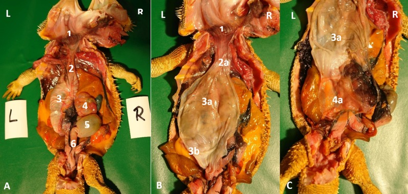
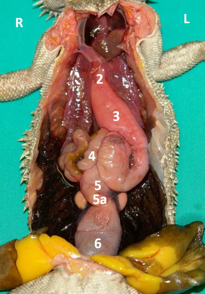
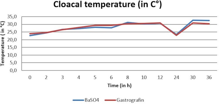
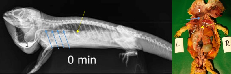
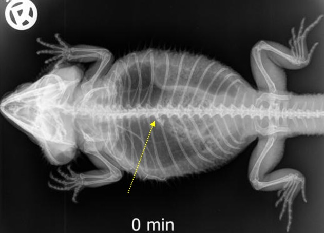
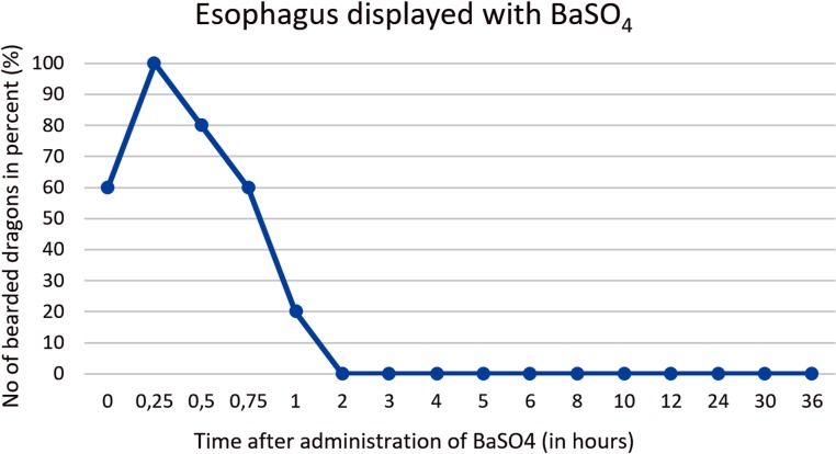
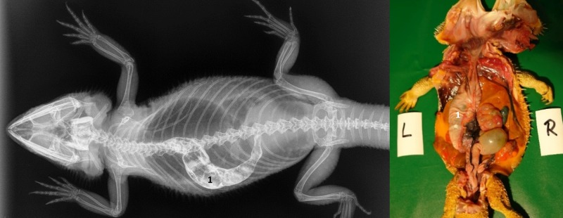
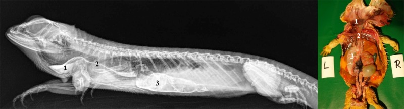
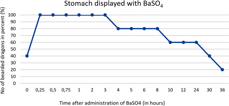
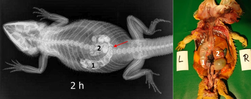
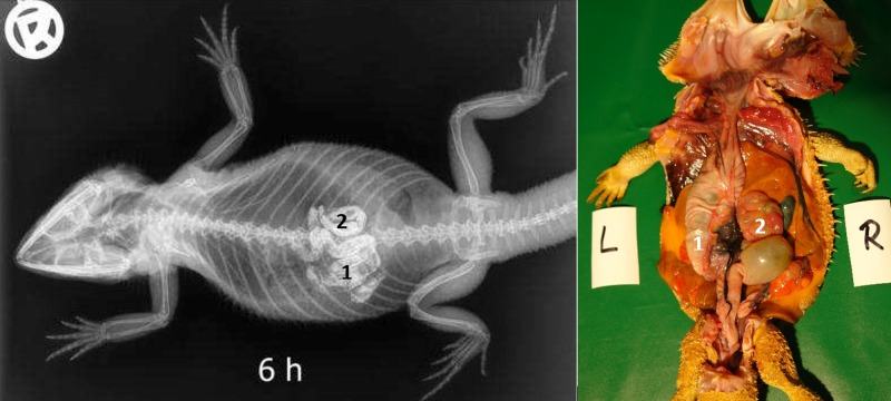
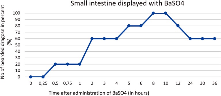
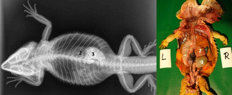
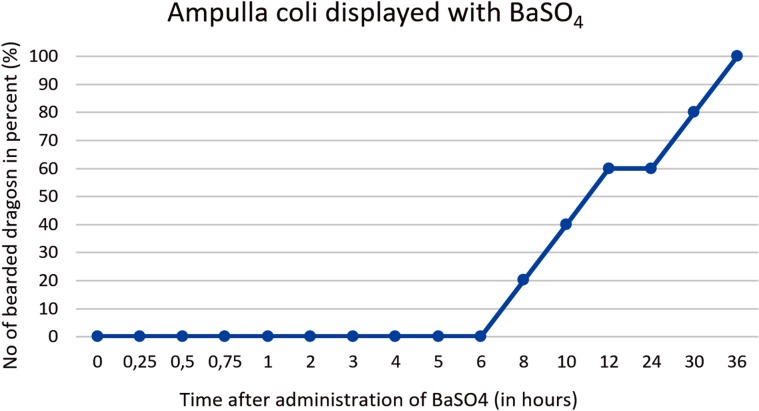
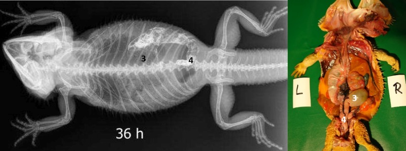
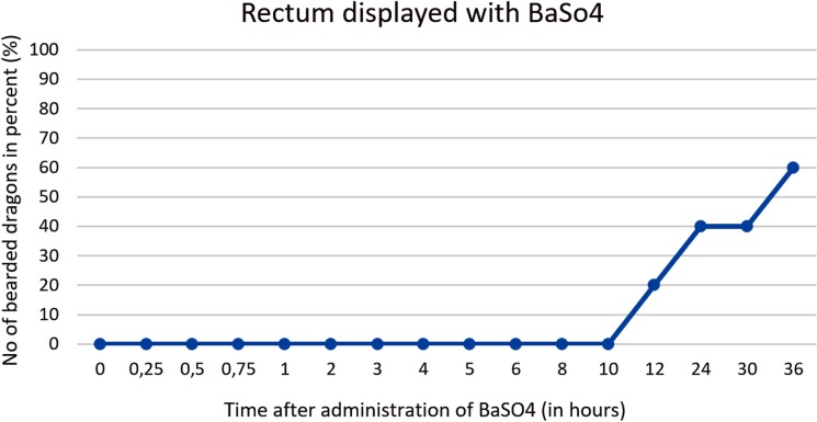
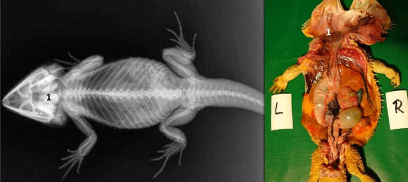
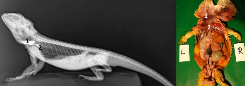
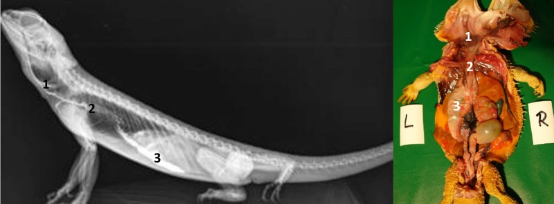
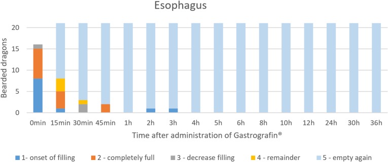
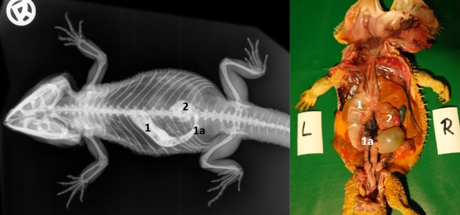
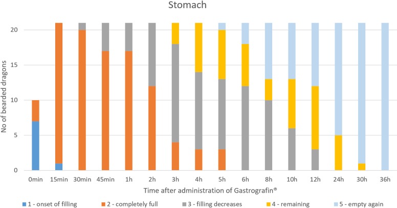
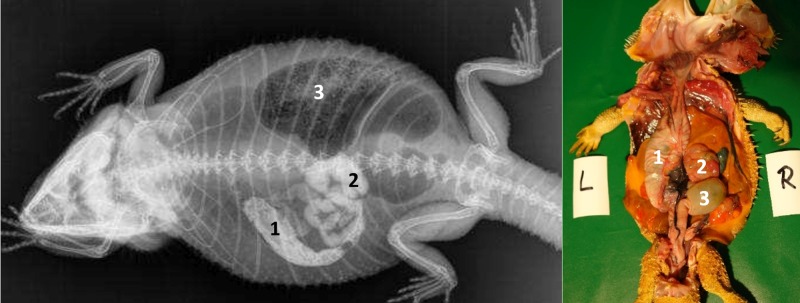
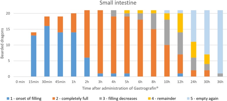
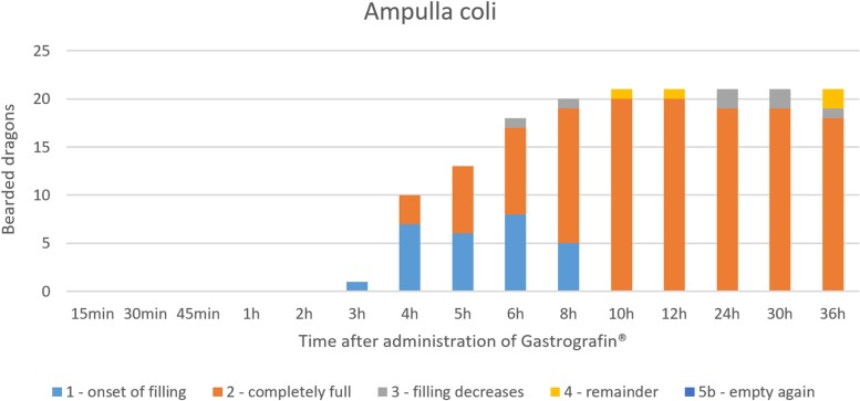
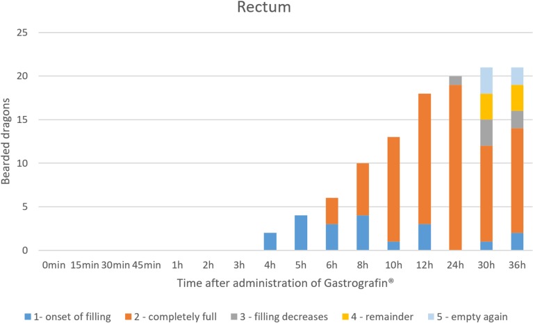
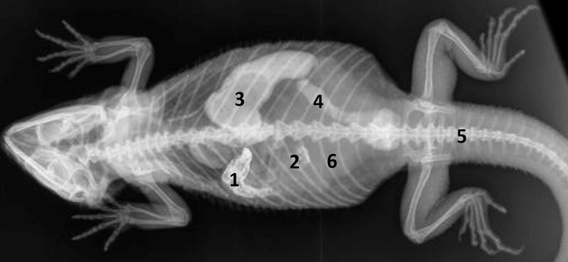
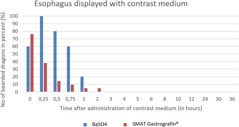
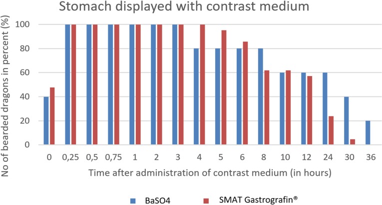
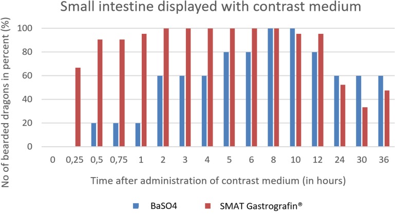
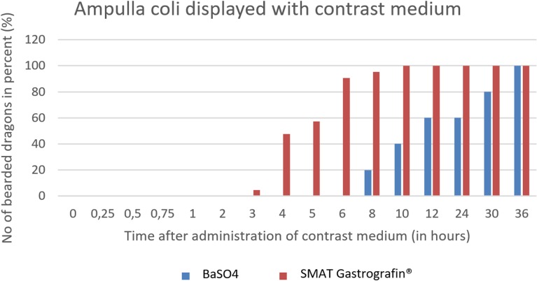
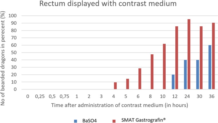
The aim of this study was to describe the specific gross and radiographic anatomy of the digestive tract of inland bearded dragons (Pogona vitticeps). Eleven bearded dragon cadavers of both sexes (6 females, 5 males) were dissected to examine, measure, and document the specific gross anatomy of the alimentary canal. Measurements collected from the cadavers included snout-vent length, total length of the alimentary canal, and the lengths of the individual sections of the gastrointestinal tract, including the esophagus, stomach, small intestine, ampulla coli, isthmus coli, rectum, and the distance from the coprodeum to the vent opening. Twenty-two healthy adult bearded dragons (13 females, 9 males) maintained under standardized husbandry conditions underwent a physical examination, blood collection, and whole-body dorsoventral and lateral survey radiographs; these animals were used to provide the radiographic images of the complete digestive tract. For the subsequent contrast passage studies, two different contrast media, barium sulfate (BaSO4, Barilux suspension) and an iodinated ionic radiocontrast agent (Sodium meglumine amidotrizoate [SMAT], Gastrografin), were used. Water-diluted Barilux suspension (dose 9 ml/kg) was administered orally to 5 bearded dragons, while Gastrografin (dose 5ml/kg) was administered orally to 21 bearded dragons. Four animals were used for both contrast media studies, but received a break of four weeks in between. Dorsoventral and laterolateral radiographs were collected at 0 (baseline), 15, 30, and 45 minutes and 1, 2, 3, 4, 5, 6, 8, 10, 12, 24, 30, and 36 hours after each contrast medium was administered. Both contrast media were found to illustrate the alimentary tracts in the adult bearded dragons. Transit time was substantially faster with SMAT, and SMAT illustrated the entire gastrointestinal tract within 36 hours; BaSO4 did not fully illustrate the gastrointestinal tract in 36 hours. These results might serve as a guideline for the interpretation of subsequent contrast studies in this lizard species.


Conflict of interest statement

The authors have declared that no competing interests exist.

Figures

Fig 1
Fig 1. Topography of the alimentary canal of a female bearded dragon, dorsal view.
Dorsal skin, muscles, skeleton, and serosa removed; lungs distracted cranially; and dorsal pelvis removed for an in situ view. L: left; R: right. A. Topography in situ: oral cavity and pharynx (1), esophagus (2), stomach (3), small intestine (4), ampulla coli (5), and rectum (6). B. Alimentary canal opened: oral cavity and pharynx (1), esophagus (2a), stomach (3a), and transverse gastric fold caused by angular incision (3b). C. Alimentary canal opened and distracted cranially to demonstrate the pyloric circular fold (3c*); stomach (3a), and duodenum (4a).
Fig 2
Fig 2. Topography of the alimentary canal of a male bearded dragon in situ, ventral view.
Ventral skin, muscles, skeleton, and serosa removed; pericardium opened ventrally and partially removed; and fat bodies distracted caudolaterally to expose the esophagus (2), stomach (3), small intestine (4), ampulla coli (5), isthmus coli (5), and rectum (6).
Fig 3
Fig 3. Arithmetic mean of body surface temperature (in°C) during the barium sulfate and Sodium/Meglumine–Amidotrizoate (SMAT, Gastrografin) contrast passage.
Fig 4
Fig 4. Arithmetic mean of cloacal temperature (in°C) during the barium sulfate and Sodium/Meglumine–Amidotrizoate (SMAT, Gastrografin) contrast passage.
Fig 5
Fig 5. Laterolateral radiograph in ventral recumbency using a horizontal beam.
Sternal recumbency on a standardized foam pad. Immediately after oral administration of BaSO4, (kV 42, mAs 2). Note the BaSO4 in the esophagus in the ventrally dependent gular area. A smaller volume of contrast media has already entered the distal esophagus (blue arrows) and is seen at the entrance (cardia) of the stomach (yellow arrow).
Fig 6
Fig 6. Dorsoventral (DV) radiograph immediately after oral administration of BaSO4.
Sternal recumbency on a standardized foam pad. Immediately after oral administration of BaSO4, (kV 42, mAs 2). The entrance of the stomach at the 6th and 7 th thoracic vertebrae is illustrated (yellow arrow).
Fig 7
Fig 7. BaSO4 contrast visibility in the esophagus over time.
Percentage of animals and frequency at measuring time point; y = number of animals in percent (%) in which contrast media could be found in the esophagus; x = time point during the barium sulfate (BaSo4) contrast passage (hours).
Fig 8
Fig 8. Dorsoventral (DV) radiograph 15 minutes after oral administration of BaSO4.
15 minutes after oral administration of BaSO4, (kV 42, mAs 2). The stomach (1) is visible as an elongated “C” and is primarily located on the left side of the coelom.
Fig 9
Fig 9. Laterolateral (LL) radiograph in ventral recumbency and using a horizontal beam.
15 minutes after oral administration of BaSO4, (kV 42, mAs 2). Note the BaSO4 in the esophagus in the ventrally dependent gular area (1), distal esophagus (2), and stomach (3).
Fig 10
Fig 10. BaSO4 contrast visibility in the stomach over time.
Percentage of animals and frequency at measuring time point; y = number of animals in percent (%) in which contrast media could be found in the stomach; x = time point during the BaSO4contrast passage (hours).
Fig 11
Fig 11. Dorsoventral (DV) radiograph 2 hours after oral administration of BaSO4.
2 hours after oral administration of BaSO4, (kV 42, mAs 2). The stomach (1) is illustrated by the presence of BaSO4; it is located on the left side of the coelomic cavity and is emptying from oral to aboral. The first loops of the small intestine (2) are displayed with BaSO4. Peristaltic movement can be observed in the small intestines (red arrow).
Fig 12
Fig 12. Dorsoventral (DV) radiograph 6 hours after oral administration of BaSO4.
6 hours after oral administration of BaSO4, (kV 42, mAs 2). Residual BaSO4 can be seen in the caudal (pyloric) stomach (1). Enhanced contrast of the loops of the small intestine (2) is observed at this time.
Fig 13
Fig 13. BaSO4 contrast visibility in the small intestines over time.
Percentage of animals and frequency at measuring time point; y = number of animals in percent (%) in which contrast media could be found in the small intestine; x = time point during the BaSO4 contrast passage (hours).
Fig 14
Fig 14. Dorsoventral (DV) radiograph 36 hours after oral administration of BaSO4.
36 hours after oral administration of BaSO4, (kV 42, mAs 2). Note residual amount of BaSO4 in the intestinal loops (2) at 36 hours. The ampulla coli (3) can be identified with BaSO4 on the right side of the coelomic cavity at the 10th thoracic vertebrae and 2 nd lumbar vertebrae.
Fig 15
Fig 15. BaSO4 contrast visibility in the ampulla coli over time.
Percentage of animals and frequency at measuring time point; y = number of animals in percent (%) in which contrast media could be found in the ampulla coli; x = time point during BaSO4 contrast passage (hours).
Fig 16
Fig 16. Dorsoventral (DV) radiograph 36 hours after oral administration of BaSO4.
36 hours after oral administration of BaSO4, (kV 42, mAs 2). The BaSO4 has passed through the ampulla coli (3) and into the rectum (4), which has an “U-shape” and is located on the right side of the coelomic cavity running parallel to the spinal column.
Fig 17
Fig 17. BaSO4 contrast visibility in the rectum over time.
Percentage of animals and frequency at measuring time point; y = number of animals in percent (%) in which contrast media could be found in the rectum; x = time point during BaSO4contrast passage (hours).
Fig 18
Fig 18. Dorsoventral (DV) radiograph immediately after oral administration of SMAT.
Immediately after oral administration of SMAT (kV 42, mAs 2). Contrast medium can be seen in the oropharynx (1).
Fig 19
Fig 19. Laterolateral (LL) radiograph in sternal recumbency using a horizontal beam immediately after oral administration of SMAT.
Immediately after oral administration of SMAT (kV 42, mAs 2). Contrast medium is visible in the oropharynx (1), with a small volume also seen entering the esophagus (2).
Fig 20
Fig 20. Laterolateral (LL) radiograph in sternal position using a horizontal beam immediately after oral administration of SMAT.
Immediately after oral administration of SMAT (kV 42, mAs 2). Contrast medium illustrates oropharynx (1), esophagus (2), and stomach (3).
Fig 21
Fig 21. SMAT contrast visibility in the esophagus over time.
Frequency at measuring time point; y = number of animals in which contrast media could be found in the esophagus; x = time point during the Sodium/Meglumine–Amidotrizoate (SMAT); (Gastrografin) contrast passage (in minutes and hours).
Fig 22
Fig 22. Dorsoventral (DV) radiograph 15 minutes after oral administration of SMAT.
15 minutes after oral administration of SMAT (kV 42, mAs 2). The stomach (1, 1a) and cranial section of the small intestine (2) are highlighted with contrast medium. The stomach is visible on the left side of the coelom and the cardia is located between the 6th and 7th thoracic vertebrae. The pylorus is visible at the first lumbar vertebrae (1a) and some contrast medium can be seen entering the small intestine (2).
Fig 23
Fig 23. SMAT contrast visibility in the stomach over time.
Frequency at measuring time point; y = number of animals in which contrast media could be found in the stomach; x = time point during the SMAT; (Gastrografin) contrast passage (in minutes and hours).
Fig 24
Fig 24. Dorsoventral (DV) radiograph 3 hours after oral administration of SMAT.
3 hours after oral administration of SMAT (kV 42, mAs 2). The stomach (1) is visible on the left side of the coelom and transition to the small intestine (2) occurs laterally between the 9th and 10th ribs. The ampulla coli (3) contains gas and sand particles.
Fig 25
Fig 25. SMAT contrast visibility in the small intestine over time.
Frequency at measuring time point; y = number of animals in which contrast media could be found in the small intestine; x = time point during the Sodium/Meglumine–Amidotrizoate (SMAT); contrast passage (in minutes and hours).
Fig 26
Fig 26. Dorsoventral (DV) radiograph 12 hours after oral administration of SMAT.
12 hours after oral administration of SMAT (kV 42, mAs 2). The caudal aspect of the stomach (pylorus) (1) is filled with residual contrast; the small intestine (2) can be seen merging into the ampulla coli (3) at the level of the 9 th thoracic vertebrae; and the rectum (4) can be seen filling with contrast.
Fig 27
Fig 27. SMAT contrast visibility in the ampulla coli over time.
Frequency at measuring time point; y = number of animals in which contrast media could be found in the ampulla coli; x = time point during the Sodium/Meglumine–Amidotrizoate (SMAT); contrast passage (in minutes and hours).
Fig 28
Fig 28. Dorsoventral (DV) radiograph 12 hours after oral administration of SMAT.
12 hours after oral administration of SMAT (kV 42, mAs 2). The stomach (1) and small intestine (2) are shifted cranially because of the prominent fat bodies. The ampulla coli (3) can be seen merging into the rectum (4), which has a width of three vertebral bodies and is far wider than the loops of the small intestine (2).
Fig 29
Fig 29. SMAT contrast visibility in the rectum over time.
Frequency at measuring time point; y = number of animals in which contrast media could be found in the rectum; x = time point during the Sodium/Meglumine–Amidotrizoate (SMAT); contrast passage (in minutes and hours).
Fig 30
Fig 30. Dorsoventral (DV) radiograph 12 hours after oral administration of SMAT.
12 hours after oral administration of SMAT (kV 42, mAs 2). Prominent ovarian follicles (6) can be seen displacing the stomach (1) and small intestines cranially (2), both of which are filled with residual contrast. The ampulla coli (3) is filled with gas and contrast medium, while the rectum (4) can be seen on the right side of the coelom before emptying into the cloaca (5).
Fig 31
Fig 31. Radiographic visualization (OR imaging) with barium sulfate (BaSO4) (in blue) in comparison with SMAT (Gastrografin) (in red).
Contrast passage time of the esophagus; percentage of animals and frequency at measuring time point; y = number of animals in percent (%) in which contrast media could be found in the esophagus; x = time point during the contrast passage (in hours).
Fig 32
Fig 32. Radiographic visualization (OR imaging) with barium sulfate (BaSO4) (in blue) in comparison with SMAT (Gastrografin) (in red) contrast passage time of the stomach.
Percentage of animals and frequency at measuring time point; y = number of animals in percent (%) in which contrast media could be found in the stomach; x = time point during the contrast passage (in hours).
Fig 33
Fig 33. Radiographic visualization (OR imaging) with barium sulfate (BaSO4) (in blue) in comparison with SMAT (Gastrografin) (in red) contrast passage time of the small intestine.
Percentage of animals and frequency at measuring time point; y = number of animals in percent (%) in which contrast media could be found in the small intestine; x = time point during the contrast passage (in hours).
Fig 34
Fig 34. Radiographic visualization (OR imaging) with barium sulfate (BaSO4) (in blue) in comparison with SMAT (Gastrografin) (in red) contrast passage time of the ampulla coli.
Percentage of animals and frequency at measuring time point; y = number of animals in percent (%) in which contrast media could be found in the ampulla coli; x = time point during the contrast passage (in hours).
Fig 35
Fig 35. Radiographic visualization (OR imaging) with barium sulfate (BaSO4) (in blue) in comparison with SMAT (Gastrografin) (in red) contrast passage time of the rectum.
Percentage of animals and frequency at measuring time point; y = number of animals in percent (%) in which contrast media could be found in the rectum; x = time point during the contrast passage (in hours).
Fig 36
Fig 36. Same bearded dragon (Bearded dragon number 10), 12 hours after peroral application of contrast media.
A. Dorsoventral radiograph of bearded dragon number 10, 12 hours post-SMAT dosing. Note the contrast in the stomach (1), small intestine (2), ampulla coli (3), and rectum (4). B. Dorsoventral radiograph of bearded dragon number 10, 12 hours post- BaSO4 dosing. Note that only the ampulla coli (3) and rectum (4) are clearly identified.

References

    1. Silverman S, Guzman DSM, Stern J, Gustavsen KA, Griffiths LG. Standardization of the two-dimensional transcoelomic echocardiographic examination in the central bearded dragon (Pogona vitticeps). J Vet Cardiol. 2016;18(2):168–78. 10.1016/j.jvc.2015.10.011 WOS:000377325200008. - DOI - PubMed
    1. Bucy DS, Guzman DS, Zwingenberger AL. Ultrasonographic anatomy of bearded dragons (Pogona vitticeps). Journal of the American Veterinary Medical Association. 2015;246(8):868–76. 10.2460/javma.246.8.868 . - DOI - PubMed
    1. Ritter JM, Garner MM, Chilton JA, Jacobson ER, Kiupel M. Gastric neuroendocrine carcinomas in bearded dragons (Pogona vitticeps). Veterinary pathology. 2009;46(6):1109–16. 10.1354/vp.09-VP-0019-K-FL . - DOI - PubMed
    1. Wright K. Two Common Disorders of Captive Bearded Dragons (Pogona vitticeps): Nutritional Secondary Hyperparathyroidism and Constipation. J Exot Pet Med. 2008;17(4):267–72.
    1. Mitchell MA. Managing the Reptile Patient in the Veterinary Hospital: Establishing a Standards of Care Model for Nontraditional Species. J Exot Pet Med. 2010;19(1 (January)):56–72.

MeSH terms