. 2019 Aug 22;14(8):e0221466.
doi: 10.1371/journal.pone.0221466. eCollection 2019.

Repeatability of quantitative measurements of retinal layers with SD-OCT and agreement between vertical and horizontal scan protocols in healthy eyes

Affiliations

Repeatability of quantitative measurements of retinal layers with SD-OCT and agreement between vertical and horizontal scan protocols in healthy eyes

Alberto Domínguez-Vicent et al. PLoS One. .

Abstract

Purpose: To evaluate the repeatability of the new spectral domain optical coherence tomography (HOCT-1F), and also to evaluate the agreement between vertical and horizontal scan protocols. In addition, we also evaluated the relation between the repeatability and age.

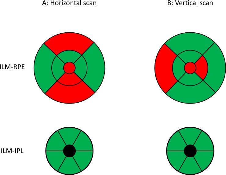
Methods: Three consecutive measurements of the inner limiting membrane-retinal pigment epithelium (ILM-RPE), inner limiting membrane-inner plexiform layer (ILM-IPL) from macular horizontal and vertical scans, and inner limiting membrane-retinal nerve fiber layer (ILM-RNFL) from optic disc horizontal scan. 159 subjects were included in the analysis. The within subject standard deviation (Sw) and the repeatability limits (Rlimit) are used to represent the repeatability of the parameters for the different sectors.

Results: The Sw for the ILM-RPE thickness was less than 3.5 μm for each sector and scan direction. The Sw values varied within the sectors and scan modes, with horizontal scan modes resulting in better values for the horizontal sectors, and vice versa. The Sw for the GCL-IPL thickness was less than 2 μm, and was similar between the vertical and horizontal scan modes for each sector map. For the optic disc scan, the Sw was not symmetric along the clock-hour map sectors, the largest Sw values were seen in the vertical sectors (8.6 μm). The mean difference between the vertical and horizontal scans was less than 2 μm for each retinal thickness sector map. Significant but weak correlation between the Sw and the subject's age was seen in both macular and optic disc scans.

Conclusions: The repeatability of the HOCT-1F to measure the ILM-RPE-, ILM-IPL- and ILM-RNFL-thickness is good. The repeatability of the ILM-RPE thickness is dependent on the scan direction, which should be taken into account when calculating retinal thickness. There is a weak correlation between the repeatability and the subject's age.


Conflict of interest statement

The authors have declared that no competing interests exist.

Figures

Fig 1
Fig 1. Schematic representation of the sectors for the retinal layer thickness.
In A ILM-RPE represents inner limiting membrane to retinal pigment epithelium thickness and numbers 1 to 9 indicate Center, inner superior, inner nasal, inner inferior, inner temporal, outer superior, outer nasal, outer inferior and outer temporal sectors. In B, ILM-IPL represents inner limiting membrane to inner plexiform layer thickness and numbers 1to 6 indicate superior, superior nasal, inferior nasal, inferior, inferior temporal and superior temporal sectors. In C, ILM-RNFL represents inner limiting membrane to retinal nerve fiber layer thickness and the numbers represent the clock hour position of the sectors. A and B are from the macular scans and C is from the optic disc scan.
Fig 2
Fig 2. Intra-device repeatability for the inner limiting membrane to retinal pigment epithelium thickness in different sectors for horizontal and vertical scans.
OT: outer temporal, IT: inner temporal, IN: inner nasal, ON: outer nasal, C: central circle, OS: outer superior, IS: inner superior, II: inner inferior, OI: outer inferior sectors.
Fig 3
Fig 3. Intra-device repeatability for the inner limiting membrane to inner plexiform layer thickness in different sectors for horizontal and vertical scans.
S: superior, SN: superior nasal, IN: inferior nasal, I: inferior, IT: inferior temporal and ST: superior temporal sectors.
Fig 4
Fig 4. Intra-device repeatability for the inner limiting membrane to retinal nerve fiber layer thickness in different clock hour sectors for horizontal scans.
The small black dots represent the within subject standard deviation and the grey circles surrounding each black point represent the repeatability limits, where the areas are scaled by a factor of 30 points for visualization purposes.
Fig 5
Fig 5. Colour coded map of the intra-device repeatability for the macula scans in different sectors.
Green: repeatability limit values within the instrument’s axial resolution, Red: repeatability limit values above the instrument’s axial resolution. ILM-RPE: inner limiting membrane to retinal pigment epithelium thickness ILM-IPL: inner limiting membrane to inner plexiform layer thickness.

References

    1. Mwanza J-C, Budenz DL. New developments in optical coherence tomography imaging for glaucoma. Curr Opin Ophthalmol. United States; 2018;29: 121–129. 10.1097/ICU.0000000000000452 - DOI - PubMed
    1. Mwanza J-C, Warren JL, Budenz DL. Utility of combining spectral domain optical coherence tomography structural parameters for the diagnosis of early Glaucoma: a mini-review. Eye Vis (London, England). England; 2018;5: 9 10.1186/s40662-018-0101-6 - DOI - PMC - PubMed
    1. Wintergerst MWM, Schultz T, Birtel J, Schuster AK, Pfeiffer N, Schmitz-Valckenberg S, et al. Algorithms for the Automated Analysis of Age-Related Macular Degeneration Biomarkers on Optical Coherence Tomography: A Systematic Review. Transl Vis Sci Technol. United States; 2017;6: 10 10.1167/tvst.6.4.10 - DOI - PMC - PubMed
    1. Denniss J, Turpin A, McKendrick AM. Relating optical coherence tomography to visual fields in glaucoma: structure-function mapping, limitations and future applications. Clin Exp Optom. Australia; 2019;102: 291–299. 10.1111/cxo.12844 - DOI - PubMed
    1. Wu Z, Weng DSD, Rajshekhar R, Thenappan A, Ritch R, Hood DC. Evaluation of a Qualitative Approach for Detecting Glaucomatous Progression Using Wide-Field Optical Coherence Tomography Scans. Transl Vis Sci Technol. United States; 2018;7: 5 10.1167/tvst.7.3.5 - DOI - PMC - PubMed

MeSH terms