Review
. 2020 Jan 11;22(1):17-30.
doi: 10.1093/neuonc/noz147.

Imaging challenges of immunotherapy and targeted therapy in patients with brain metastases: response, progression, and pseudoprogression

Affiliations
Review

Imaging challenges of immunotherapy and targeted therapy in patients with brain metastases: response, progression, and pseudoprogression

Norbert Galldiks et al. Neuro Oncol. .

Abstract

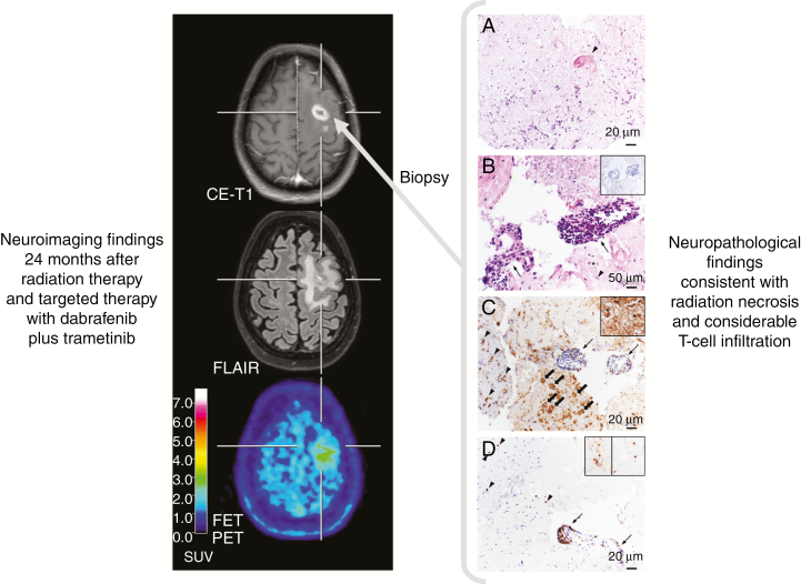
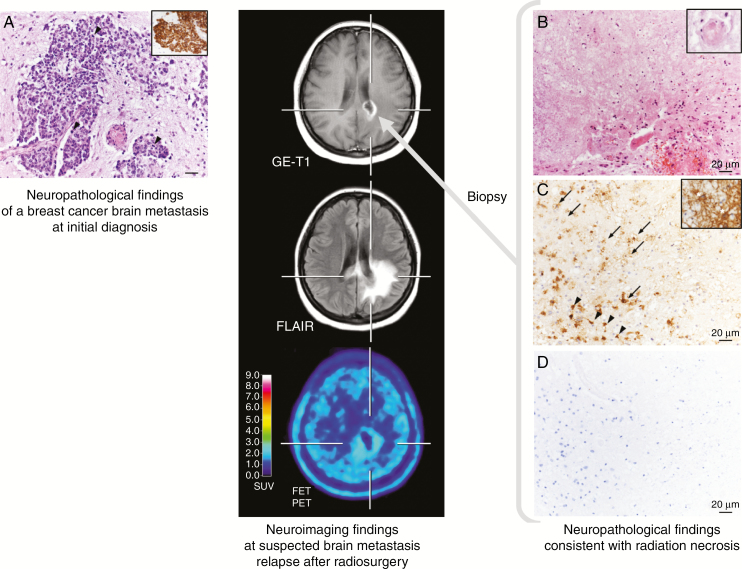
The advent of immunotherapy using immune checkpoint inhibitors (ICIs) and targeted therapy (TT) has dramatically improved the prognosis of various cancer types. However, following ICI therapy or TT-either alone (especially ICI) or in combination with radiotherapy-imaging findings on anatomical contrast-enhanced MRI can be unpredictable and highly variable, and are often difficult to interpret regarding treatment response and outcome. This review aims at summarizing the imaging challenges related to TT and ICI monotherapy as well as combined with radiotherapy in patients with brain metastases, and to give an overview on advanced imaging techniques which potentially overcome some of these imaging challenges. Currently, major evidence suggests that imaging parameters especially derived from amino acid PET, perfusion-/diffusion-weighted MRI, or MR spectroscopy may provide valuable additional information for the differentiation of treatment-induced changes from brain metastases recurrence and the evaluation of treatment response.

Keywords: FET PET; brain metastasis; immune checkpoint inhibitors; lung cancer; melanoma; radiomics.


Figures

Fig. 1
Fig. 1
Radiation necrosis and chronic inflammation in a patient with brain metastases of a BRAF-mutated malignant melanoma who had been treated with whole-brain radiation therapy and concurrently with dabrafenib plus trametinib. Twenty-four months later, the contrast-enhanced MRI suggests brain metastasis recurrence (left panel), whereas the FET PET shows only an insignificant uptake, consistent with treatment-related effects. Neuropathological findings obtained following stereotactic biopsy revealed besides signs of radiation necrosis a considerable infiltration of intra- and perivascular T cells (right panel). (A) Hyaline, eosinophilic necrosis with only single leukocytes and cell detritus. A necrotic vessel wall is hyalinized and thickened (arrowhead). Hematoxylin and eosin (H&E) staining; original magnification x200. (B) Adjacent to necrosis, small fragments of vital brain parenchyma harbor activated microglial cells (arrowhead) and reactive astrocytes (asterisk). Two blood vessels are heavily infiltrated by lymphocytes (arrows). Tumor cells are absent (insert). H&E staining; original magnification x500; insert: immunohistochemistry with monoclonal mouse anti-HMB45 (DCS Diagnostics) and slight counterstaining with hemalum; original magnification, x200. (C) Adjacent to the inflamed blood vessels (arrows), foamy CD68+ macrophages are in the process of resorption of necrosis (block arrows). In the brain parenchyma, microglial cells (arrowheads) and astrocytes (insert, asterisks) are activated. Immunohistochemistry with monoclonal mouse anti-CD68 (DCS Diagnostics) and slight counterstaining with hemalum; original magnification, x200; insert: immunohistochemistry with monoclonal mouse anti–glial fibrillary acidic protein (BioGenex) and slight counterstaining with hemalum; original magnification, x500. (D) CD3+ T cells are the major population of intra- and perivascular infiltrates (arrow). Both CD4+ (left insert) and CD8+ (right insert) T cells contribute to the infiltrates. Immunohistochemistry with monoclonal rabbit anti-CD3 (DCS Diagnostics) and slight counterstaining with hemalum; original magnification, x200; inserts: immunohistochemistry with monoclonal mouse anti-CD4 (left, BioGenex) and with monoclonal rabbit anti-CD8 (right, DCS Diagnostics), slight counterstaining with hemalum; original magnification, x400.
Fig. 2
Fig. 2
Radiation necrosis in a patient with brain metastases secondary to a breast cancer (ductal carcinoma, HER2 negative, estrogen and progesterone receptor positive) (left panel). Five months after external fractionated radiation therapy, contrast-enhanced MRI suggests BM relapse (middle panel). In contrast, FET PET shows no increased metabolic activity, indicating treatment-related changes. Neuropathological findings obtained following stereotactic biopsy were consistent with radiation necrosis (right panel). (A) Epithelial, pleomorphic tumor with increased mitotic activity (arrowheads) in the brain parenchyma expressing cytokeratin (CK) 8 (insert) at initial diagnosis. Hematoxylin and eosin (H&E) staining; original magnification x200. Insert: immunohistochemistry with monoclonal mouse anti-CK8 (BioGenex) and slight counterstaining with hemalum; original magnification, x100. (B) Hyaline, eosinophilic necrosis with only single leukocytes. A necrotic vessel wall is hyalinized and thickened (insert). Adjacent vital brain parenchyma shows reactive alterations with activated microglial cells and reactive astrocytes. H&E staining; original magnification x200; insert: H&E staining; original magnification, x500. (C) Necrosis is infiltrated by foamy macrophages (arrows). In the brain parenchyma, microglial cells (arrowheads) and astrocytes (insert, asterisks) are activated. Immunohistochemistry with monoclonal mouse anti–major histocompatibility complex class I antigen (DCS Diagnostics) and slight counterstaining with hemalum; original magnification x200; insert: immunohistochemistry with monoclonal mouse anti–glial fibrillary acidic protein (BioGenex) and slight counterstaining with hemalum; original magnification, x500. (D) Epithelial tumor cells were absent from necrosis and vital brain parenchyma. Immunohistochemistry with monoclonal mouse anti-CK8 (BioGenex) and slight counterstaining with hemalum; original magnification, x200.

References

    1. Hodi FS, O’Day SJ, McDermott DF, et al. . Improved survival with ipilimumab in patients with metastatic melanoma. N Engl J Med. 2010;363(8):711–723. - PMC - PubMed
    1. Kluger HM, Chiang V, Mahajan A, et al. . Long-term survival of patients with melanoma with active brain metastases treated with pembrolizumab on a phase II trial. J Clin Oncol. 2019;37(1):52–60. - PMC - PubMed
    1. Tawbi HA, Forsyth PA, Algazi A, et al. . Combined nivolumab and ipilimumab in melanoma metastatic to the brain. N Engl J Med. 2018;379(8):722–730. - PMC - PubMed
    1. Long GV, Atkinson V, Lo S, et al. . Combination nivolumab and ipilimumab or nivolumab alone in melanoma brain metastases: a multicentre randomised phase 2 study. Lancet Oncol. 2018;19(5):672–681. - PubMed
    1. Stokes WA, Binder DC, Jones BL, et al. . Impact of immunotherapy among patients with melanoma brain metastases managed with radiotherapy. J Neuroimmunol. 2017;313:118–122. - PubMed

Publication types

MeSH terms

Substances