. 2019 Nov;189(11):2258-2268.
doi: 10.1016/j.ajpath.2019.07.011. Epub 2019 Aug 19.

Proresolving Mediators LXB4 and RvE1 Regulate Inflammation in Stromal Cells from Patients with Shoulder Tendon Tears

Affiliations

Proresolving Mediators LXB4 and RvE1 Regulate Inflammation in Stromal Cells from Patients with Shoulder Tendon Tears

Stephanie G Dakin et al. Am J Pathol. 2019 Nov.

Abstract

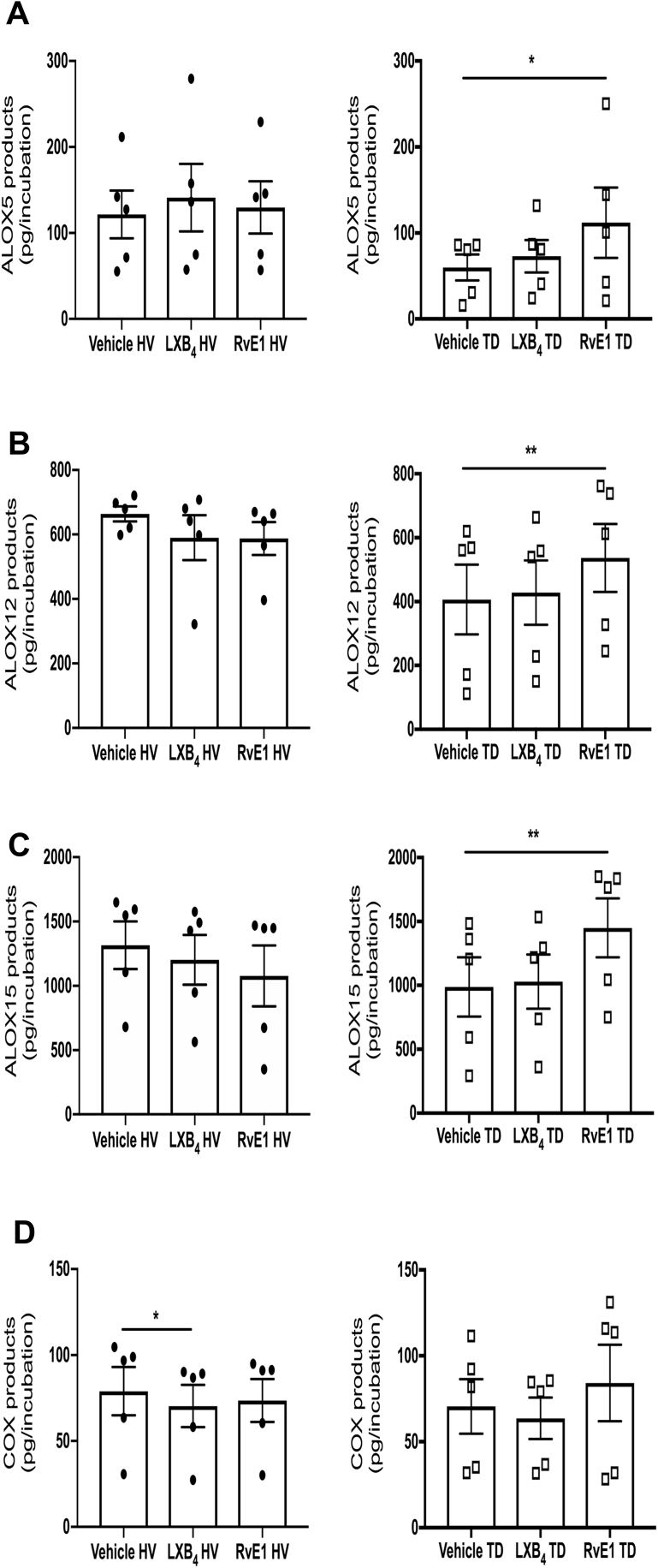
Tendon stromal cells isolated from patients with chronic shoulder rotator cuff tendon tears have dysregulated resolution responses. Current therapies do not address the biological processes concerned with persistent tendon inflammation; therefore, new therapeutic approaches that target tendon stromal cells are required. We examined whether two specialized proresolving mediators (SPMs), lipoxin B4 (LXB4) and resolvin E1 (RvE1), modulate the bioactive lipid mediator profiles of IL-1β-stimulated tendon cells derived from patients with shoulder tendon tears and healthy volunteers. We also examined whether LXB4 or RvE1 treatments moderated the proinflammatory phenotype of tendon tear stromal cells. Incubation of IL-1β-treated patient-derived tendon cells in LXB4 or RvE1 up-regulated concentrations of SPMs. RvE1 treatment of diseased tendon stromal cells increased 15-epi-LXB4 and regulated postaglandin F2α. LXB4 or RvE1 also induced expression of the SPM biosynthetic enzymes 12-lipoxygenase and 15-lipoxygenase. RvE1 treatment up-regulated the proresolving receptor human resolvin E1 compared with vehicle-treated cells. Incubation in LXB4 or RvE1 moderated the proinflammatory phenotype of patient-derived tendon tear cells, regulating markers of tendon inflammation, including podoplanin, CD90, phosphorylated signal transducer and activator of transcription 1, and IL-6. LXB4 and RvE1 counterregulate inflammatory processes in tendon stromal cells, supporting the role of these molecules as potential therapeutics to resolve tendon inflammation.


Figures

Figure 1
Figure 1
Lipoxin B4 (LXB4) up-regulates specialized proresolving mediator (SPM) concentrations in IL-1β–stimulated tendon stromal cells. Tendon stromal cells were derived from healthy volunteers (H) and patients with shoulder tendon tears [diseased (D)]. Cells were incubated with 10 nmol/L LXB4 or vehicle for 24 hours at 37°C then with 10 ng mL−1 of IL-1β for 24 hours. Lipid mediators (LMs) were identified and quantified using LM profiling. Two-dimensional (2D) score plot (A) and corresponding SD loading plot (B) of LM-SPMs from human tendon–derived stromal cell incubations isolated from the H group incubated with IL-1β and 10 nmol/L LXB4 or vehicle only. 2D score plot (C) and corresponding 2D loading plot (D) of LM-SPMs from human tendon–derived stromal cell incubations isolated from the D group incubated with IL-1β and 10 nmol/L LXB4 or vehicle only. Cumulative concentrations of proresolving mediators [docosahexaenoic acid–derived resolvin D (RvD), protectins (PD), maresin (MaR), n-3 docosapentaenoic acid (DPA) –derived RvDn-3 DPA, PDn-3 DPA, MaRn-3 DPA, EPA-derived RvE, and arachadonic acid–derived LX] in IL-1β–stimulated tendon stromal cell incubations in the presence of 10 nmol/L LXB4 or vehicle for 24 hours. LXB4 up-regulated concentrations of SPMs compared with vehicle only in H (E) and D (F) tendon stromal cells. Results are representative of n = 5 donors per group. **P < 0.01, ***P < 0.001. TD, tendon disease.
Figure 2
Figure 2
Resolvin E1 (RvE1) increases specialized proresolving mediator (SPM) levels in IL-1β–stimulated tendon stromal cells. Tendon stromal cells were derived from healthy volunteers (H) and patients with shoulder tendon tears [diseased (D)]. Cells were incubated with 10 nmol/L RvE1 or vehicle for 24 hours at 37°C and then with 10 ng mL−1 of IL-1β for 24 hours. Lipid mediators (LMs) were identified and quantified using LM profiling. Two-dimensional (2D) score plot (A) and corresponding 2D loading plot (B) of LM-SPM from human tendon–derived stromal cell incubations isolated from healthy volunteers (H) incubated with IL-1β and 10 nmol/L RvE1 or vehicle only. 2D score plot (C) and corresponding 2D loading plot (D) of LM-SPM from human tendon–derived stromal cell incubations isolated from the D group incubated with IL-1β and 10 nmol/L RvE1 or vehicle only. Cumulative concentrations of proresolving mediators [docosahexaenoic acid–derived resolvin D (RvD), protectins (PD), maresin (MaR), n-3 docosapentaenoic acid (DPA) –derived RvDn-3 DPA, PDn-3 DPA, MaRn-3 DPA, EPA-derived RvE, and arachadonic acid–derived LX] in IL-1β–stimulated tendon stromal cell incubations in the presence of 10 nmol/L RvE1 or vehicle for 24 hours. RvE1 up-regulated concentrations of SPMs compared with vehicle only in the H (E) and D (F) tendon stromal cells. Differentially regulated lipid mediators in IL-1β–stimulated diseased tendon stromal cell incubations in the presence of 10 nmol/L RvE1 or vehicle for 24 hours of RvE1 treatment up-regulated 15-epi-LXB4 (G) and reduced prostaglandin F2α (PGF2α) levels (H) compared with vehicle controls. Data are expressed as means ± SEM. n = 5 donors per group. *P < 0.05, **P < 0.01, and ***P < 0.001. TD, tendon disease.
Figure 3
Figure 3
Lipoxin B4 (LXB4) and resolvin E1 (RvE1) induce specialized proresolving mediator (SPM) biosynthetic enzymes and regulate the proresolving receptor for chemerin and resolvin E1 (ChemR23)/human resolvin E1 (ERV1) in tendon stromal cells. Tendon stromal cells were derived from patients with shoulder tendon tears (TD; n = 6) or healthy volunteers (HV; n = 6). Cells were incubated with 10 nmol/L LXB4, 10 nmol/L RvE1, or vehicle for 24 hours at 37°C then with 10 ng mL−1 of IL-1β for 24 hours. A: Incubation in LXB4 significantly induces ALOX15 mRNA in both TD and HV cells compared with respective vehicle controls. Incubation in RvE1 significantly induces ALOX15 mRNA in both TD and HV cells compared with respective vehicle controls. Gene expression is normalized to β-actin. Representative images of immunocytochemistry for the SPM biosynthetic enzymes 15-lipoxygenase (ALOX15) (green) and 12-lipoxygenase (ALOX12) (violet) in IL-1β–stimulated HV (B) and TD tendon stromal cells (C) incubated in 10 nmol/L LXB4, 10 nmol/L RvE1, or vehicle control for 24 hours. Cyan represents POPO-1 nuclear counterstain. D: IL-1β–stimulated TD cells show increased ERV1 mRNA expression compared with respective HV cells. Gene expression is normalized to β-actin. E: Representative images of immunocytochemistry for proresolving receptors lipoxin A4 receptor (ALX) (green), human resolvin E1 (ERV1) (red), and leukotriene B4 receptor (BLT1) (violet) in IL-1β–stimulated diseased tendon stromal cells incubated in 10 nmol/L LXB4, 10 nmol/L RvE1, or vehicle control for 24 hours. Cyan represents POPO-1 nuclear counterstain. Bars indicate median values. n = 3 donors. Scale bars = 20 μm (B, C, and E). *P < 0.05, **P < 0.01.
Figure 4
Figure 4
Lipoxin B4 (LXB4) and resolvin E1 (RvE1) moderate the proinflammatory phenotype of tendon stromal cells. Tendon stromal cells were derived from patients with shoulder tendon tears (TD) or healthy volunteers (HV). A: Representative images of immunocytochemistry for established markers of tendon inflammation, including podoplanin (PDPN) (green), phosphorylated signal transducer and activator of transcription (pSTAT)-1 (green), and IL-6 (red) in IL-1β–stimulated diseased tendon stromal cells incubated in 10 nmol/L LXB4, 10 nmol/L RvE1, or vehicle control for 24 hours. Cyan represents POPO-1 nuclear counterstain. B: Enzyme-linked immunosorbent assay of IL-6 protein secretion from IL-1β–stimulated diseased tendon cells incubated in the presence and absence of 10 nmol/L LXB4 or 10 nmol/L RvE1. LXB4 or RvE1 reduces IL-6 levels compared with vehicle only. C: Representative images of immunocytochemistry for PDPN, pSTAT-1, and IL-6 in IL-1β–stimulated healthy stromal cells incubated in 10 nmol/L LXB4, 10 nmol/L RvE1, or vehicle control for 24 hours. Cyan represents POPO-1 nuclear counterstain. D: LXB4 or RvE1 reduces IL-6 levels in HV incubations compared with vehicle only. E: mRNA expression of markers of tendon inflammation, including IL-6, and the fibroblast activation markers PDPN and CD90 in IL-1β–stimulated TD cells incubated in 10 nmol/L LXB4, 10 nmol/L RvE1, or vehicle control for 24 hours. Incubation in LXB4 or RvE1 reduces IL6, PDPN, and CD90 mRNA expression compared with vehicle controls. Gene expression is normalized to β-actin. Data are expressed as means ± SEM. Bars indicate median values. n = 3 donors (A and C); n = 5 donors (B and D); n = 6 donors (E). Scale bars = 20 μm (A and C). *P < 0.05, **P < 0.01.
Figure 5
Figure 5
Effects of Lipoxin B4 (LXB4) and resolvin E1 (RvE1) on protein phosphokinase signaling in diseased tendon stromal cells. Densitometric analysis was acquired using ImageJ software version 1.47 to identify the effects of incubating IL-1β–treated tendon disease (TD) cells in 10 nmol/L LXB4 or RvE1 on protein phosphokinase signaling pathways JNK1/2/3, Lyn, signal transducer and activator of transcription (STAT)-3, STAT6, and p70s6 kinase. Data are expressed as means ± SEM. n = 3 donors per group relative to respective vehicle control treated cells.
Supplemental Figure S1
Isotype control staining of patient-derived tendon stromal cells. Representative confocal immunofluorescence images showing merged images of stromal cells isolated from patients with shoulder tendon tears, stained with isotype control antibodies for mouse IgG1, IgG2a, IgG2b, and rabbit IgG fractions. Cyan represents POPO-1 nuclear counterstain. Scale bar = 20 μm.
Supplemental Figure S2
Biosynthesis of specialized proresolving mediators in tendon stromal cells. A: mRNA expression of enzymes implicated in the biosynthesis of specialized proresolving mediator (SPMs; ALOX15, ALOX12), inflammation-initiating eicosanoids (PTGS2), and the degradation of SPMs and inflammation-initiating eicosanoids (HPGD) in tendon stromal cells from patients with shoulder tendon tears and healthy volunteers (HVs). Incubation in lipoxin B4 (LXB4) significantly induces ALOX15 mRNA in both tendon disease (TD) and HV cells compared with respective vehicle controls. Incubation in resolvin E1 (RvE1) significantly induces ALOX15 mRNA in both TD and HV cells compared with respective vehicle controls. Gene expression is normalized to β-actin. B: Summary of lipid mediator biosynthetic pathways. Bars indicate median values. *P < 0.05 versus respective vehicle control. PD, protectin; RvT, 13-series resolvin; MaR, maresin; RvE, E-series resolvins; LX, lipoxins; LT, leukotrienes; PG, prostaglandins; Tx, thromboxane; ALOX, lipoxygenase; COX, cyclooxygenase; CYP, cytochrome P450.
Supplemental Figure S3
Differential regulation of specialized proresolving mediator (SPM) biosynthetic enzyme activity by resolvin E1 (RvE1) and lipoxin B4 (LXB4) in patient-derived and healthy (H) volunteer tendon stromal cells. Tendon stromal cells were derived from the H group (n = 5 donors) and patients with shoulder tendon tears or the diseased (D) group (n = 5 donors). Cells were incubated with 10 nmol/L RvE1, 10 nmol/L LXB4, or vehicle for 24 hours at 37°C then with 10 ng mL−1 of IL-1β for 24 hours. Monohydroxy fatty acids–produced SPM biosynthetic enzymes were identified and quantified using liquid chromatography with tandem mass spectrometry–based lipid mediator profiling. Cumulative concentrations of the 5-lipoxygenase (ALOX5) products 7-hydroxydocosahexaenoic acid, 7-hydroxydocosapentaenoic acid, 5-hydroxyeicosapentaenoic acid, and 5-hydroxyeicosatetraenoic acid (A); the 12-lipoxygenase (ALOX12) products 14-hydroxydocosahexaenoic acid, 14-hydroxydocosapentaenoic acid, 12-hydroxyeicosapentaenoic acid, and 12-hydroxyeicosatetraenoic acid (B); the 15-lipoxygenase (ALOX15) products 17-hydroxydocosahexaenoic acid, 17-hydroxydocosapentaenoic acid, 15-hydroxyeicosapentaenoic acid, and 15-hydroxyeicosatetraenoic acid (C); and cyclooxygenase (COX) products 17-hydroxydocosahexaenoic acid and 17-hydroxydocosapentaenoic acid (D). Data are expressed as means ± SEM. *P < 0.05, **P < 0.01 versus respective vehicle control (Friedman's test followed by Dunn's post hoc test).
Supplemental Figure S4
Effects of lipoxin B4 (LXB4) and resolvin E1 (RvE1) on protein kinase expression in diseased tendon stromal cells. The image showing human phosphokinase array (ARY003B; R&D Systems, Minneapolis, MN) was used to detect phosphorylated proteins in lysates of IL-1β–stimulated diseased tendon stromal cells in the presence of 10 nmol/L LXB4, RvE1, or vehicle for 24 hours. Data are shown from an 8-minute exposure. Image is representative of data from incubated cells derived from one donor. Quantitative analysis of data from three donors is shown in Figure 5. Human phosphokinase array coordinates are listed in the R&D Systems product insert Proteome Profiler Array, Human Phospho-Kinase Array Kit. Minneapolis, MN. Colored circles show phosphorylated proteins of interest shown in Figure 5.

Comment in

References

    1. Global Burden of Disease Study 2013 Collaborators Global, regional, and national incidence, prevalence, and years lived with disability for 301 acute and chronic diseases and injuries in 188 countries, 1990-2013: a systematic analysis for the Global Burden of Disease Study 2013. Lancet. 2015;386:743–800. - PMC - PubMed
    1. Thangarajah T., Pendegrass C.J., Shahbazi S., Lambert S., Alexander S., Blunn G.W. Augmentation of rotator cuff repair with soft tissue scaffolds. Orthop J Sports Med. 2015;3 2325967115587495. - PMC - PubMed
    1. Minagawa H., Yamamoto N., Abe H., Fukuda M., Seki N., Kikuchi K., Kijima H., Itoi E. Prevalence of symptomatic and asymptomatic rotator cuff tears in the general population: from mass-screening in one village. J Orthop. 2013;10:8–12. - PMC - PubMed
    1. Carr A.J., Cooper C.D., Campbell M.K., Rees J.L., Moser J., Beard D.J., Fitzpatrick R., Gray A., Dawson J., Murphy J., Bruhn H., Cooper D., Ramsay C.R. Clinical effectiveness and cost-effectiveness of open and arthroscopic rotator cuff repair [the UK Rotator Cuff Surgery (UKUFF) randomised trial] Health Technol Assess. 2015;19:1–218. - PMC - PubMed
    1. Dean B.J., Franklin S.L., Murphy R.J., Javaid M.K., Carr A.J. Glucocorticoids induce specific ion-channel-mediated toxicity in human rotator cuff tendon: a mechanism underpinning the ultimately deleterious effect of steroid injection in tendinopathy? Br J Sports Med. 2014;48:1620–1626. - PubMed

Publication types

MeSH terms