. 2020 Feb;140(2):338-347.e5.
doi: 10.1016/j.jid.2019.07.701. Epub 2019 Aug 19.

Base Editor Correction of COL7A1 in Recessive Dystrophic Epidermolysis Bullosa Patient-Derived Fibroblasts and iPSCs

Affiliations

Base Editor Correction of COL7A1 in Recessive Dystrophic Epidermolysis Bullosa Patient-Derived Fibroblasts and iPSCs

Mark J Osborn et al. J Invest Dermatol. 2020 Feb.

Abstract

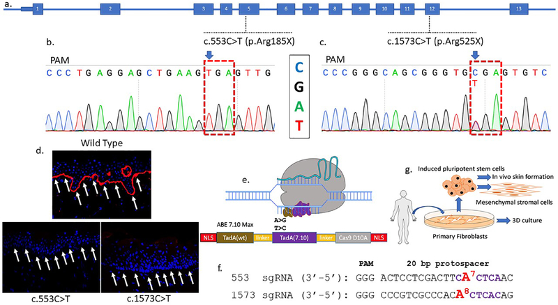
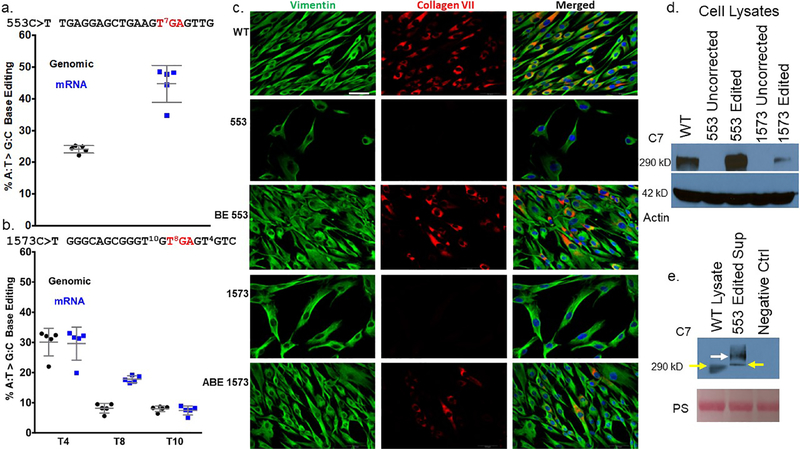
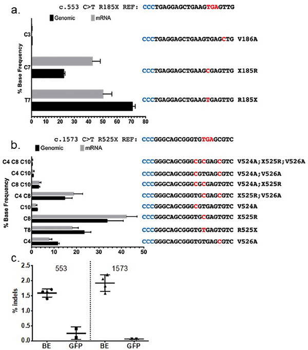
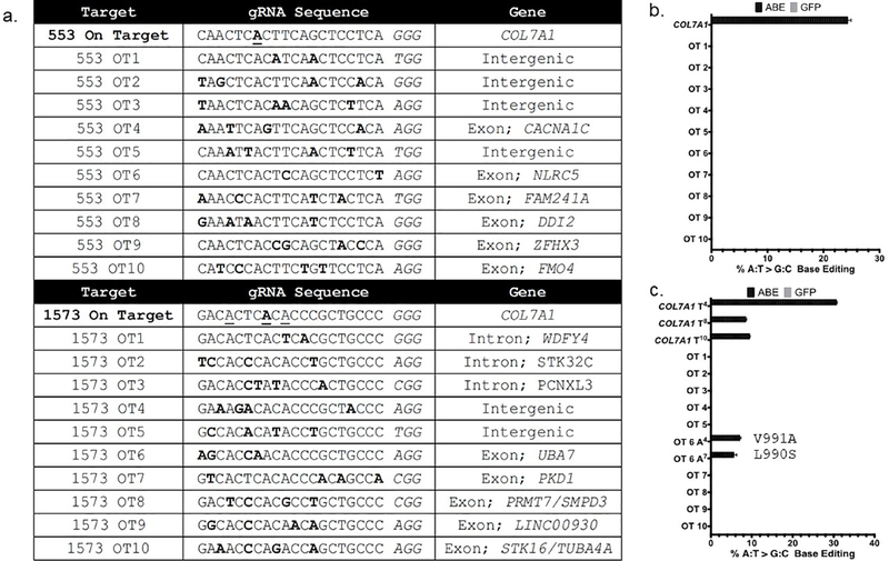
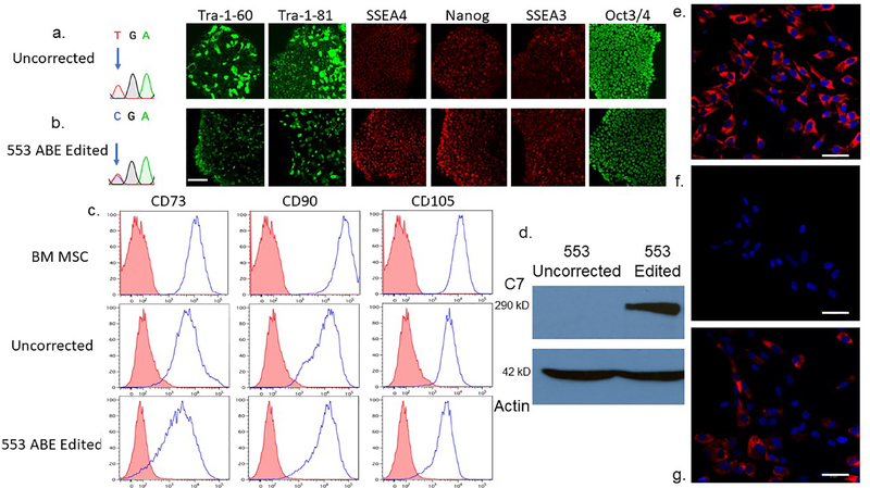
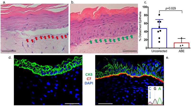
Genome editing represents a promising strategy for the therapeutic correction of COL7A1 mutations that cause recessive dystrophic epidermolysis bullosa (RDEB). DNA cleavage followed by homology-directed repair (HDR) using an exogenous template has previously been used to correct COL7A1 mutations. HDR rates can be modest, and the double-strand DNA breaks that initiate HDR commonly result in accompanying undesired insertions and deletions (indels). To overcome these limitations, we applied an A•T→G•C adenine base editor (ABE) to correct two different COL7A1 mutations in primary fibroblasts derived from RDEB patients. ABE enabled higher COL7A1 correction efficiencies than previously reported HDR efforts. Moreover, ABE obviated the need for a repair template, and minimal indels or editing at off-target sites was detected. Base editing restored the endogenous type VII collagen expression and function in vitro. We also treated induced pluripotent stem cells (iPSCs) derived from RDEB fibroblasts with ABE. The edited iPSCs were differentiated into mesenchymal stromal cells, a cell population with therapeutic potential for RDEB. In a mouse teratoma model, the skin derived from ABE-treated iPSCs showed the proper deposition of C7 at the dermal-epidermal junction in vivo. These demonstrate that base editing provides an efficient and precise genome editing method for autologous cell engineering for RDEB.


Conflict of interest statement

CONFLICT OF INTEREST.

G.A.N. has filed patents relating to base editor use.

D.R.L. is a consultant and cofounder of Editas Medicine, Pairwise Plants, and Beam Therapeutics, companies that use genome editing.

B.R.B. is cofounder of Tmunity.

Figures

Figure 1.
Figure 1.. COL7A1 base editing experimental design.
a COL7A1 exons are numbered and the mutations targeted in this study are shown. One patient harbors a homozygous C:G>T:A mutation at nucleotide position c.553 causing an arginine to stop codon mutation in exon 5 at amino acid residue 185. The second patient was diagnosed as a compound heterozygote with a c.1573 C>T mutation in exon 12 at amino acid position 525 leading to a premature termination codon. The second allele has a c.2005C>T (R669X) mutation. b and c Sanger sequencing chromatograms of primary fibroblast samples from homozygous RDEB patients with Arg>X mutations. The sequence shown in the chromatograms corresponds to the sgRNA sequence and the NGG protospacer adjacent motif (PAM) sequence is indicated (anti-sense). d Skin biopsy immunofluorescence. Full thickness punch skin biopsies were obtained from a healthy control and the c.553 C>T and c.1573 C>T RDEB patients. Each sectioned tissue was stained with an equivalent amount of a polyclonal anti-C7 antibody and analyzed by confocal microscopy. C7 stains in red and white arrows show the DEJ in wild type but not RDEB samples. e Adenine base editor architecture. N- and C-terminal nuclear localization signals flank the E. coli TadA wildtype (WT) and evolved adenine deaminases that are separated by an XTEN linker and fused to the S. pyogenes Cas9 D10A nickase. Following Cas9 binding, the deaminases can act on the single-stranded DNA displaced by the protospacer. f c.553 and c.1573 sgRNA sequences with the target base numbered and shown in red. Purple lettering shows the ABE activity window between nucleotides corresponding to positions 4–8 of the protospacer (counting the 5’ nucleotide as position 1). g RDEB primary fibroblasts and induced pluripotent stem cells (iPSC) were obtained and corrected by electroporation of base editor mRNA and targeting sgRNAs. Fibroblasts were used in 3D organotypic cultures in vitro. Corrected iPSC were used as a platform for mesenchymal stromal cell derivation and in vivo teratoma formation that gave rise to ectoderm derived skin.
Figure 2.
Figure 2.. c.553 COL7A1 base editing in RDEB primary cells.
a and b Quantification of DNA base editing by deep sequencing. Genomic DNA and mRNA were sequenced on an Illumina MiSeq to determine the frequency of A:T>G:C editing of a c.553 cells and b c.1573 cells. Values and error bars are the mean and standard deviation from five experimental replicates. sgRNA sequences for each mutation are shown and the red lettered ‘TGA’ represents the nonsense mutation codon. Superscript numbers represent the target base for mutation correction or bystander editing and are numbered relative to the 5’ start of the sgRNA. c Immunofluorescence of primary fibroblasts. White boxes embedded in the images on the left identify samples in that row. Labels at top identify the antigen stained for in that column. Edited and uncorrected cells were stained simultaneously with equivalent amounts of anti-vimentin and anti-collagen type VII polyclonal antibodies. The images for each fluorescent channel were merged with DAPI nuclear stain that is shown at right. Images are representative of three independent experiments. Scale bar=50 μm (lower right in WT vimentin image). d and e C7 Western blotting. d Cell lysates from uncorrected 553 and 1573 cells were analyzed in parallel with base edited 553 and 1573 cells using a polyclonal anti-C7 antibody. Wild type (WT) cells are from a healthy donor. The C7 lane shows the ~290 kD C7 band and actin was used as a loading control. e Secreted C7 from cell supernatant. C7 was detected in the supernatant of c.553 edited cells that were plated in serum free media. WT is a healthy donor lysate sample and negative control (ctrl) is from cells that have a COL7A1 mutation that inactivates the gene. Pro-collagen type VII is shown at 290 kD with yellow arrows. A larger molecular weight species is shown with a white arrow representing the non-reduced C7 ultrastructural trimeric polypeptide. The Ponceau S loading control is shown and is labeled ‘PS.’
Figure 3.
Figure 3.. Allele frequencies following ABE treatment.
a COL7A1 c.553 C>T allelic analysis. The mean percentage of each individual edited DNA and mRNA sequence is shown. Amino acid changes and base alterations corresponding to the observed outcomes are shown at right (3’−5’) with PAM (anti-sense) colored in blue. b Allele distribution in c.1573 C>T cells following base editing. The mutation reference sequence is shown at top. A bar graph of edited cells and the frequency of alleles observed in genomic or mRNA derived cDNA is shown. At right are the individual allele sequences (3’−5’) identified following base editing, with altered bases highlighted in red and PAM in blue. Allelic variants occurring at less than 0.2% frequency, approximately the frequency of sequencing errors, were not included in the analysis and therefore the values for the graphs are <100%. c COL7A1 locus insertions and deletions from base editing. The percentage of deep sequencing reads with indels are shown for treated (BE) and control cells transfected with GFP for the c.553 and c.1573 mutation genomic DNA, respectively. Data for each are from 5 replicates and mean and standard deviation are graphed.
Figure 4.
Figure 4.. Off target analysis.
a CRISPOR identified off target (OT) sites for the c.553 and c.1573 guide RNAs and associated genomic OT loci. Letters in bold represent mismatches between the on and off target sites. The underlined bases in the on target sequences are those capable of being edited by ABE. b and c High throughput sequencing to assess OT base editing in c.553 or c.1573 cells. Data are from three independent biological replicates of edited or control cells treated with GFP mRNA and error bars show standard deviation.
Figure 5.
Figure 5.. RDEB induced pluripotent stem cell base editing and directed mesenchymal stromal cell differentiation and characterization.
a and b iPSC editing. Sanger chromatogram of uncorrected c.553 C>T; R553X iPSC and a representative base edited clone are shown with arrow showing the mutant/target base. B Pluripotency immunofluorescence marker analysis. Antibodies against the pluripotency markers: podocalyxin TRA-1–60/TRA-1–81, Stage-specific embryonic antigen 4 (SSEA4), SSEA3, Nanog and OCT3/4 were used to detect expression levels. c Mesenchymal stromal cell characterization. Adult bone marrow from normal donors or iPSC derived MSCs were analyzed for the cell surface markers CD73, CD90, and CD105. The isotype staining control peaks are shown in pink. d iPSC derivative MSC Western blot analysis. MSCs derived from uncorrected c.553 C>T iPSCs were compared with those corrected by ABE in a Western blot using a polyclonal anti-C7 antibody. A ~290 kD band is shown with a 42 kD actin loading control below. e-g C7 immunostaining of chamber slides containing e wild type, bone marrow derived MSCs, f unedited 553 patient RDEB iPSC-derived MSCs, and g ABE edited 553 iPSC derivative MSCs. All cells were stained at the same time with the equivalent amount of polyclonal anti-C7 primary and secondary antibodies. Scale bar=50 μm.
Figure 6.
Figure 6.. In vitro three dimensional organotypic culture and in vivo expression of type VII collagen.
a-c 3D organotypic culture. Uncorrected or base edited fibroblasts from c.553 patient cells were layered with transformed keratinocytes on a supportive matrix and stained by hematoxylin and eosin. a Uncorrected fibroblast cell 3D culture. The red arrows show the detached epithelial layer due to a fragile dermal epidermal junction that is structurally deficient when the culture contains uncorrected fibroblasts. b ABE corrected cell 3D culture. Improved structural integrity without epithelial layer detachment (green arrows) was observed when ABE corrected cells with restored C7 were employed. c Epithelium detachment quantification. The percent of detached epithelia observed by microscopy and scored by an expert was quantified for eight and three experimental replicates for uncorrected and ABE corrected fibroblasts, respectively. Mean and standard deviation are graphed and p value from Student’s t-test are shown. d-e In vivo teratoma. iPSC were injected into immune deficient mice and representative images of d COL7A1 defective and e base edited iPSC teratoma derived skin equivalents are shown. Both RDEB null and base edited samples were stained with polyclonal anti-human type VII collagen (red) and anti-human cytokeratin (CK5; green) antibodies as well as DAPI nuclear stain. Scale bars=50 μm. At inset, lower right is the base edited nucleotide that was observed following amplification of human COL7A1 DNA from the in vivo teratoma tissue.

References

    1. Bonafont J, Mencia A, Garcia M, Torres R, Rodriguez S, Carretero M, et al. Clinically Relevant Correction of Recessive Dystrophic Epidermolysis Bullosa by Dual sgRNA CRISPR/Cas9-Mediated Gene Editing. Mol Ther 2019;27(5):986–98. - PMC - PubMed
    1. Bornert O, Kuhl T, Bremer J, van den Akker PC, Pasmooij AM, Nystrom A. Analysis of the functional consequences of targeted exon deletion in COL7A1 reveals prospects for dystrophic epidermolysis bullosa therapy. Mol Ther 2016;24(7):1302–11. - PMC - PubMed
    1. Chen M, Costa FK, Lindvay CR, Han YP, Woodley DT. The recombinant expression of full-length type VII collagen and characterization of molecular mechanisms underlying dystrophic epidermolysis bullosa. J Biol Chem 2002;277(3):2118–24. - PubMed
    1. Christiano AM, Anton-Lamprecht I, Amano S, Ebschner U, Burgeson RE, Uitto J. Compound heterozygosity for COL7A1 mutations in twins with dystrophic epidermolysis bullosa: a recessive paternal deletion/insertion mutation and a dominant negative maternal glycine substitution result in a severe phenotype. Am J Hum Genet 1996;58(4):682–93. - PMC - PubMed
    1. Clement K, Rees H, Canver MC, Gehrke JM, Farouni R, Hsu JY, et al. CRISPResso2 provides accurate and rapid genome editing sequence analysis. Nat Biotechnol 2019;37(3):224–6. - PMC - PubMed

Publication types

MeSH terms